Jump to content
In this section we have spot diagnoses posted on a daily basis since June 2010, now over 4000! You can review the archived cases and read the suggested diagnoses by users and the final comment by the contributors.
Case are uploaded each week day by 10 am UK time with the correct diagnosis will generally be posted at 8 pm UK time. Why not view the most recent spot diagnosis and proffer a diagnosis?

Case Number : Case 2761 - 4 February 2021 Posted By: Saleem Taibjee

Please read the clinical history and view the images by clicking on them before you proffer your diagnosis.
Submitted Date :
   (0 reviews)

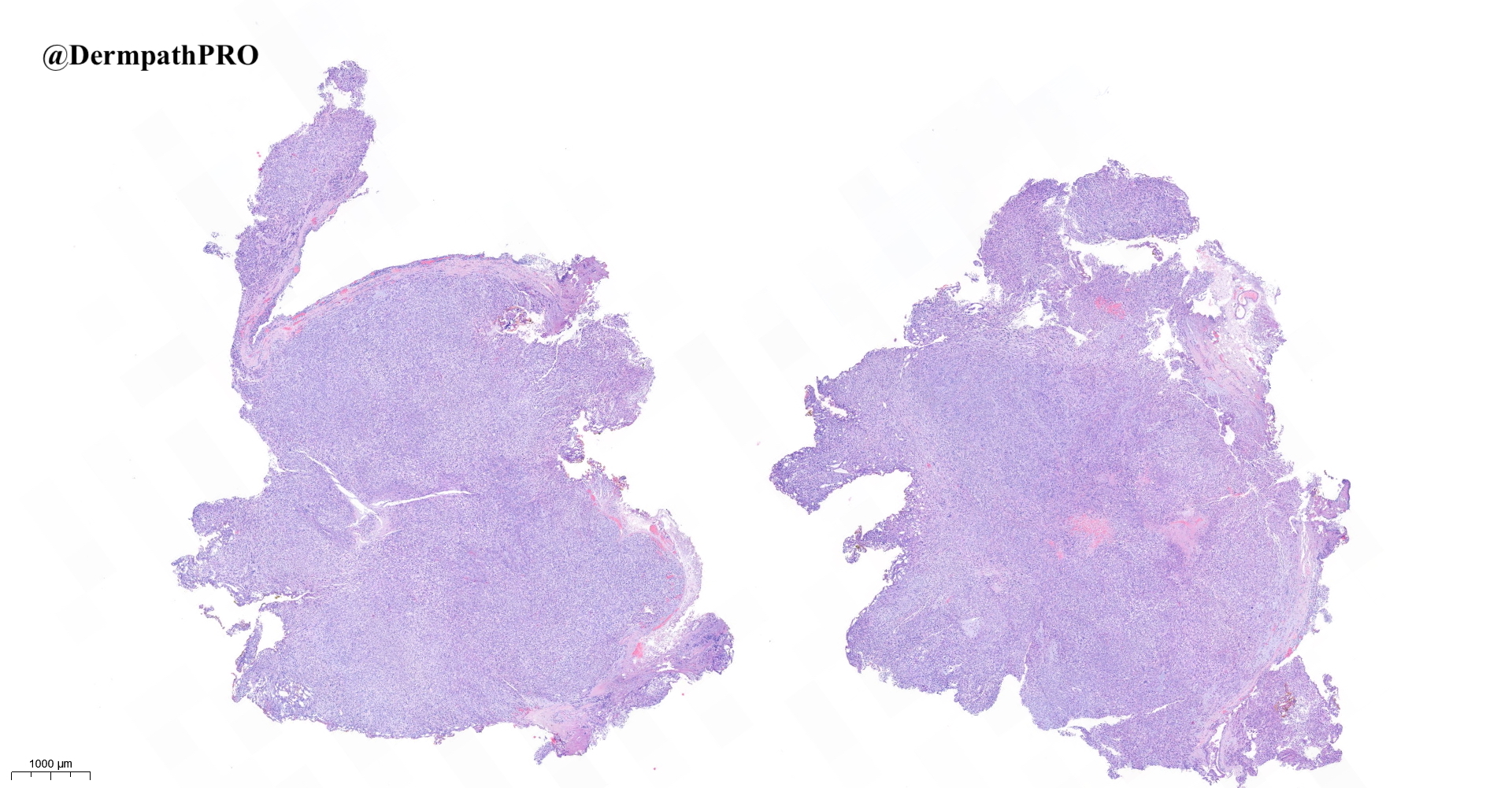
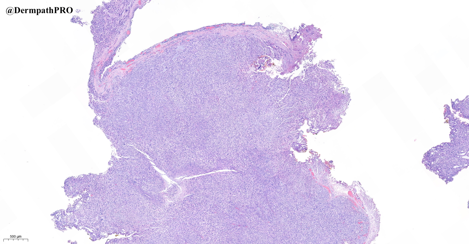
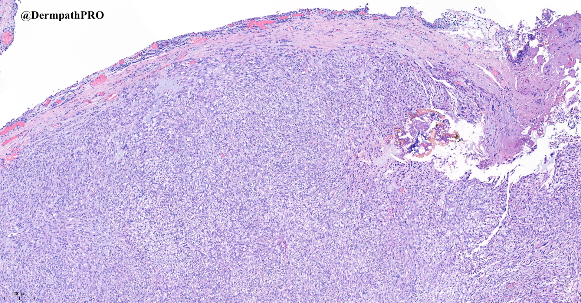
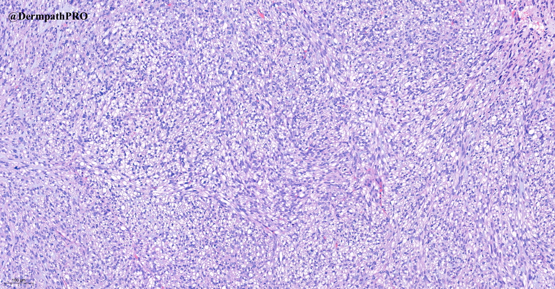
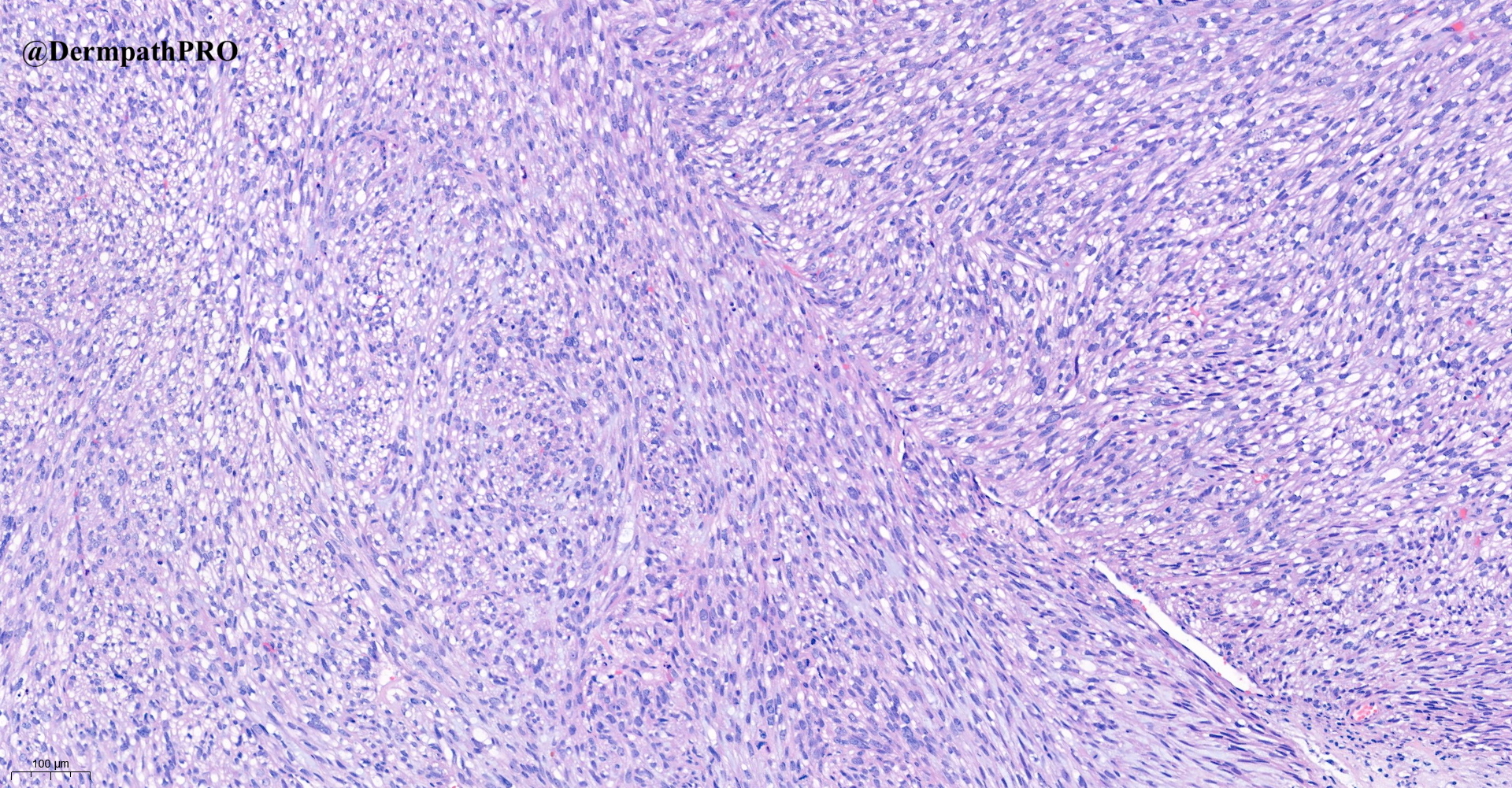
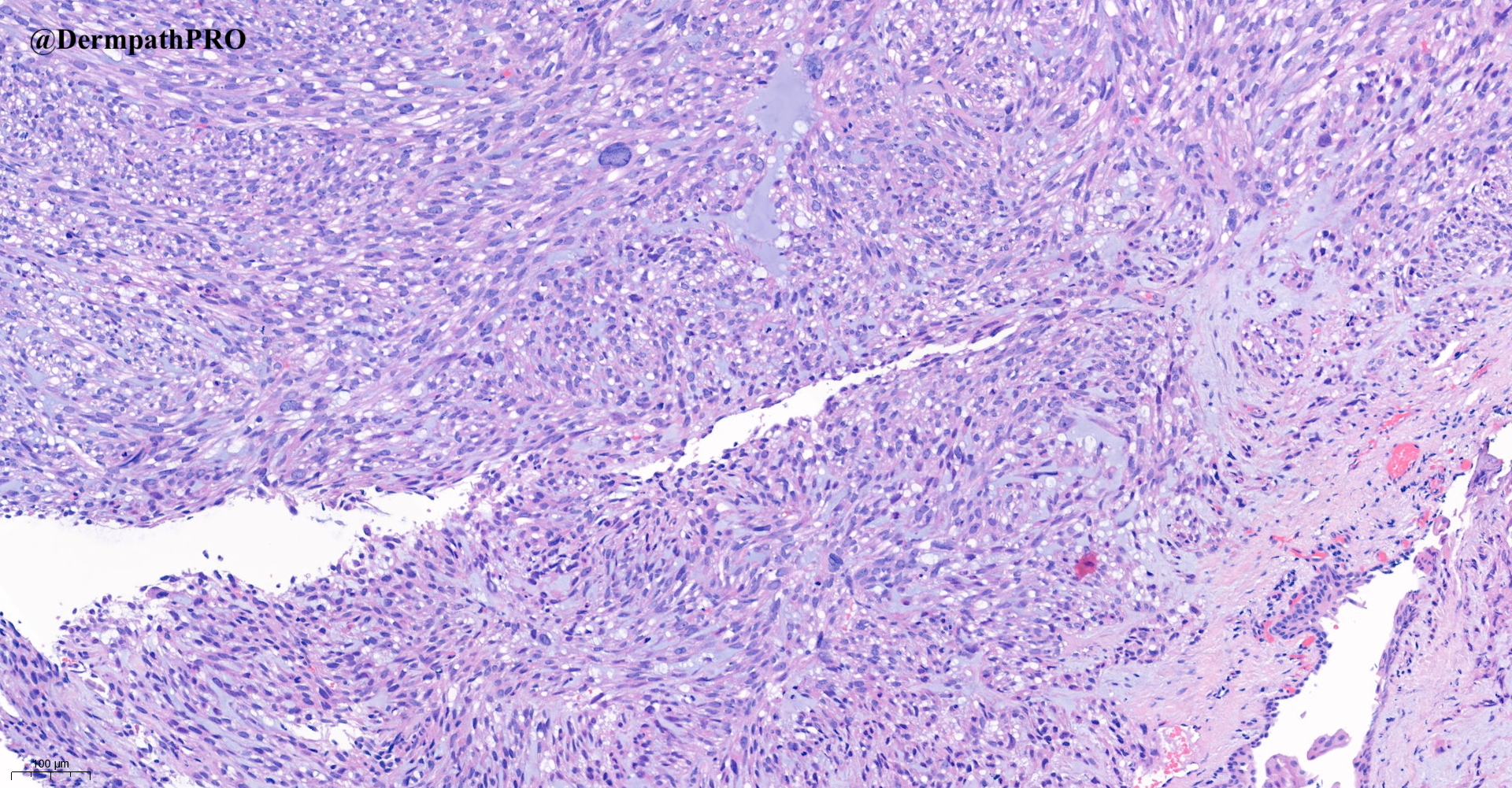
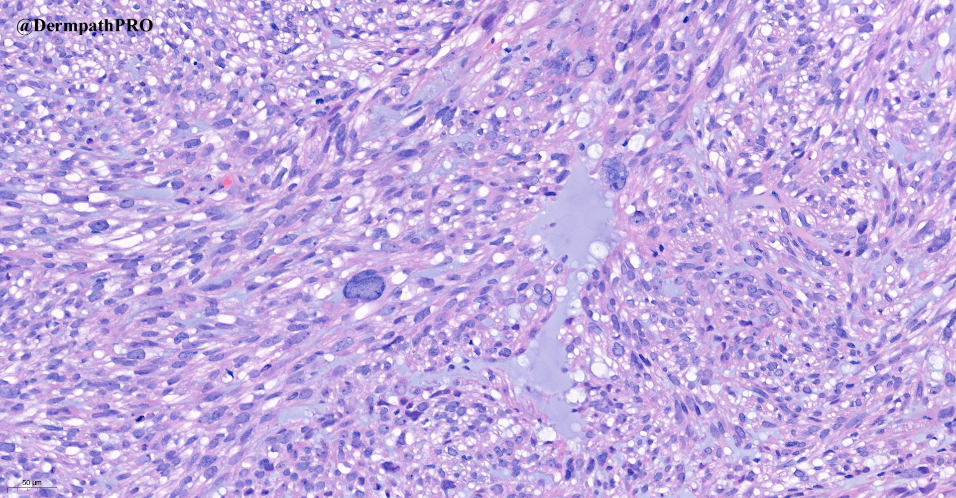
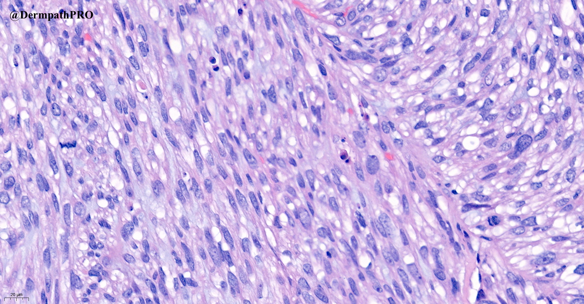
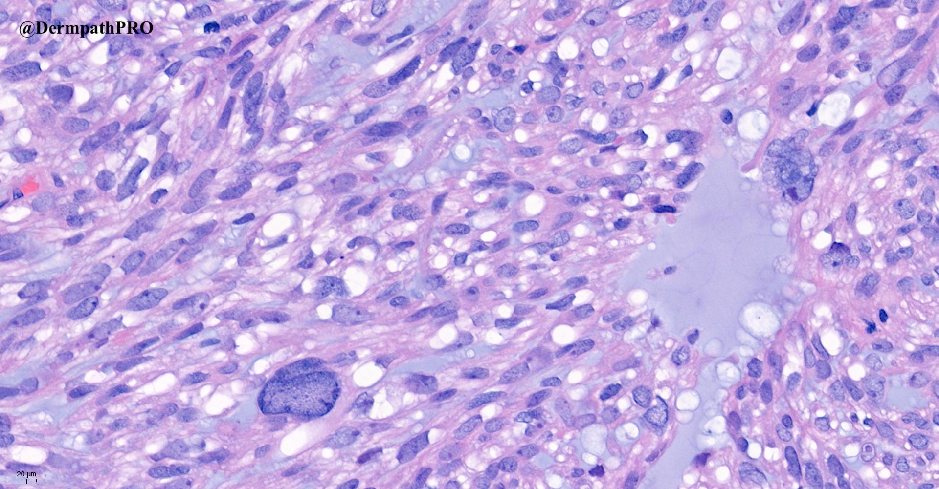
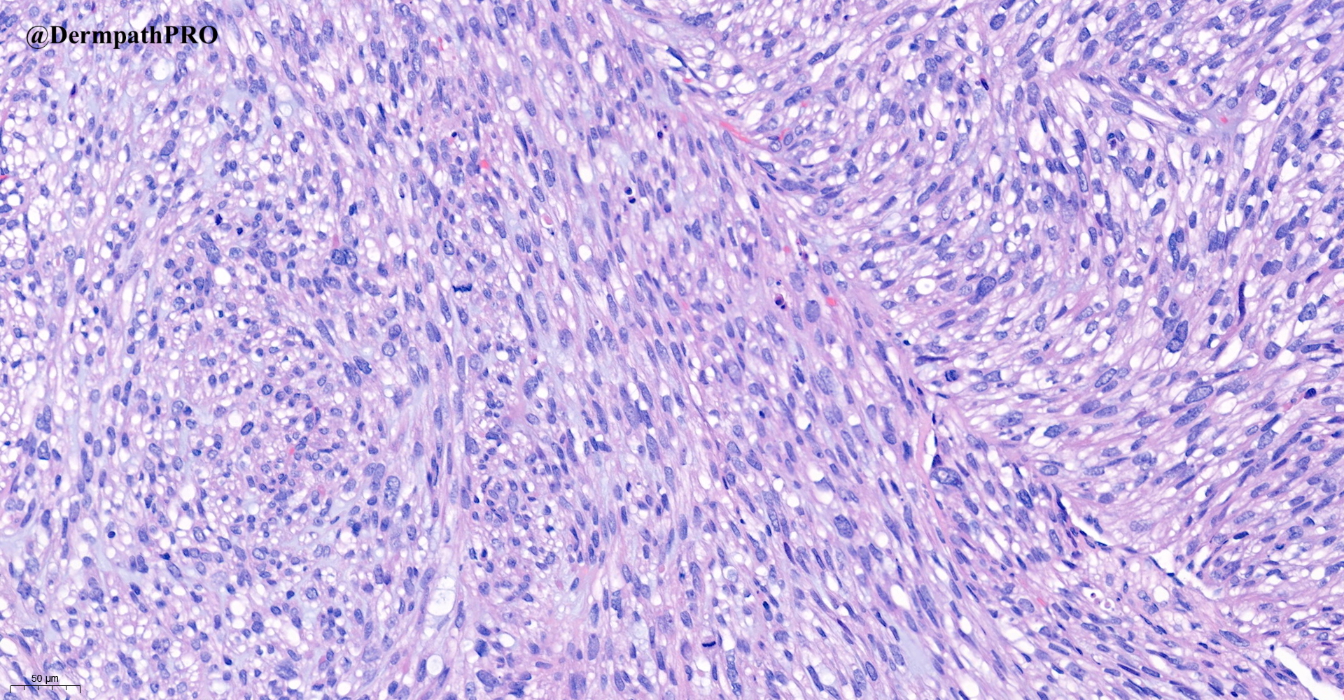
86F, lesion right lower eyelid.

  Report Record

User Feedback


Richard Logan

Posted

A mitotically active, myxoid spindle cell tumour showing storiform patterning and monster cells.  At this age it is much more likely to be a malignant tumour than a reactive process such as nodular fasciitis. How about something rare like a myxoid liposarcoma?

Obviously immunohistochemistry is vital here.

Share this comment


Link to comment
share_externally

Leiomyosarcoma ( ?metastatic ). 
Also thought of Pecoma, but too cigar shape nucleus, as expected in smooth muscle tumors. 

Share this comment


Link to comment
share_externally

Anurag gupta

Posted

Tumor shows fascicles and storiform pattern with paranuclear vacuoles and cigar shaped nuclei. Above features suggest leiomyosarcoma.

Share this comment


Link to comment
share_externally

Saleem Taibjee

Posted

Here is the pankeratin (MNF116) stain

S21-189_5-2-2021_08-43-04_2.0x MNF116 labelled.jpg

S21-189_5-2-2021_08-43-04_20.0x MNF116  labelled.jpg


Share this comment


Link to comment
share_externally

Carmen Lisievici

Posted

Considering the IHC, I would start thinking of something more in the lines of a sarcomatoid carcinoma, maybe even a squamous cell carcinoma.


Share this comment


Link to comment
share_externally

Eman El-Nabarawy

Posted

25 minutes ago, Carmen Lisievici said:

Considering the IHC, I would start thinking of something more in the lines of a sarcomatoid carcinoma, maybe even a squamous cell carcinoma.


Agree.

Share this comment


Link to comment
share_externally

Alex-Ventura-Leon

Posted

Despite PanK positivity I still favor Leiomyosarcoma. Of course we need more IHC (Desmin, p40, SMA, Sox10, CD10)

Share this comment


Link to comment
share_externally

Saleem Taibjee

Posted

OK, well done. So I didn't put you off the scent with the keratin stain. Desmin was positive (patchy), H-caldesmon strongly and diffusely positive (see below).

This was indeed metastatic leiomyosarcoma.

The case was brought to me by one of the junior pathologists in our team. My first thought on encountering the slide was to suggest to review the patient's previous medical history (it is amazing how this is frequently forgotten), and lo-and-behold it transpired that the patient was already known to Oncology with metastatic leiomyosarcoma!

Hence, I suggested a very limited initial immunopanel of pankeratin, desmin, and S100 (the latter was negative). Of course, it is interesting to see how keratin stains can be misleading, we know the pitfall that keratin can sometimes be positive in a variety of situations including melanoma, angiosarcoma. But this is the first time I have come across this with leiomyosarcoma.

BW, Saleem

S21-189_5-2-2021_08-39-28_2.0x Desmin labelled.jpg

S21-189_5-2-2021_08-39-28_10.0x Desmin labelled.jpg

S21-189_5-2-2021_08-33-35_2.0x H-caldesmon labelled.jpg


Share this comment


Link to comment
share_externally


Join the conversation

You can post now and register later. If you have an account, sign in now to post with your account.

Guest
Add a comment...

×   Pasted as rich text.   Paste as plain text instead

  Only 75 emoji are allowed.

×   Your link has been automatically embedded.   Display as a link instead

×   Your previous content has been restored.   Clear editor

×   You cannot paste images directly. Upload or insert images from URL.

Loading...

×
×
  • Create New...