Jump to content
In this section we have spot diagnoses posted on a daily basis since June 2010, now over 4000! You can review the archived cases and read the suggested diagnoses by users and the final comment by the contributors.
Case are uploaded each week day by 10 am UK time with the correct diagnosis will generally be posted at 8 pm UK time. Why not view the most recent spot diagnosis and proffer a diagnosis?

Case Number : Case 2762 - 5 February 2021 Posted By: Dr. Richard Carr

Please read the clinical history and view the images by clicking on them before you proffer your diagnosis.
Submitted Date :
   (0 reviews)

M80. “Spot diagnosis”


  Report Record

User Feedback


Richard Logan

Posted

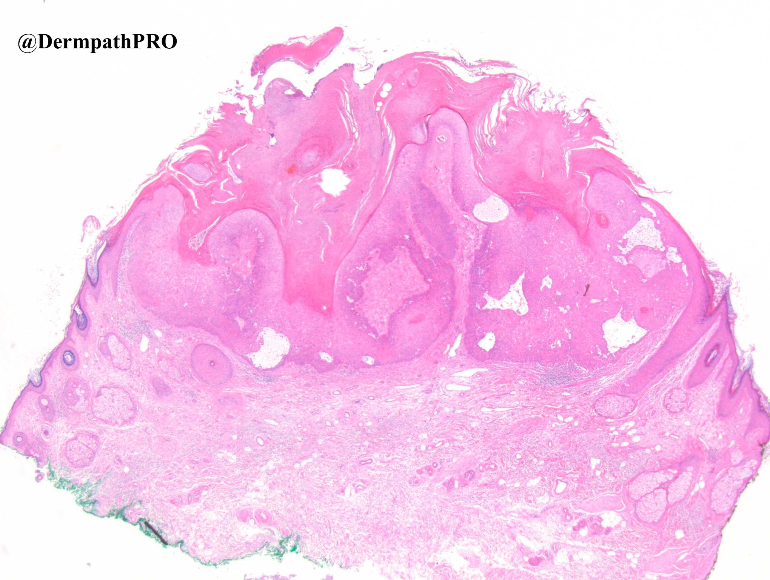
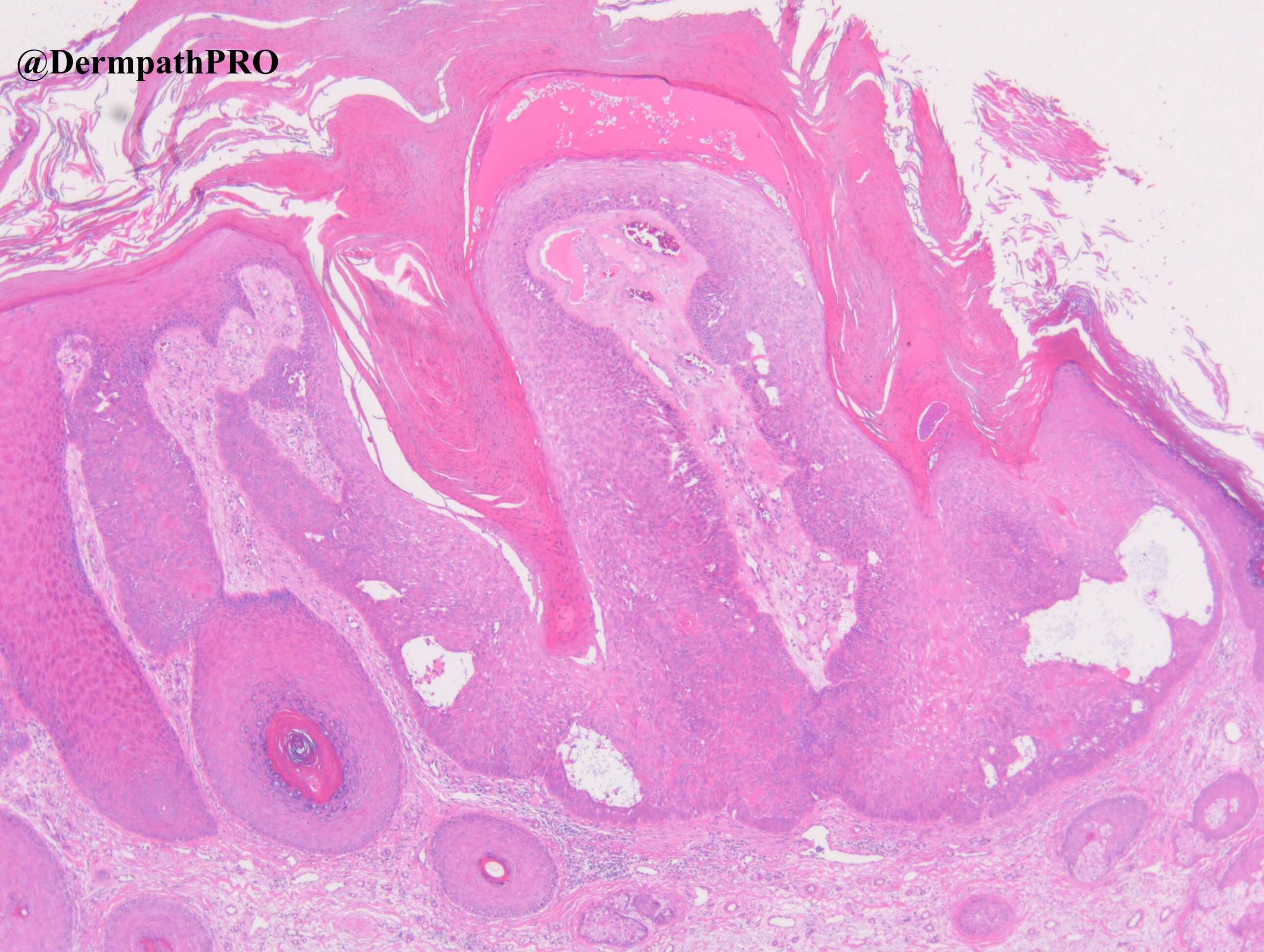
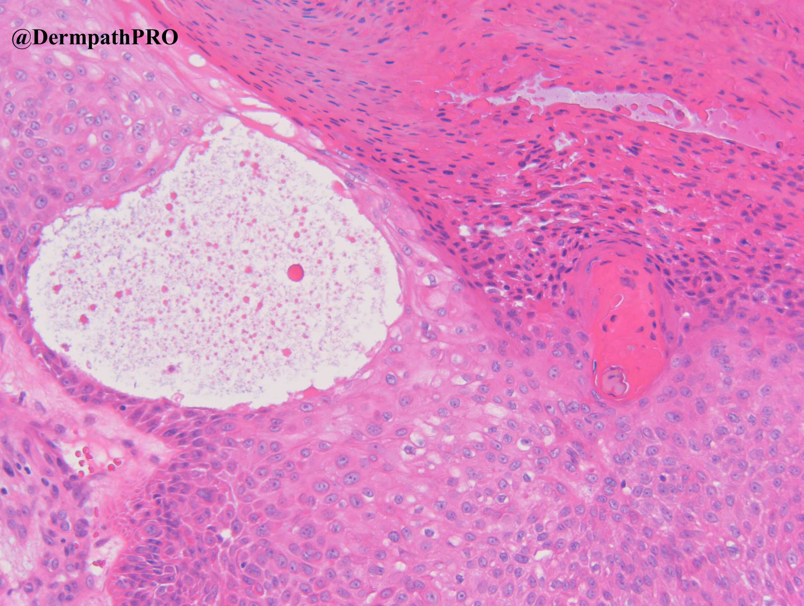
Using Dr. Carr's proposed scoring system to distinguish between keratoacanthoma and follicular squamous carcinoma (see case 2707 posted 20th November 2020) I scored this at 12/32 (in the absence of EVG stain).  That pushes me towards a diagnosis of follicular squamous carcinoma.

Share this comment


Link to comment
share_externally

Meenakshi Batrani

Posted

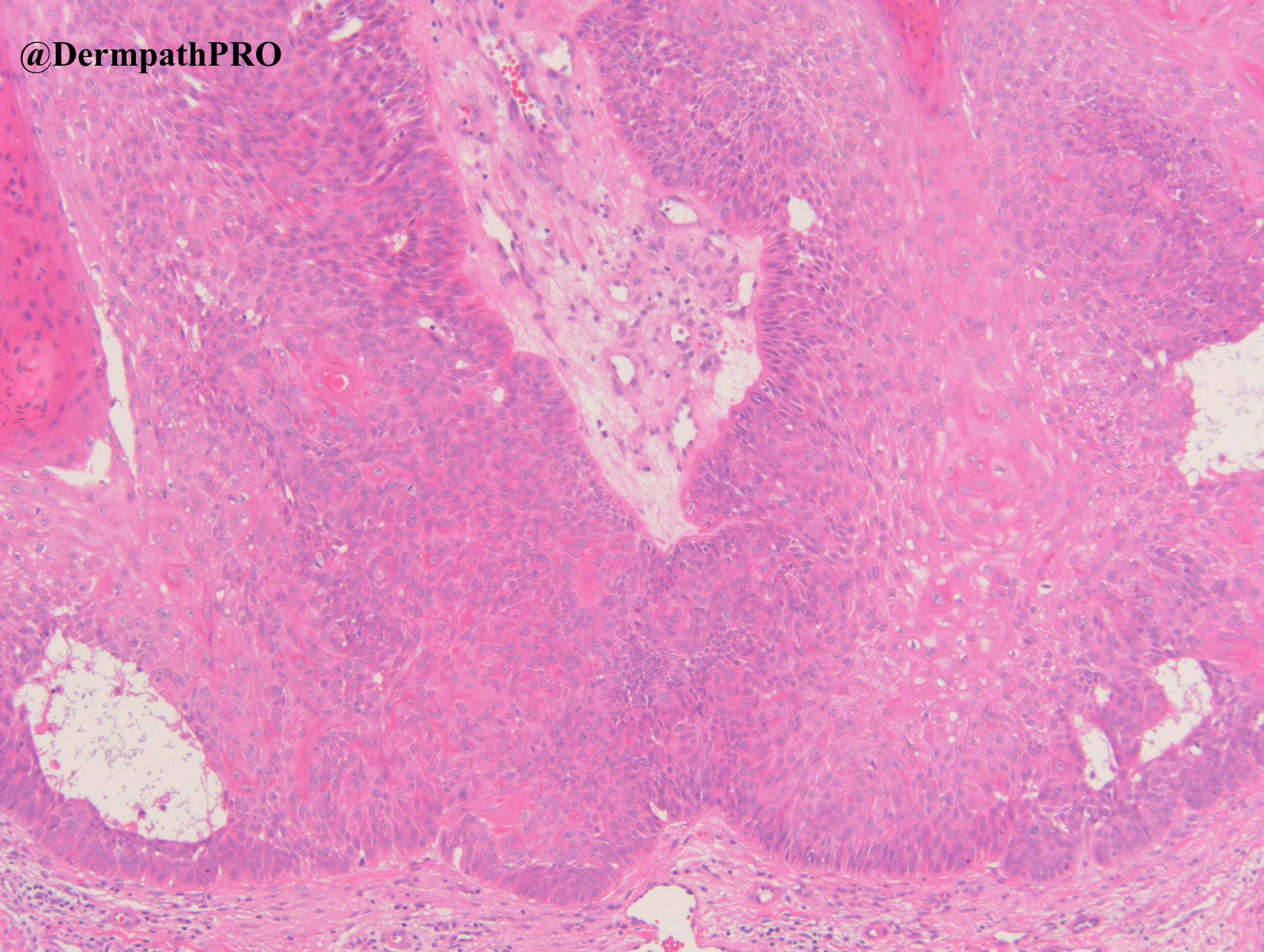
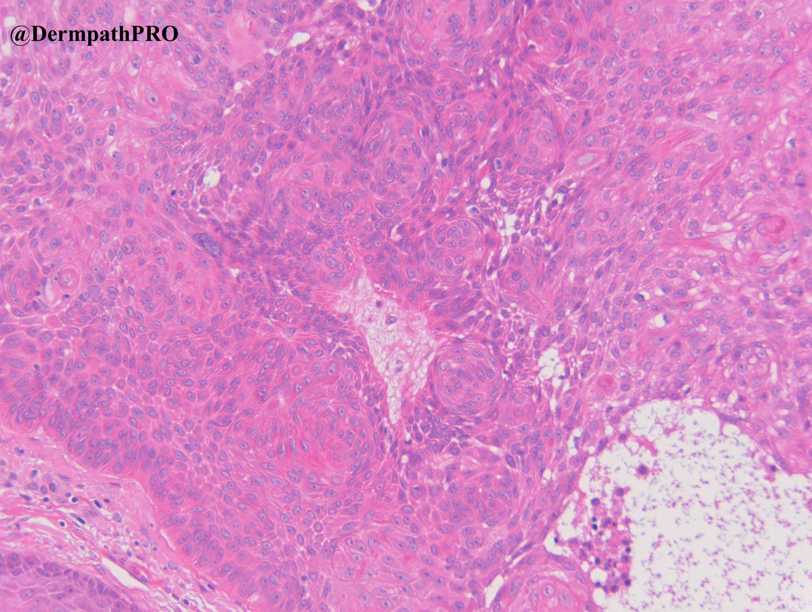
I see multiple squamous eddies, making me think of inverted follicular keratosis. 

Share this comment


Link to comment
share_externally

Eman El-Nabarawy

Posted

Keratoacanthoma-like inverted follicular keratosis.

Share this comment


Link to comment
share_externally

Alex-Ventura-Leon

Posted

Follicular SCC for me. Pushing borders

Share this comment


Link to comment
share_externally

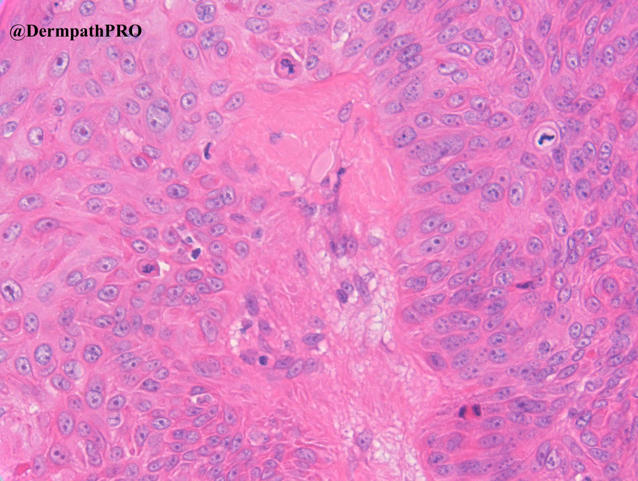
It’s a very low grade malignancy, of course. And an inverted follicular keratosis pattern could fit better an irritated SK...but mucin secretion and some atypical mitosis make me think of fSCC, pushing borders. 

Share this comment


Link to comment
share_externally

Dr. Richard Carr

Posted

Good discussion which highlights the differential nicely. What about the IHC interpretation?

Share this comment


Link to comment
share_externally

Alex-Ventura-Leon

Posted

P53 is wild type (that would favor KA)

P16 is null type (that would favor SCC)

No intraepithelial collagen trapping (that would favor SCC)

At least that is my intrepretation

Share this comment


Link to comment
share_externally

Dr. Richard Carr

Posted

Okay thanks for adding these additional comments. 

p16 we'd expect to show a nice checkerboard / mosaic pattern in a proliferative lesion that appears to be from the central face of an elderly man. I did not show but there was moderate solar elastosis. We have quite a bit of lesion and it's a complete null pattern. For me this is incompatible with a benign diagnosis although one has to be aware of context a non-inflamed benign lesion on non-sun-damaged skin for example might have constitutively low expression of p16 (like some completely banal naevi).

p53 here to me is markedly up-regulated and I suspect this is wild type protein trying to compensate for the loss of p16 function. I'd probably interpret this pattern as aberrantly high rather than a pure wild type / reactive pattern. 

Given the null p16 this sways me towards a follicular SCC, the completely circumscript borders and lack of entrapment on EVG for me are compatible with an in situ lesion (for practical purposes i.e. considered to lack metastatic potential).

In this case the differential is more IFK v's FSCC with IFK-like features rather than KA so the scoring is not really appropriate. An IFK or SEBK with some KA-like features will give  high scores. I have to say this case is highly challenging without the IHC. With the IHC we can make a pretty confident diagnosis.

Share this comment


Link to comment
share_externally

Meenakshi Batrani

Posted

17 hours ago, Dr. Richard Carr said:

Okay thanks for adding these additional comments. 

p16 we'd expect to show a nice checkerboard / mosaic pattern in a proliferative lesion that appears to be from the central face of an elderly man. I did not show but there was moderate solar elastosis. We have quite a bit of lesion and it's a complete null pattern. For me this is incompatible with a benign diagnosis although one has to be aware of context a non-inflamed benign lesion on non-sun-damaged skin for example might have constitutively low expression of p16 (like some completely banal naevi).

p53 here to me is markedly up-regulated and I suspect this is wild type protein trying to compensate for the loss of p16 function. I'd probably interpret this pattern as aberrantly high rather than a pure wild type / reactive pattern. 

Given the null p16 this sways me towards a follicular SCC, the completely circumscript borders and lack of entrapment on EVG for me are compatible with an in situ lesion (for practical purposes i.e. considered to lack metastatic potential).

In this case the differential is more IFK v's FSCC with IFK-like features rather than KA so the scoring is not really appropriate. An IFK or SEBK with some KA-like features will give  high scores. I have to say this case is highly challenging without the IHC. With the IHC we can make a pretty confident diagnosis.

Thanks Dr. Carr for posting these challenging cases and the relevant interpretations. It is a great learning for us. 

Share this comment


Link to comment
share_externally

DR NADINE BURKE

Posted

ive had a go with the IHC-it is really helpful and works!

Share this comment


Link to comment
share_externally


Join the conversation

You can post now and register later. If you have an account, sign in now to post with your account.

Guest
Add a comment...

×   Pasted as rich text.   Paste as plain text instead

  Only 75 emoji are allowed.

×   Your link has been automatically embedded.   Display as a link instead

×   Your previous content has been restored.   Clear editor

×   You cannot paste images directly. Upload or insert images from URL.

Loading...

×
×
  • Create New...