Jump to content
In this section we have spot diagnoses posted on a daily basis since June 2010, now over 4000! You can review the archived cases and read the suggested diagnoses by users and the final comment by the contributors.
Case are uploaded each week day by 10 am UK time with the correct diagnosis will generally be posted at 8 pm UK time. Why not view the most recent spot diagnosis and proffer a diagnosis?

Case Number : Case 2766 - 11 February 2021 Posted By: Saleem Taibjee

Please read the clinical history and view the images by clicking on them before you proffer your diagnosis.
Submitted Date :
   (0 reviews)

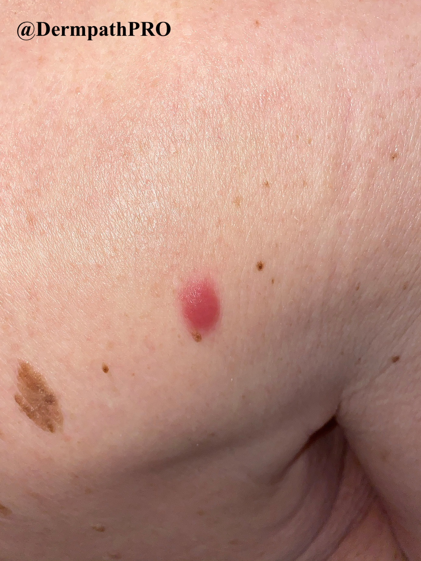
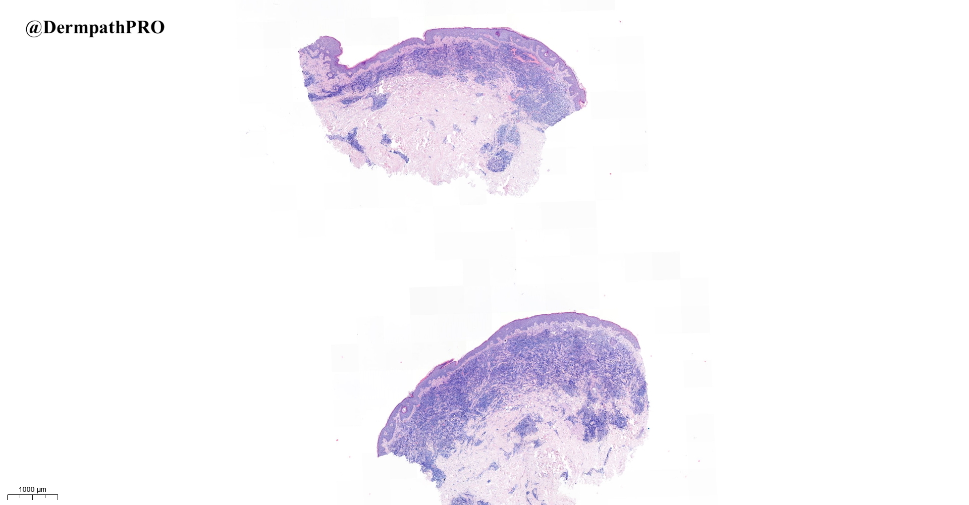
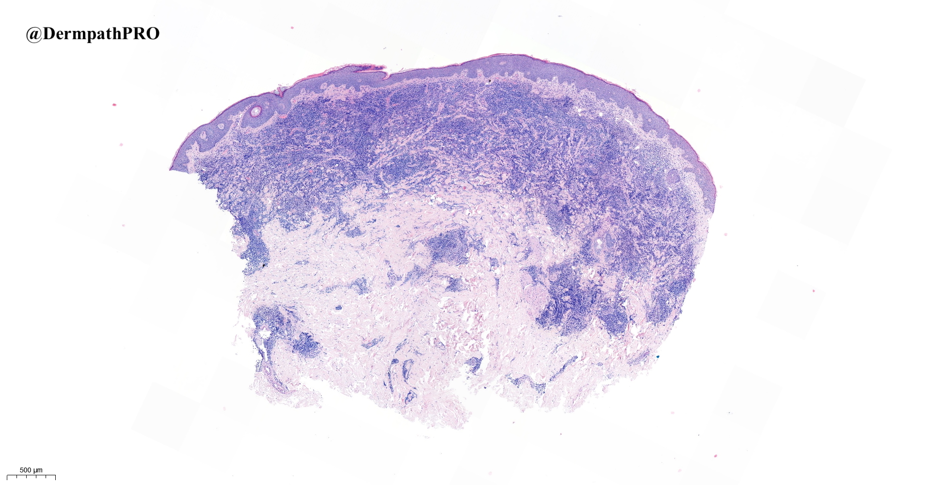
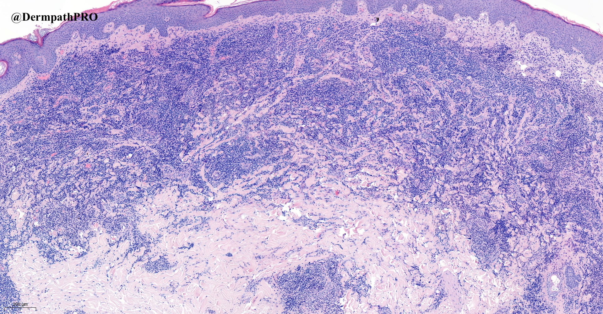
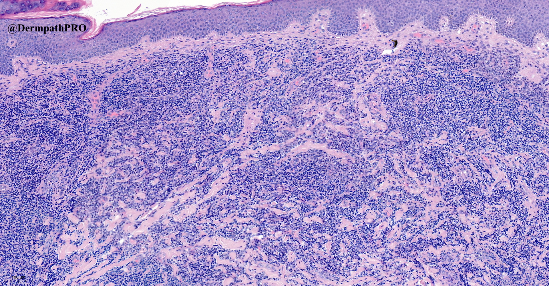
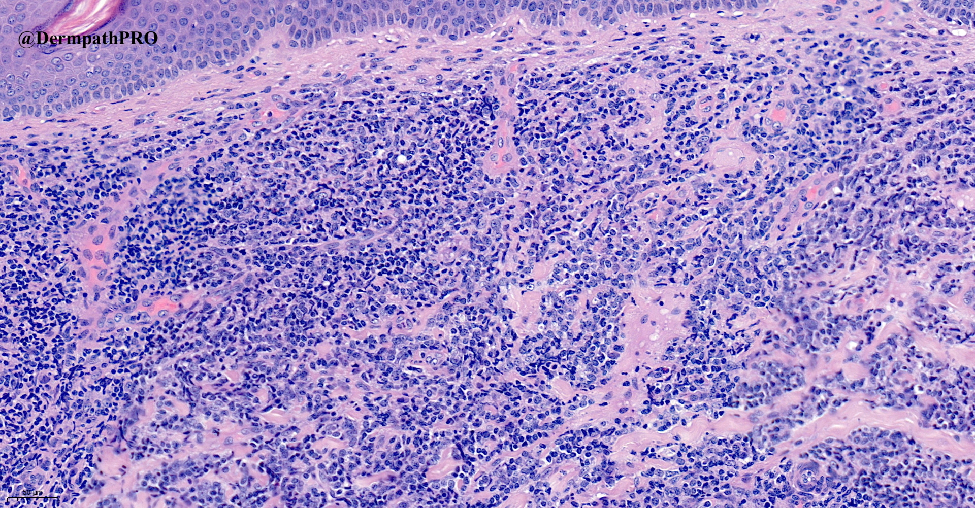
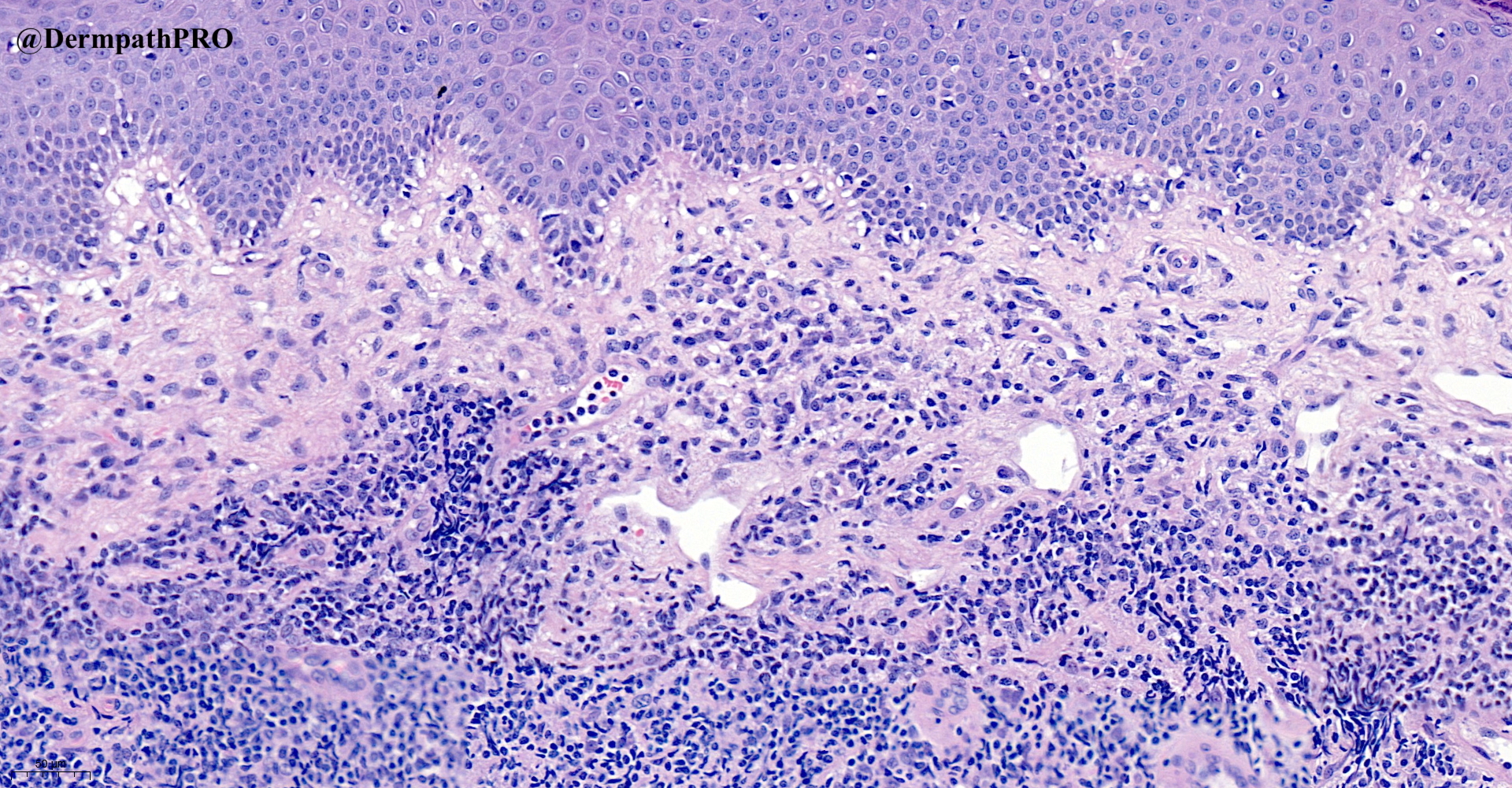
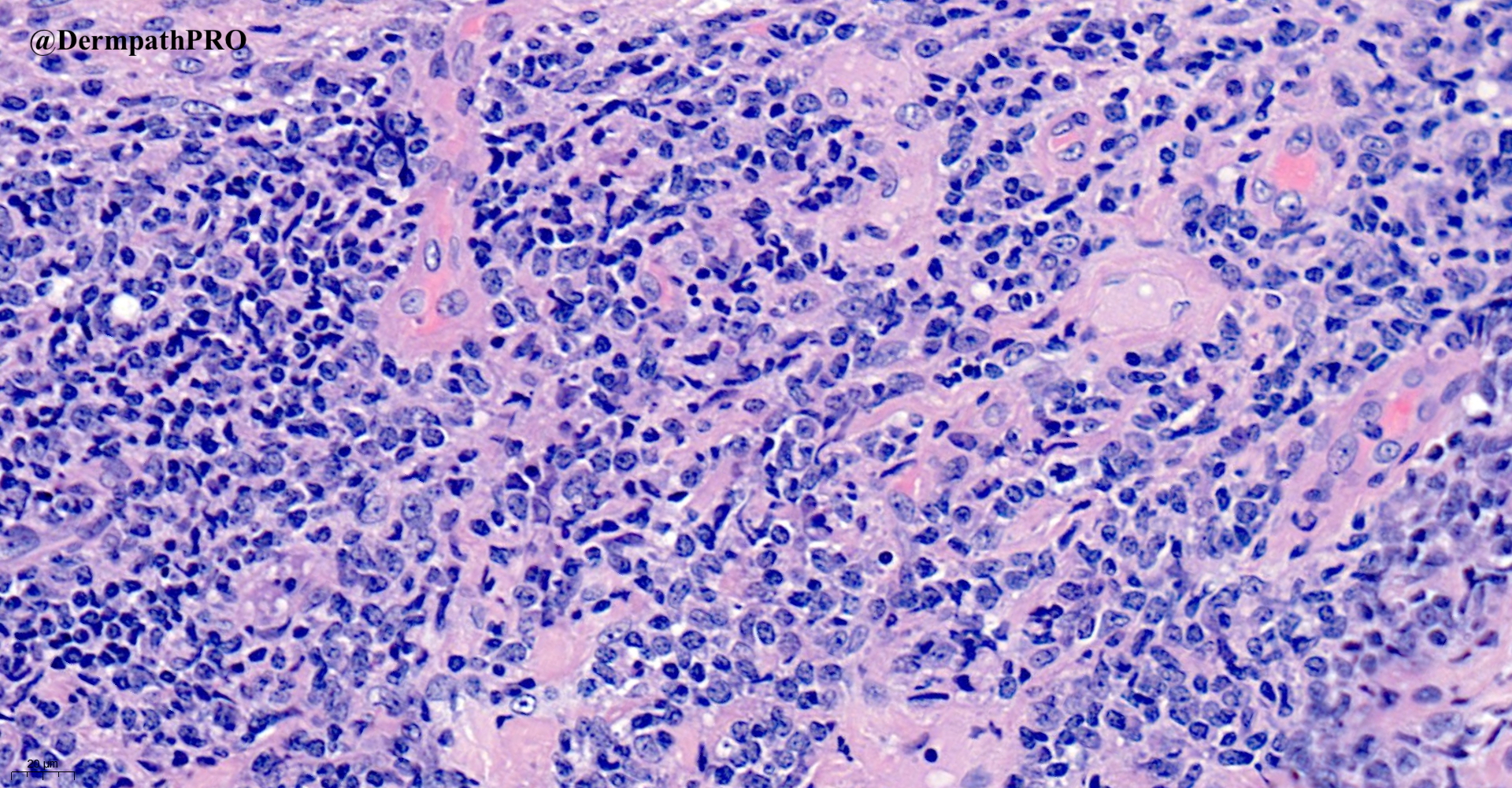
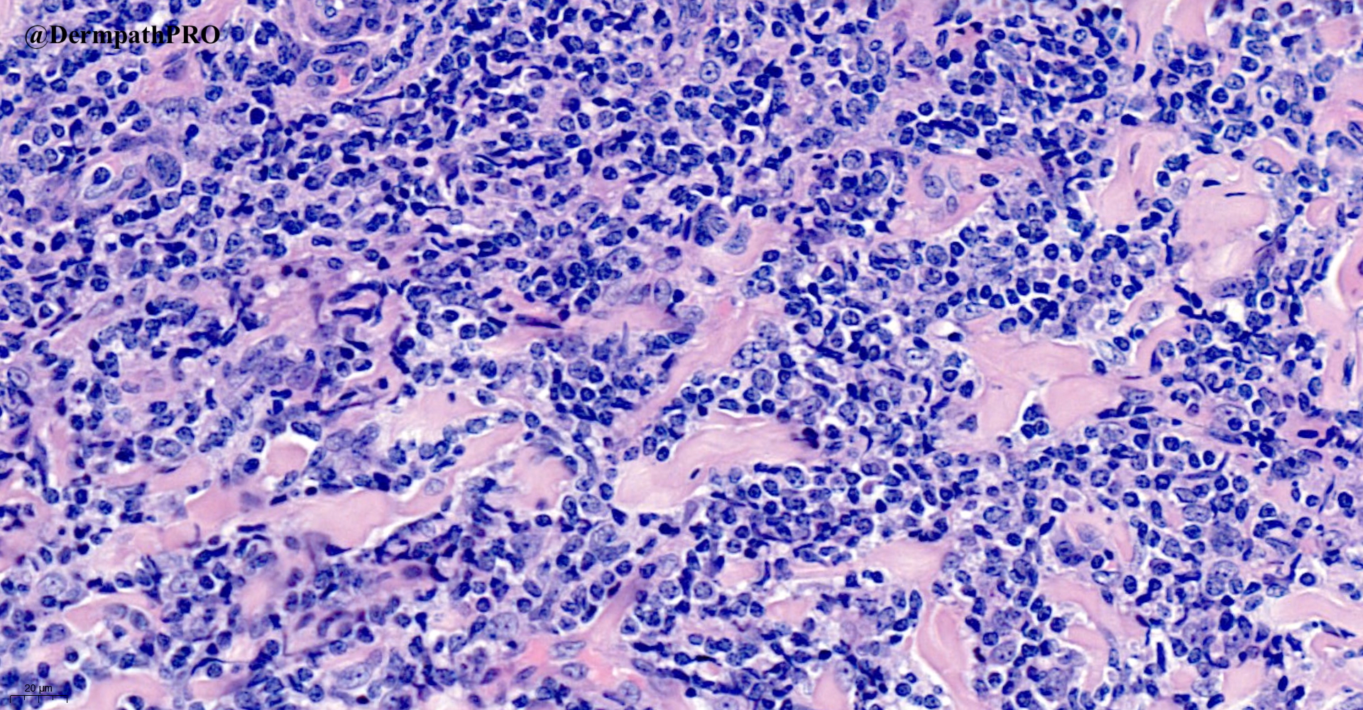
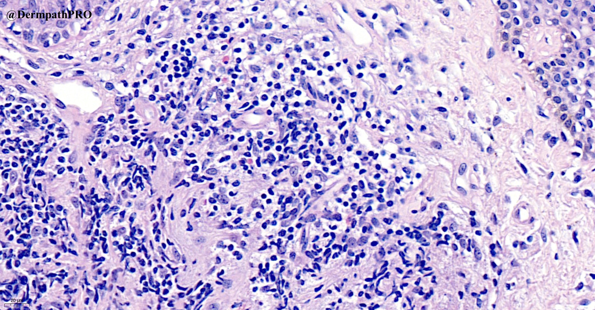
85F, punch biopsy right lateral chest – solitary red nodule, duration uncertain


  Report Record

User Feedback


Richard Logan

Posted

B-cell lymphoma pattern.  However, there is quite a lot of collagen dissection, so I would also wonder about leukaemia cutis or metastasis.  Clinical correlation and immunohistochemistry crucial.

Share this comment


Link to comment
share_externally

Saman Fatah

Posted

Primary cutaneous low grade B-cell lymphoma particularly marginal zone Vs CD4+ small/medium sized T-cell LPD (Lymphoproliferative Disorder) depending on further IHC, light chain and clonality results.

Share this comment


Link to comment
share_externally

Alex-Ventura-Leon

Posted

59 minutes ago, Saman Fatah said:

Primary cutaneous low grade B-cell lymphoma particularly marginal zone Vs CD4+ small/medium sized T-cell LPD (Lymphoproliferative Disorder) depending on further IHC, light chain and clonality results.

Agree. I have the same impression. Slightly favor Marginal Zone Lymphoma

Share this comment


Link to comment
share_externally

Richard Logan

Posted

Mixed T- and B-cell infiltrate with apparent predominance of T-helper cells.  Is this a pseudolymphoma?

Share this comment


Link to comment
share_externally

Carmen Lisievici

Posted

Maybe CD4+ small/medium lymphoproliferative disorder?

Share this comment


Link to comment
share_externally

Saman Fatah

Posted

Any immunos for follicular helper T-cell markers especially PD-1 and evidence of light chain restriction if performed? 

Share this comment


Link to comment
share_externally

Alex-Ventura-Leon

Posted

Primary cutaneous CD4 + Small/Medium T cell Lymphopriferative disorder

Share this comment


Link to comment
share_externally

Saleem Taibjee

Posted

Many thanks to Eduardo Calonje for his expert opinion in this case, and for also arranging TCR gene rearrangement studies.

This did show clonality (TCRB DB-JB: Clonal).

There was no evidence of light chain restriction on Kappa/Lambda. However, PD-1 in particular was quite impressive (ICOS and CXCL13 less so). The CD123 also highlighted a significant subpopulation of cells. However, this case would not be typical of plasmacytoid dendritic cell tumour, which in my experience tends to show a more monotonous infiltrate (rather than mixed).

See immunostains below.

Hence the favoured diagnosis was indeed primary cutaneous CD4 + small/medium T cell lymphoproliferative disorder.

Eduardo also drew my attention to this recent case series. Beltzung F, et al. Primary Cutaneous CD4+ Small/Medium T-Cell Lymphoproliferative Disorders. Am J Surg Pathol 2020;44:862–872). In their series, the authors found PD1 and ICOS had quite similar expression rates (median=20% and 30%, respectively), but were not expressed by the same lymphocytes: PD1 was mostly expressed by atypical medium-to-large cells, whereas ICOS was also expressed by small lymphocytes. ICOS sometimes (n=14) showed a double staining intensity, with a stronger expression on the medium and large lymphocytes. The distribution of PD1+ and ICOS+ lymphocytes was also different: PD1+ cells tended to form clusters or “rosettes” around large lymphocytes in 63.8% of the cases. ICOS+ lymphocytes were more often sparse (86.0%) than grouped in clusters or in “rosettes” (14.0%). The proportion of CXCL13+ lymphocytes was low (median=5% [1 to 10]).

BW Saleem

06646_5.0x PD1 labelled.jpg

06646_20.0x PD1 labelled.jpg

06646_20.0x ICOS labelled.jpg

06646_10.0x CXCL13 CXCL13.jpg

06646_5.0x CD123 labelled.jpg
 

Share this comment


Link to comment
share_externally


Join the conversation

You can post now and register later. If you have an account, sign in now to post with your account.

Guest
Add a comment...

×   Pasted as rich text.   Paste as plain text instead

  Only 75 emoji are allowed.

×   Your link has been automatically embedded.   Display as a link instead

×   Your previous content has been restored.   Clear editor

×   You cannot paste images directly. Upload or insert images from URL.

Loading...

×
×
  • Create New...