Jump to content
In this section we have spot diagnoses posted on a daily basis since June 2010, now over 4000! You can review the archived cases and read the suggested diagnoses by users and the final comment by the contributors.
Case are uploaded each week day by 10 am UK time with the correct diagnosis will generally be posted at 8 pm UK time. Why not view the most recent spot diagnosis and proffer a diagnosis?

Case Number : Case 2769 - 16 February 2021 Posted By: Uma Sundram

Please read the clinical history and view the images by clicking on them before you proffer your diagnosis.
Submitted Date :
   (0 reviews)

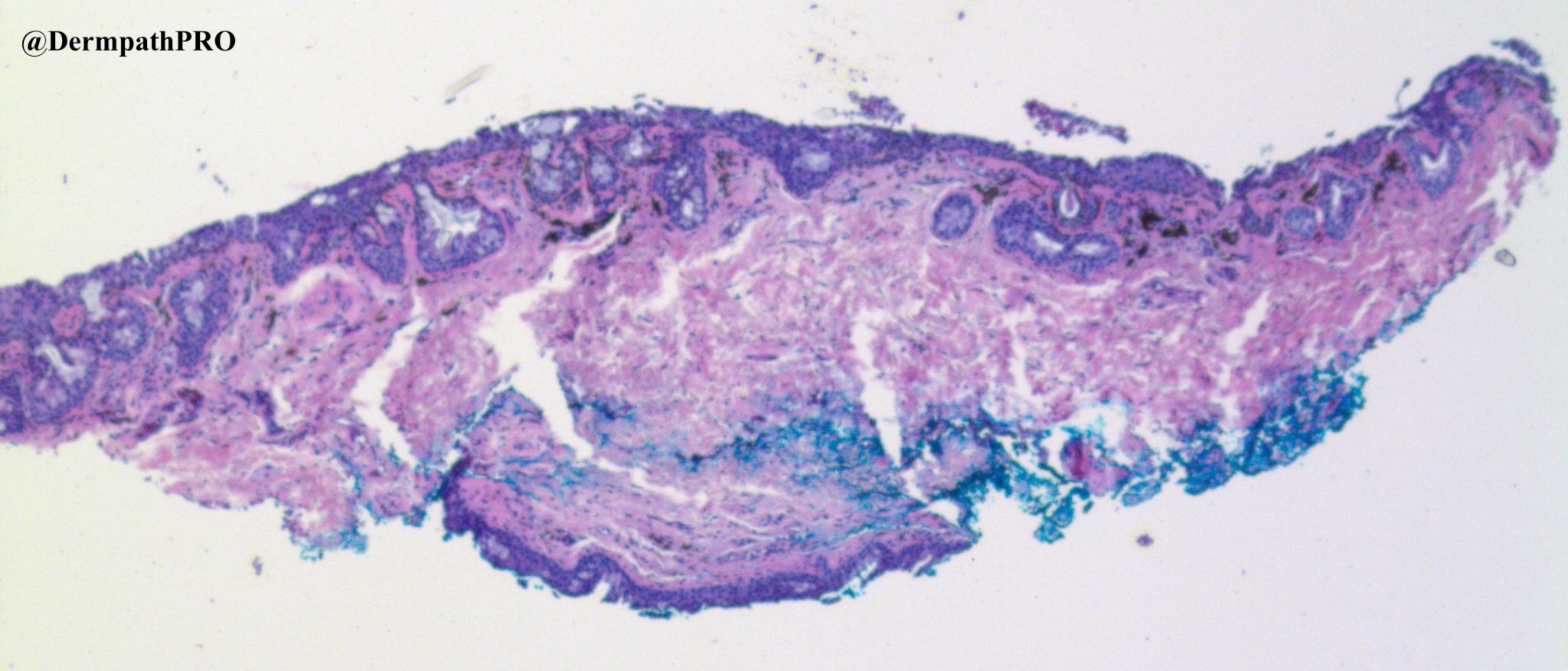
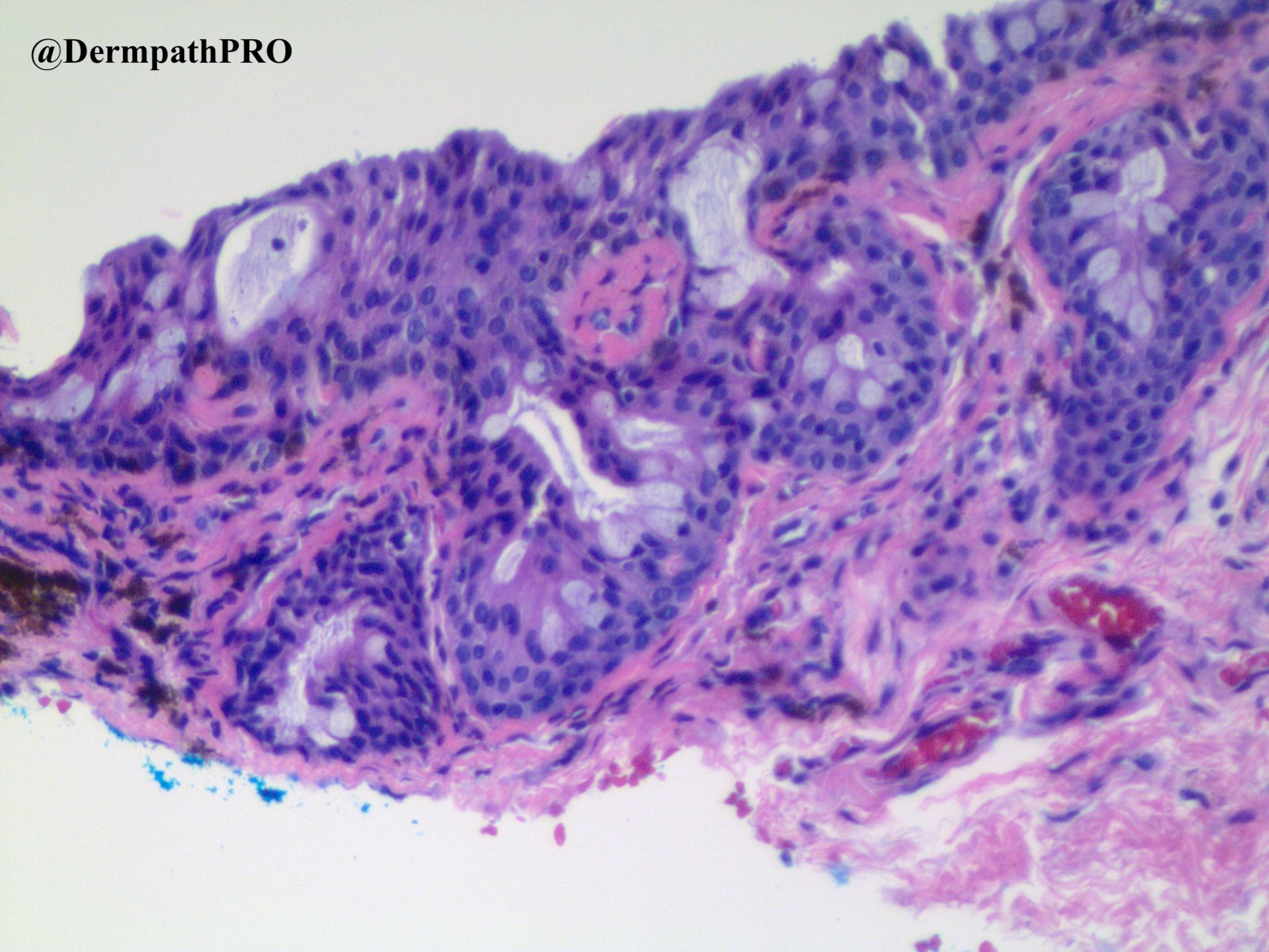
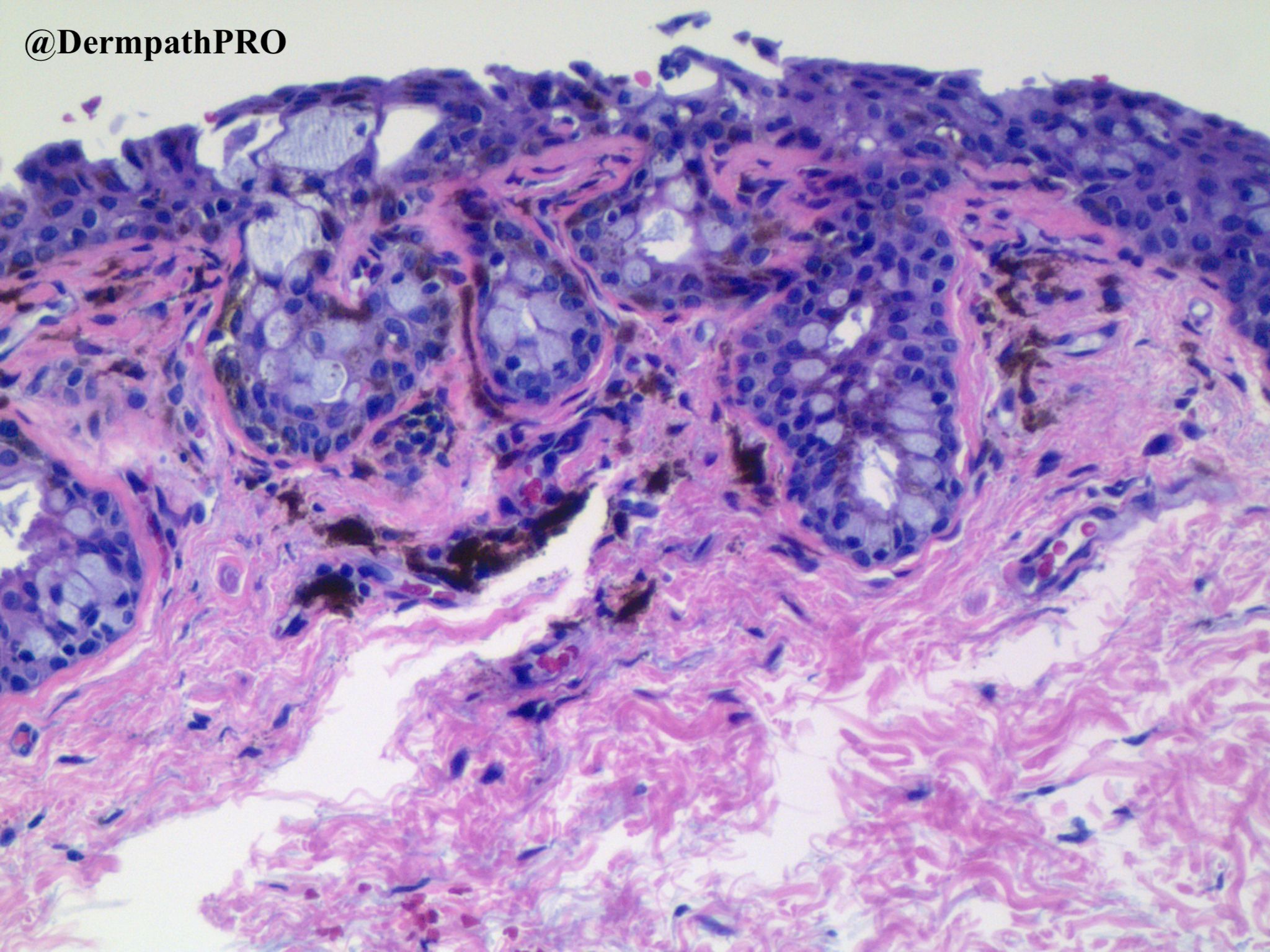
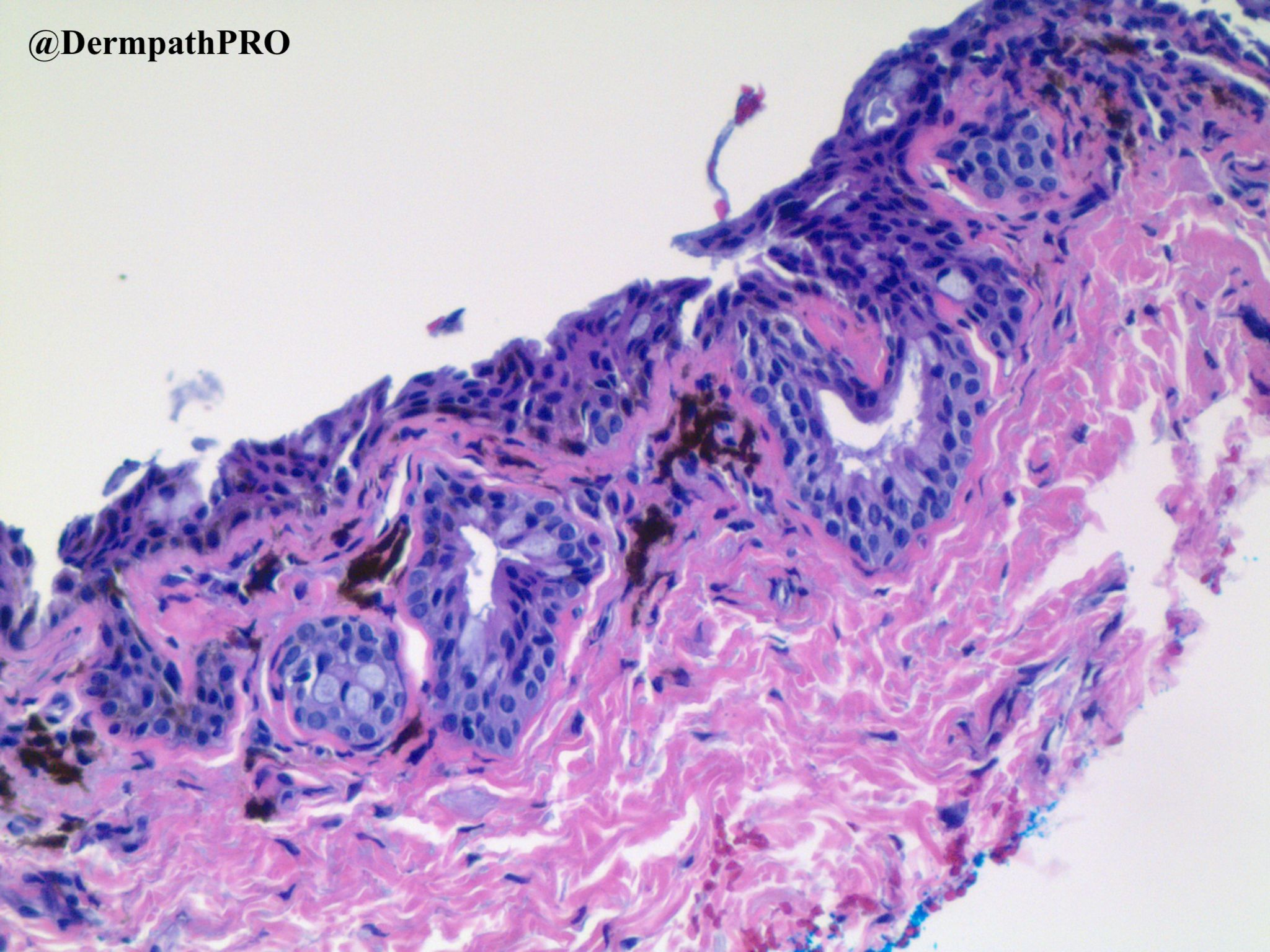
59 year old female with pigmented lesion, right lower eyelid conjunctiva.


  Report Record

User Feedback


Richard Logan

Posted

Pemphigus. 

Not sure of significance of pigmentation however.  Possibly chronicity?

Share this comment


Link to comment
share_externally

Dr. Richard Carr

Posted

PAM most likely, no atypia. Needs a Sox10 or melanA

Share this comment


Link to comment
share_externally

Leila Ahmed

Posted

Is the patient dark skinned? Could it be a benign melanosis? If fair skinned individual then PAM without atypia as suggested by Dr Carr.

Share this comment


Link to comment
share_externally

Richard Logan

Posted

I have betrayed my inexperience with interpreting conjunctival histology.  Is the surface disruption normal for conjunctival epithelium?

Share this comment


Link to comment
share_externally

Dr. Richard Carr

Posted

It's actually helpful to others to see your comments Richard as probably others will have similar ponderings. Yes looks normal pseudostratified conjunctival epithelium with scattered goblet cells. History is not good for pemphigus.

Share this comment


Link to comment
share_externally

Richard Logan

Posted

Thanks Richard. 

Yes the history doesn't suggest pemphigus, but then we are not always told the full story in this arena!

Might this appearance be compatible with a naevus of Ito with bulbar involvement?  I presented such a case to the RSM in the 1980's!

Share this comment


Link to comment
share_externally

Uma Sundram

Posted

Hi everyone, I figured conjunctiva histology is something we can all learn from. It's hard to tell from the pictures, but there are some avoid melanocytes as well. We finally landed on combined nevus. I did need an expert's help, however.

Share this comment


Link to comment
share_externally


Join the conversation

You can post now and register later. If you have an account, sign in now to post with your account.

Guest
Add a comment...

×   Pasted as rich text.   Paste as plain text instead

  Only 75 emoji are allowed.

×   Your link has been automatically embedded.   Display as a link instead

×   Your previous content has been restored.   Clear editor

×   You cannot paste images directly. Upload or insert images from URL.

Loading...

×
×
  • Create New...