Jump to content
In this section we have spot diagnoses posted on a daily basis since June 2010, now over 4000! You can review the archived cases and read the suggested diagnoses by users and the final comment by the contributors.
Case are uploaded each week day by 10 am UK time with the correct diagnosis will generally be posted at 8 pm UK time. Why not view the most recent spot diagnosis and proffer a diagnosis?

Case Number : Case 2771 - 18 February 2021 Posted By: Saleem Taibjee

Please read the clinical history and view the images by clicking on them before you proffer your diagnosis.
Submitted Date :
   (0 reviews)

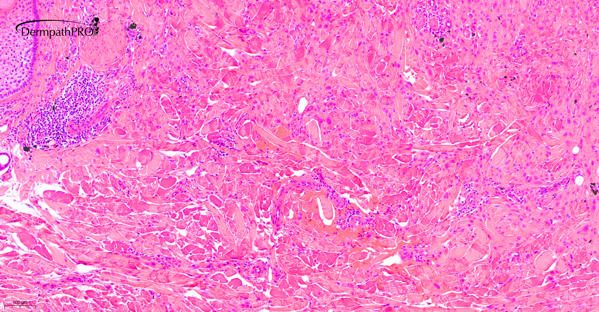
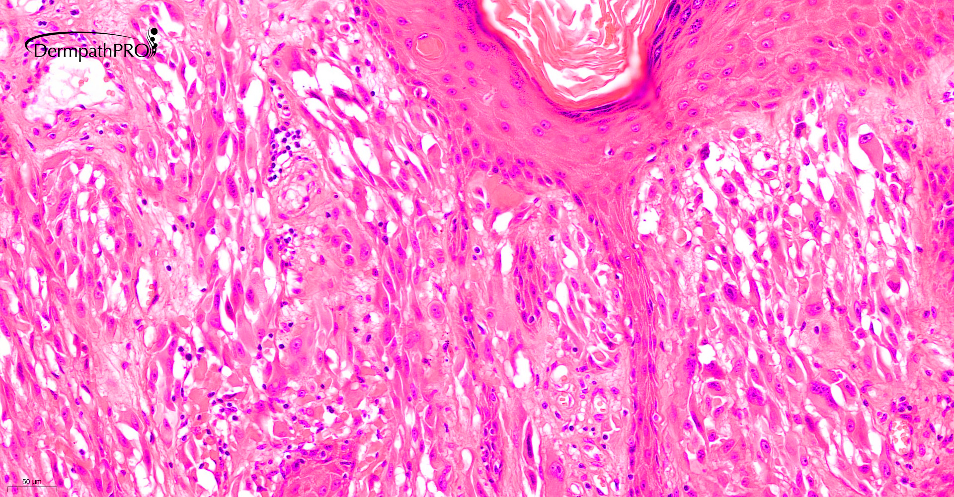
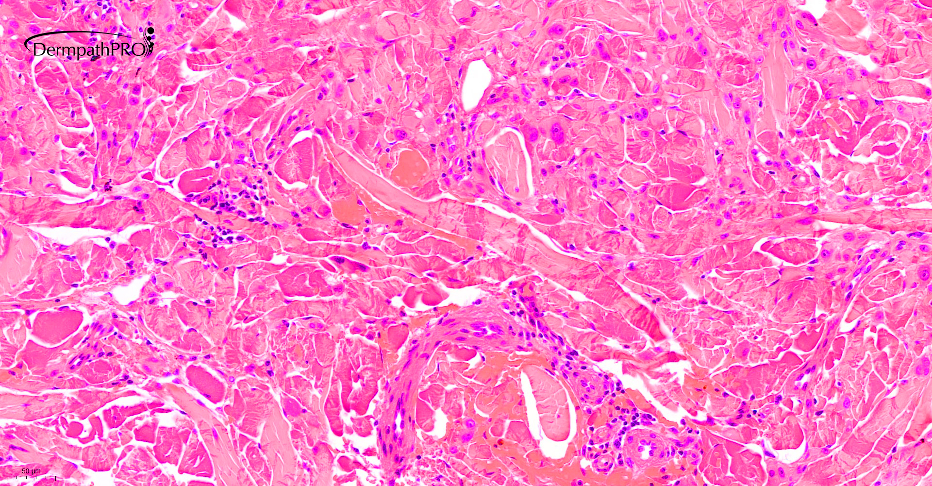
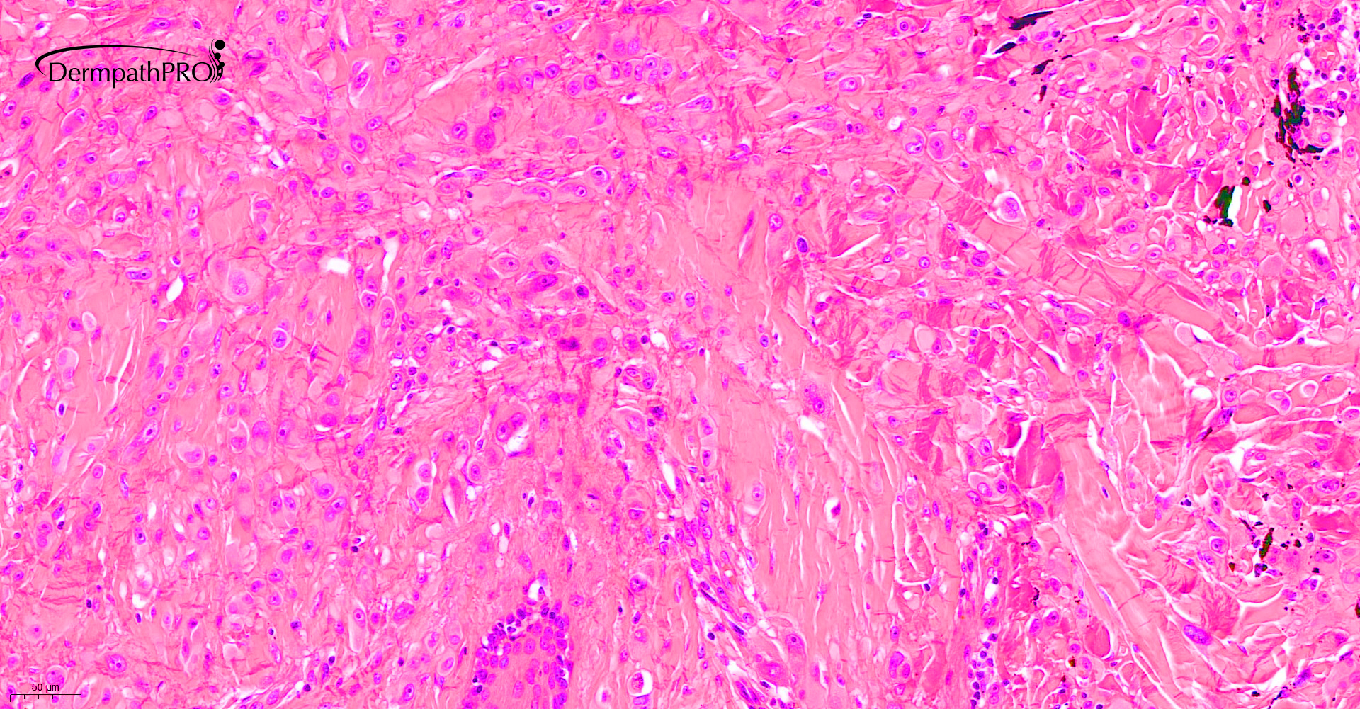
32M, lesion right leg


  Report Record

User Feedback


Richard Logan

Posted

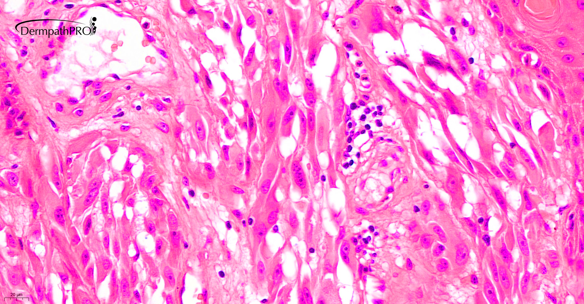
I think this is a peduculated, papillomatous Spitz naevus, with spindle cells predominating.

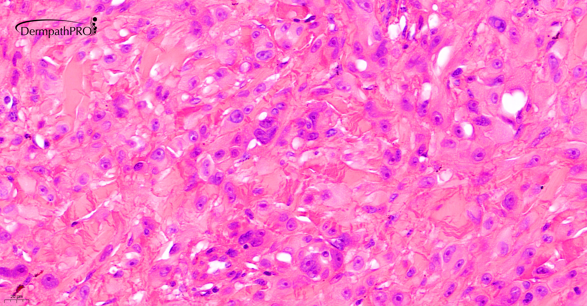
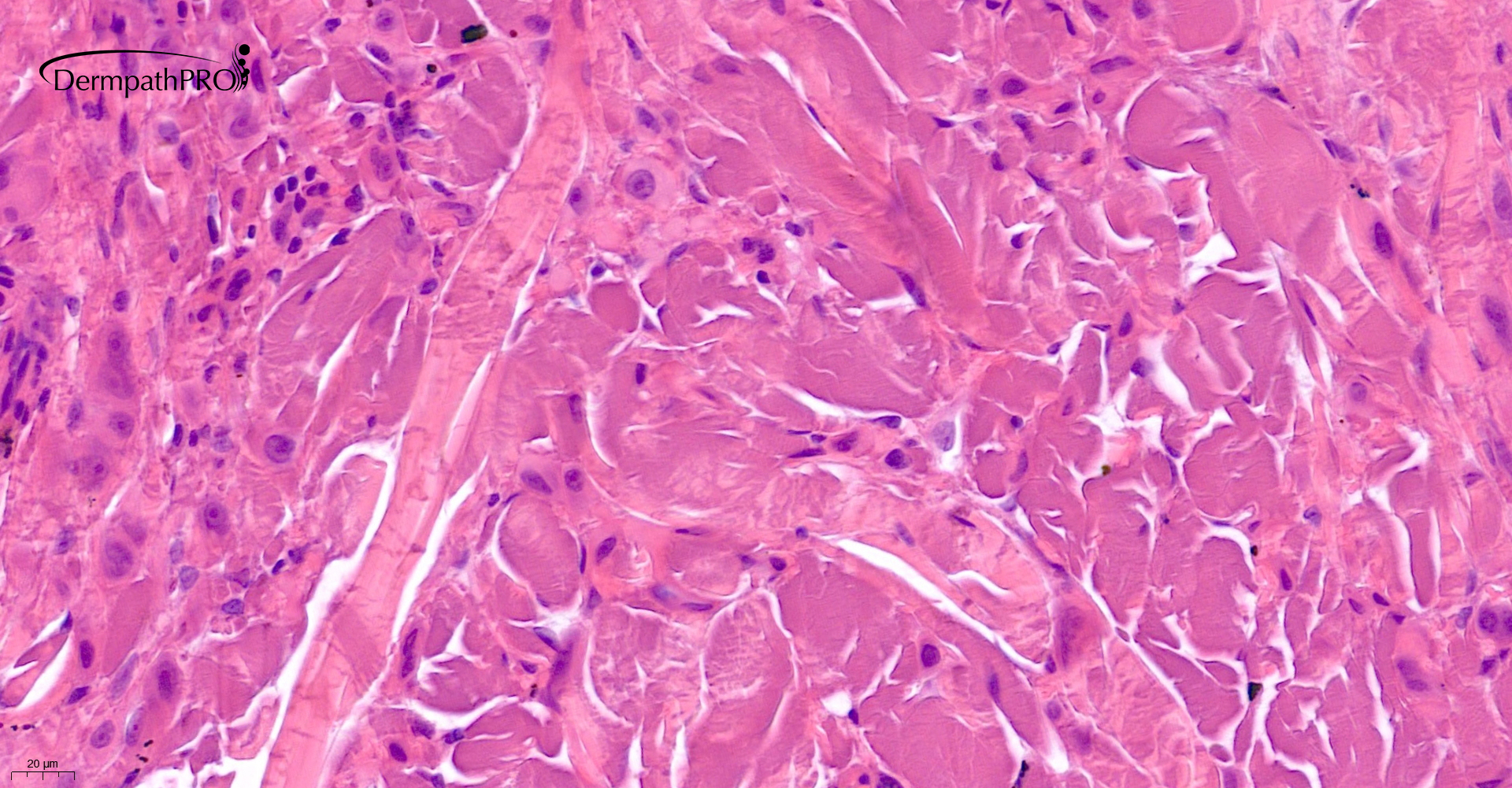
There is an interesting basophilic, tinctural alteration in the dermal collagen fibres which might be calcium deposition.  Image 4 shows small foci of black granular material which might be tattoo pigment, or calcium.

Share this comment


Link to comment
share_externally

Alex-Ventura-Leon

Posted

Polipoid Melanocitic lesion with some spindle and epithelioid features. I'm thinking in BAPoma. 

Share this comment


Link to comment
share_externally

Saleem Taibjee

Posted

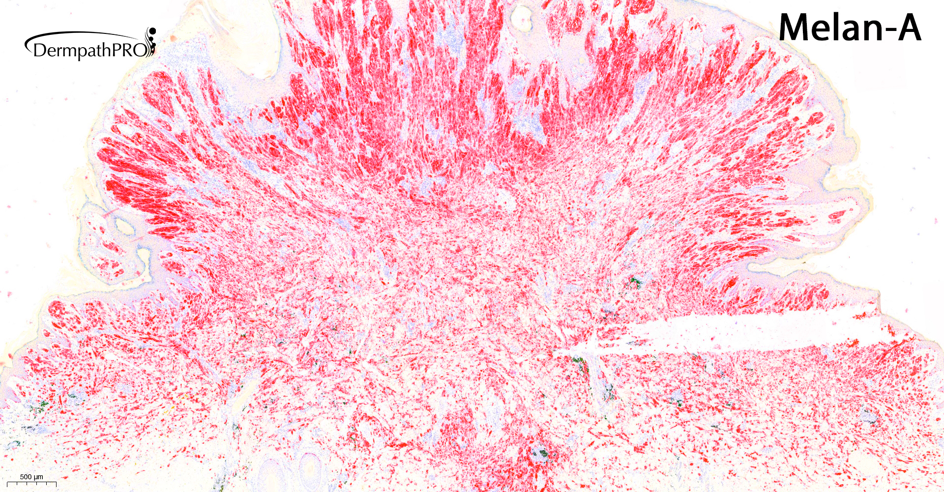
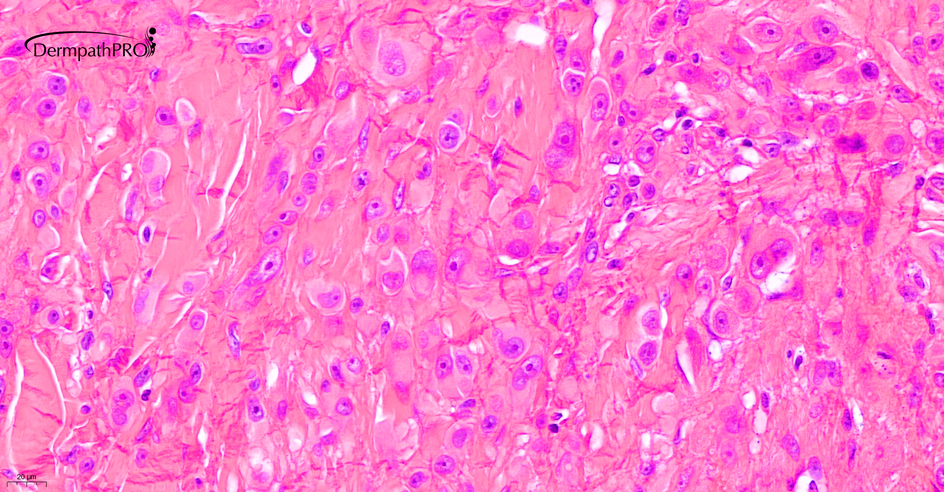
some of the images seem to be missed off. see below

03520_0.7x.jpg

03520_1.5x.jpg

03520_5.0x.jpg

03520_10.0x.jpg


Share this comment


Link to comment
share_externally

Richard Logan

Posted

These extra view reinforce my opinion that this is a Spitz naevus.

The pigment is quite widespread in the mid dermis.  Looks more like tattoo than calcium.

Share this comment


Link to comment
share_externally

Domenico Mesiano

Posted

It seems a bit asymmetrical, no ulceration or significant mitotic activity. I am  curious  to see P16, ki-67 and HMB45.

Share this comment


Link to comment
share_externally

Alex-Ventura-Leon

Posted

43 minutes ago, Xiao Mu said:

Alk mutated spitz nevus.

Yes, that´s a interesting option

Share this comment


Link to comment
share_externally

ALK mutated SN shows usually an intradermal fascicular growth pattern. So I don’t think this is an alk mutated spitzoid lesion,  however agree with Spitz Nevus. 


Share this comment


Link to comment
share_externally

There is an infiltrative growth pattern in between collagen and elastic fibers, as expected in a Nevus. Melanoma growth is usually pushing out elastic and collagen fibers. 

Share this comment


Link to comment
share_externally

Saleem Taibjee

Posted

Hi. Thanks for the comments on this case.

Yes, I thought it was a rather fascicular Spitz lineage tumour. And there is some ALK positivity, as shown below.

I can see that there is a clear consensus for benign in the comments. I did share this case with a couple of other dermatopathologists, and in view of the rather cellular nature of the lesion, adult age group, the questionable cytological maturation (still quite epithelioid and pleomorphic at the base), we all favoured designation as Atypical Spitz (but still favouring a benign course).

Somewhat reassuringly, there is minimal mitosis, and p16 is preserved with low Ki67 and negative PRAME.

As some of you pointed out, there is tattoo pigment.

But as Richard points out in the first comment (but not really elaborated upon after that), there is this curious basophilic alteration of the collagen, as shown in the last image above. This seemed confined to the base of the tumour. I do not recall seeing anything quite like this before in the context of a melanocytic lesion, and I wonder if anyone else has? The von Kossa (not shown) is negative.

BW

Saleem

DigitalSlide_B5M_1S_1_5.0x ALKb.1 labelled.jpg

DigitalSlide_B5M_1S_1_10.0x ALKb.1 labelled.jpg

03520_2.0x p16 labelled.jpg

03520_5.0x Ki67 labelled.jpg

03520_5.0x HMB45 labelled.jpg

03520_2.0x PRAME labelled.jpg


Share this comment


Link to comment
share_externally


Join the conversation

You can post now and register later. If you have an account, sign in now to post with your account.

Guest
Add a comment...

×   Pasted as rich text.   Paste as plain text instead

  Only 75 emoji are allowed.

×   Your link has been automatically embedded.   Display as a link instead

×   Your previous content has been restored.   Clear editor

×   You cannot paste images directly. Upload or insert images from URL.

Loading...

×
×
  • Create New...