Jump to content
In this section we have spot diagnoses posted on a daily basis since June 2010, now over 4000! You can review the archived cases and read the suggested diagnoses by users and the final comment by the contributors.
Case are uploaded each week day by 10 am UK time with the correct diagnosis will generally be posted at 8 pm UK time. Why not view the most recent spot diagnosis and proffer a diagnosis?

Case Number : Case 2737 - 1 January 2021 Posted By: Dr. Richard Carr

Please read the clinical history and view the images by clicking on them before you proffer your diagnosis.
Submitted Date :
   (0 reviews)

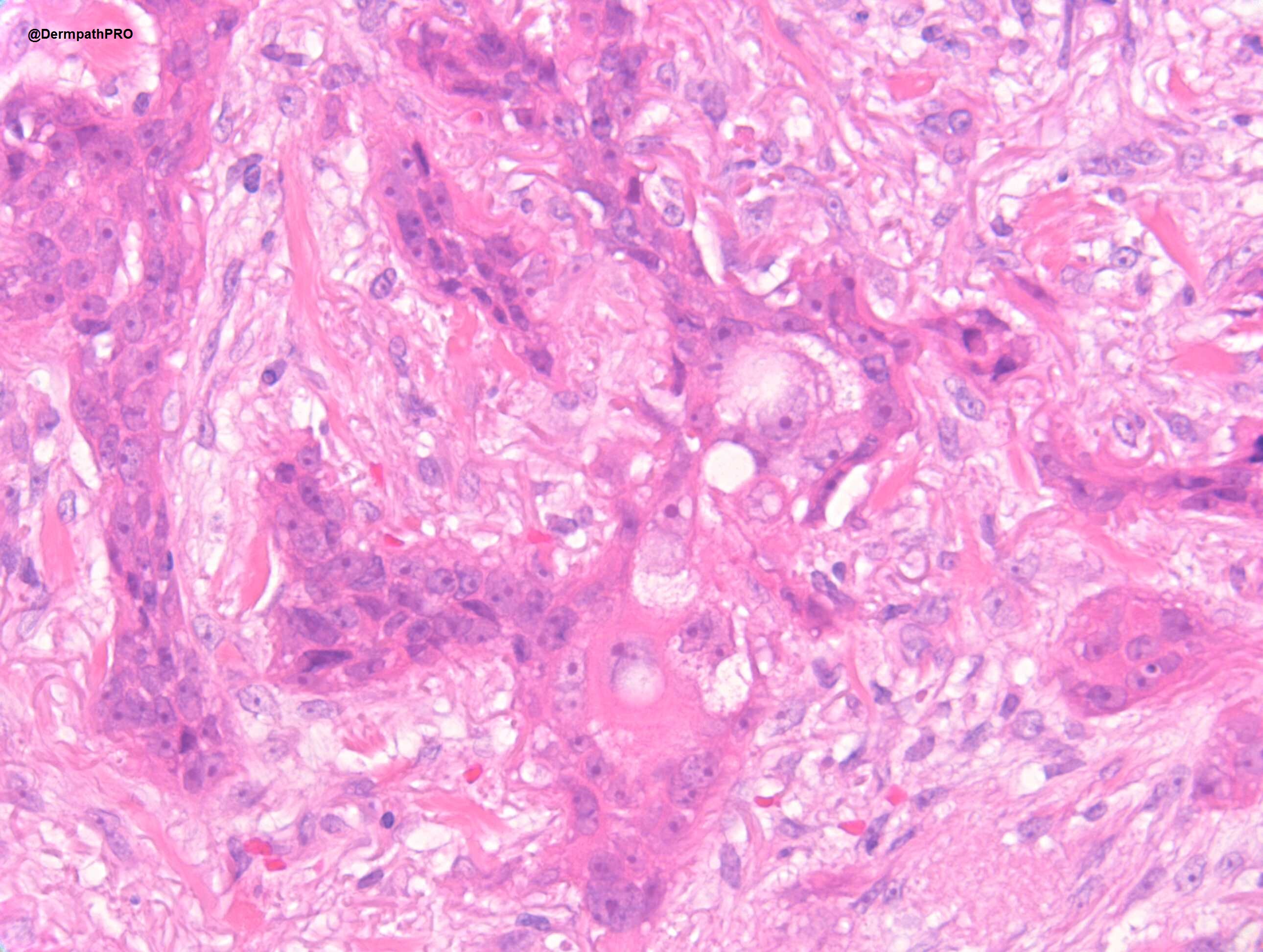
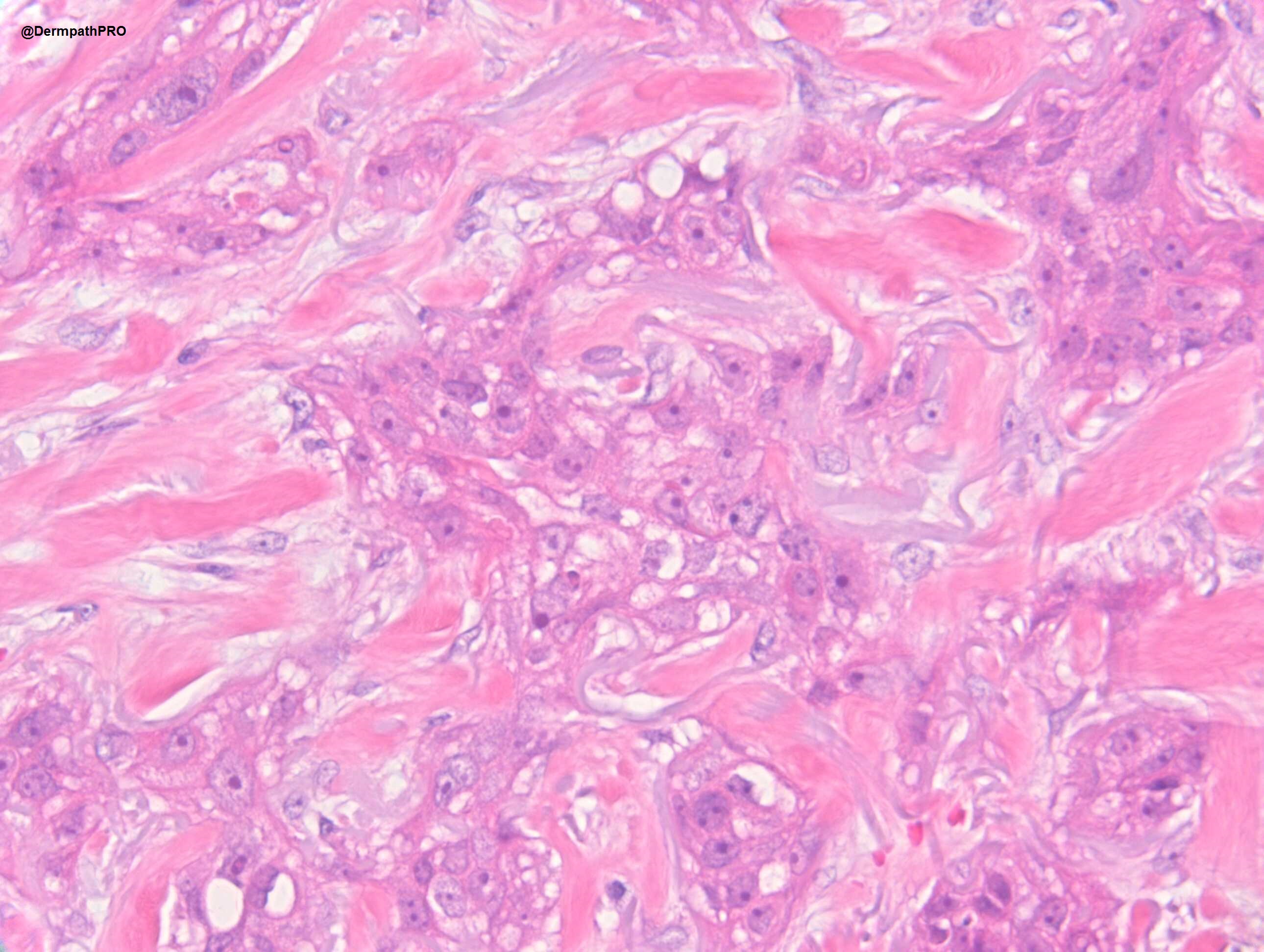
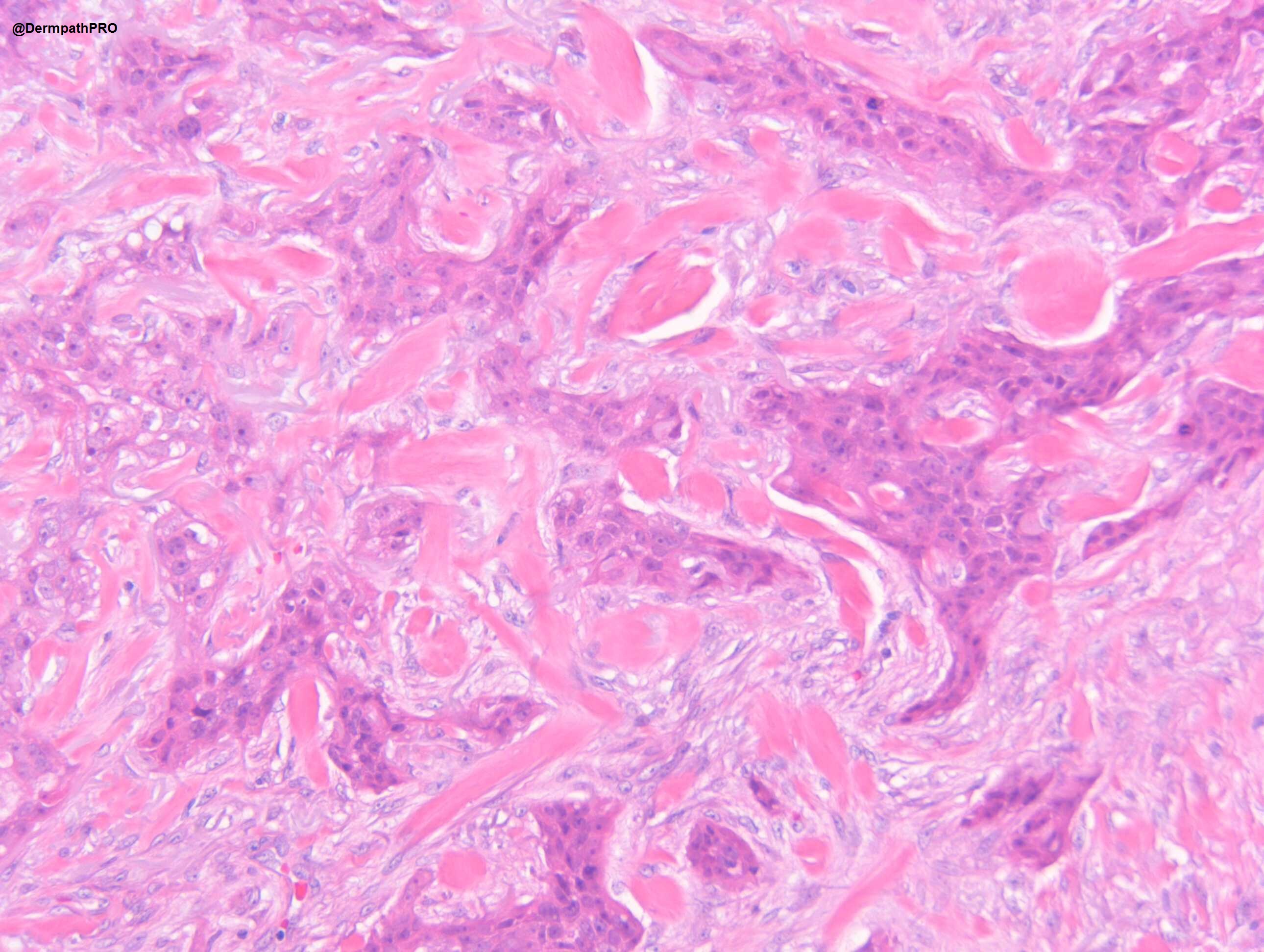
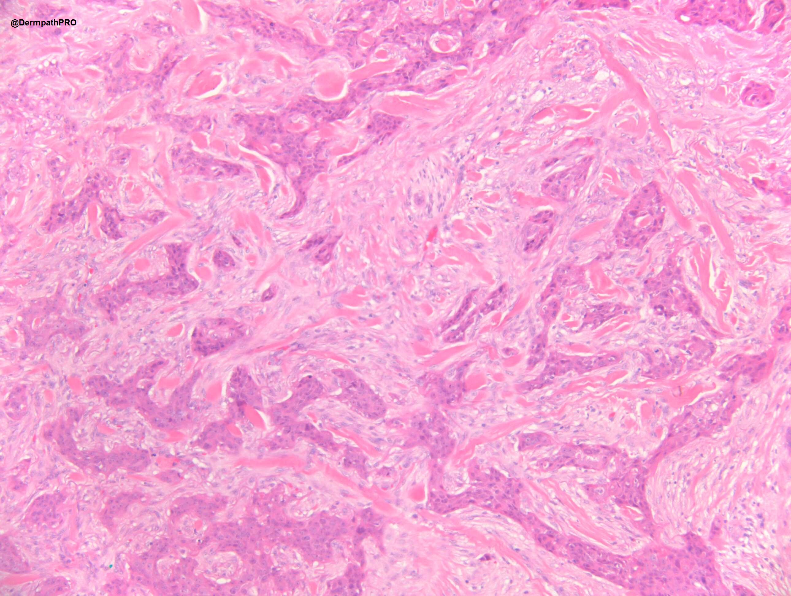
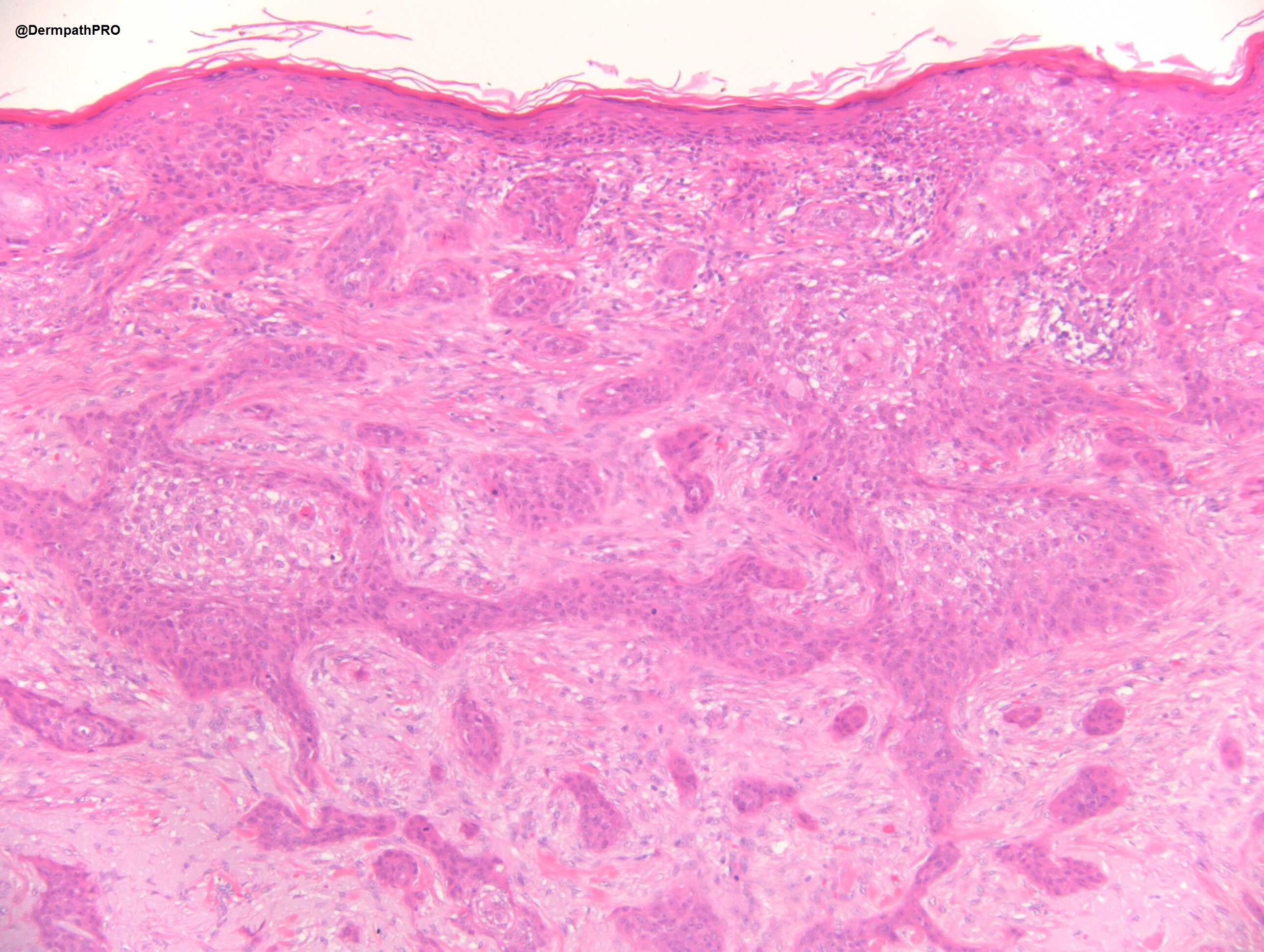
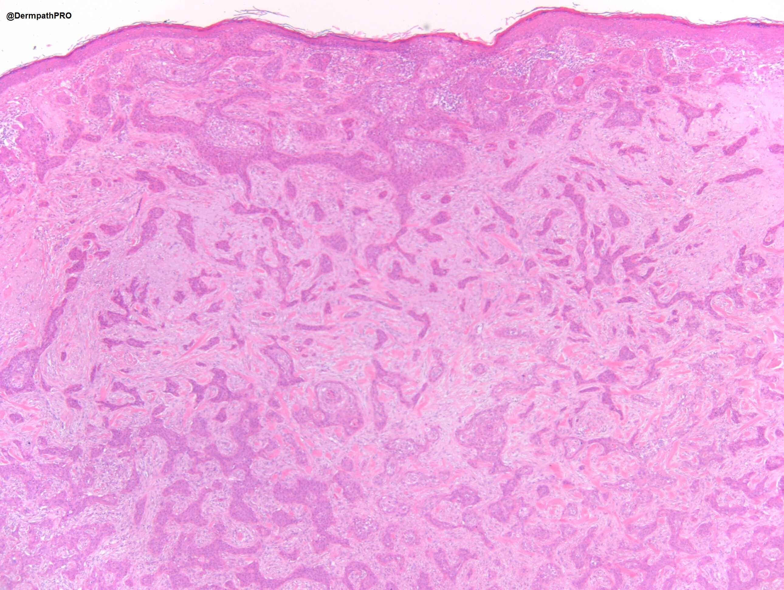
M80 Cheek. ?BCC


  • Like 4
  Report Record

User Feedback


Richard Logan

Posted

At first I wondered whether this was some form of sclerotic metastasis, but the last two images clearly show the tumour arising from the epidermis.  The overall growth pattern is suggestive of an infiltrative BCC inducing a fibrotic dermal resonse, but the tumour cells, at least as reproduced here, don't look particularly basophilic, nor is there any retraction artefact or peripheral palisading.  In places the tumour cells show cytoplasmic vacuolation and/or a bubbly appearance.

I don't know what to call this.  Knowing Richard Carr's interest in squamous carcinoma, I am inclined towards a variant of SCC, and would be interested to see some immunhistochemistry.

Share this comment


Link to comment
share_externally

nick turnbull

Posted

I initially thought desmoplastic scc  or basisquam but I can see what looks like ducts. I thought porocarcinoma also but the sclerotic collagen reminds me of a MAC. I think I vote MAC. Happy New Years to all. 

Share this comment


Link to comment
share_externally

Leila Ahmed

Posted

Are the IHC showing sebaceous differentiation? 
BCC with sebaceous differentiation?? Sebaceous carcinoma looks different.

Share this comment


Link to comment
share_externally

In porocarcinoma the neoplastic aggregates are, usually, close to each other ( stroma is scarce ). But this is the only not very suitable feature for that diagnosis. All the other ones--including IHC, two types of cells, poroid and cuticular, irregularly shaped aggregates, desmoplastic and sclerotic stroma, mitosis ( fig 3 ) and a moderate pleomorphism--are consistent with Porocarcinoma. 

Share this comment


Link to comment
share_externally

Alex-Ventura-Leon

Posted

I would add to the differentials Eccrine Squamous Ductal Carcinoma

Share this comment


Link to comment
share_externally

Dr. Richard Carr

Posted

Great discussion and thanks to those who contributed at this time of year. In this area I suspect I'm a lumper. As long as it's not a BCC (and therefore of minimal risk for metastasis) I'd lump all tumours like this and grade them based on the degree of pleomorphism and mitotic activity. Whether there is focal ductal or sebaceous differentiation is probably academic. Interestingly I've just spent the afternoon looking at the new UK clinical guidelines for SCC and bizarrely spindle cell, adenosquamous, metaplastic/sarcomatoid & desmoplastic SCC have been labelled as "very high risk" which is completely not my experience as they are often rather indolent and low grade although I accept they may have invaded a long way (if left for many years). Certainly cytologically low grade lesions I've practically never seen metastasise despite them being "infiltrative" in pattern and extensive.

I did not see clear cut ductal differentiation (necrotic cells that "drop out" can mimic small lumina, and one can see degenerative vacuolisation) so I think I called this SCC but as I say I think that is academic. I chose this case to show that very rarely an SCC may have a morphoeic / keloidal stroma as this usually is a good clue to an infiltrative BCC (that often lack obvious palisading, retraction and mucin in the retraction space). I always do BerEP4 / EMA in such cases if there is no classical BCC. This case probably surprised me in being an SCC (or porocarcinoma or adenosquamous or non-low grade squamoid eccrine ductal carcinoma etc etc). In BCC that basaloid epithelium is always completely EMA negative and the BerEP4 is strongest in the peripheral cells (this case clearly shows the opposite pattern).


Share this comment


Link to comment
share_externally

nice case. actually I think it is pretty good for the entity "squamoid eccrine ductal carcinoma". With the top area showing connection to the epidermis and showing clearcut squamoid features while the deep lesion showing more compressed infiltrating strands with focal ductal differentiation. 

Share this comment


Link to comment
share_externally

Dr. Richard Carr

Posted

The original description of that entity (SEDC) was a very low grade lesion akin to MAC and sclerosing eccrine ductal carcinoma. The recently published series by Calonje and colleagues moved away from a purely low grade designation and the WHO 4th seems to have adopted this too but I note it's synonymous with adenosquamous carcinoma. For me I only use squamoid eccrine ductal carcinoma according to the original authors as a low grade indolent lesion (and the ductal part should be clear cut) and I personally prefer adenosquamous for the others (that will include tumours that others will call porocarcinoma I suspect). As I say mainly academic as it's the distinction from BCC and then the grading that are probably more important. Obviously very indolent and low grade lesions can extend widely, if left to grow other many years, and may require margin control surgery. 

Share this comment


Link to comment
share_externally


Join the conversation

You can post now and register later. If you have an account, sign in now to post with your account.

Guest
Add a comment...

×   Pasted as rich text.   Paste as plain text instead

  Only 75 emoji are allowed.

×   Your link has been automatically embedded.   Display as a link instead

×   Your previous content has been restored.   Clear editor

×   You cannot paste images directly. Upload or insert images from URL.

Loading...

×
×
  • Create New...