Jump to content
In this section we have spot diagnoses posted on a daily basis since June 2010, now over 4000! You can review the archived cases and read the suggested diagnoses by users and the final comment by the contributors.
Case are uploaded each week day by 10 am UK time with the correct diagnosis will generally be posted at 8 pm UK time. Why not view the most recent spot diagnosis and proffer a diagnosis?

Case Number : Case 2746 - 14 January 2021 Posted By: Saleem Taibjee

Please read the clinical history and view the images by clicking on them before you proffer your diagnosis.
Submitted Date :
   (0 reviews)

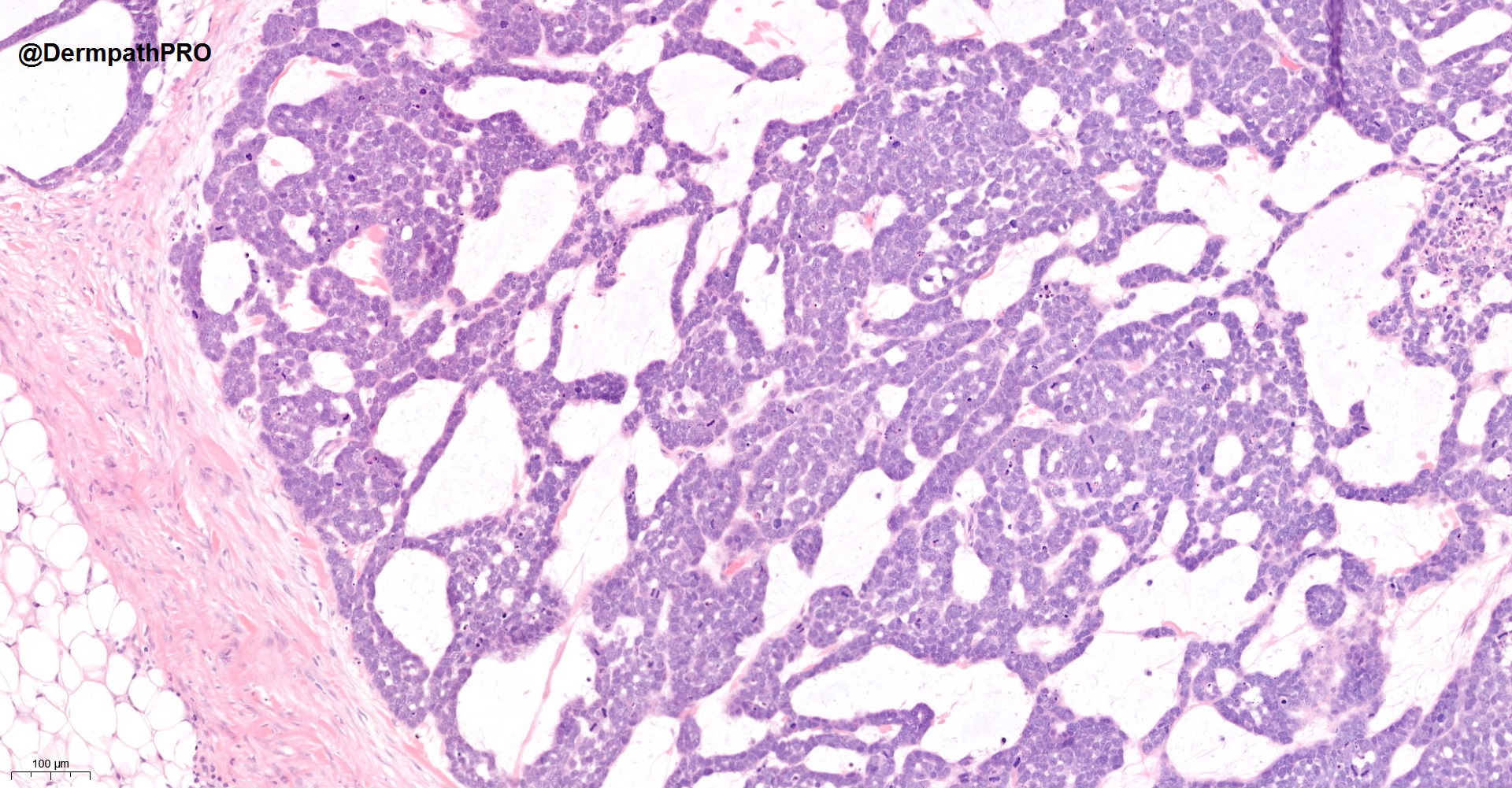
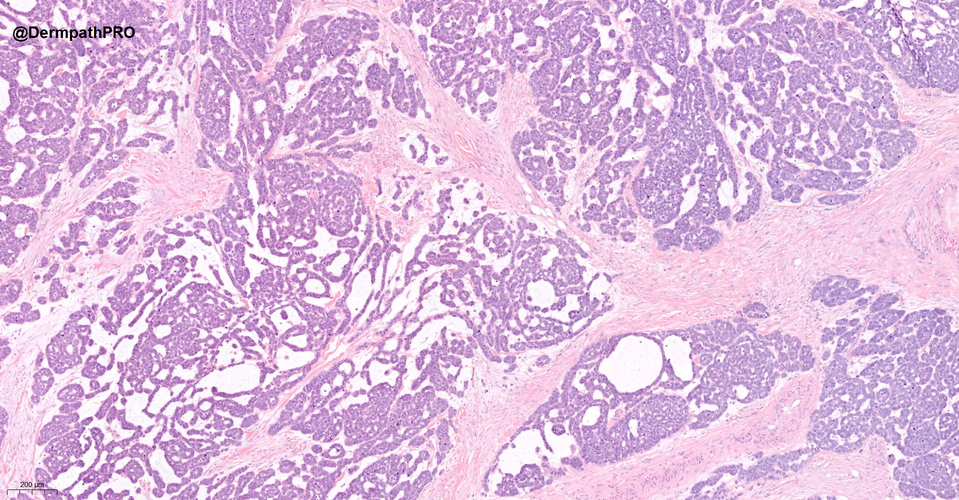
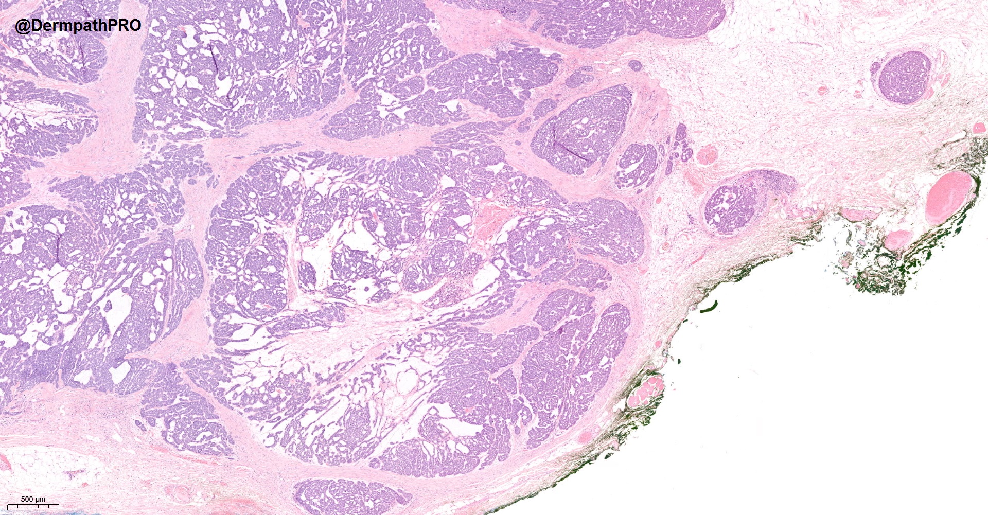
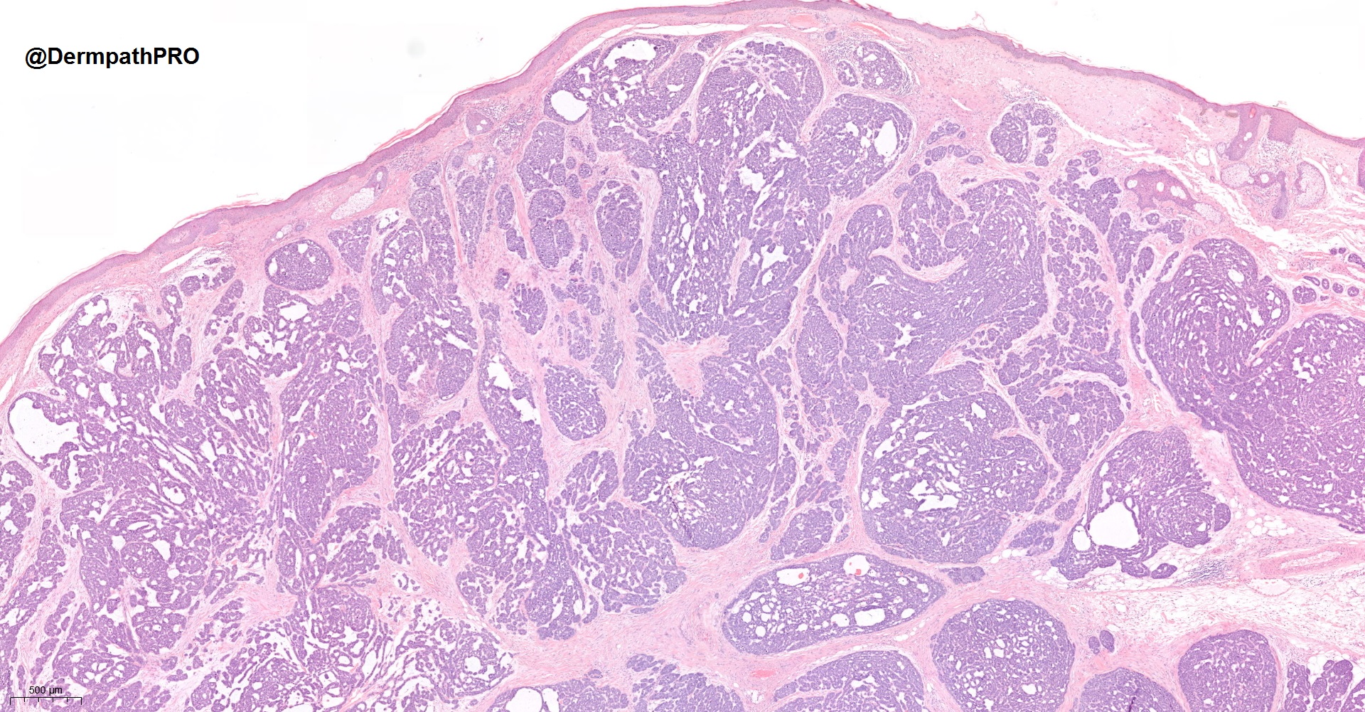
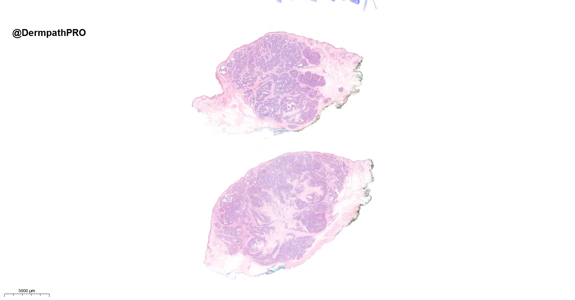
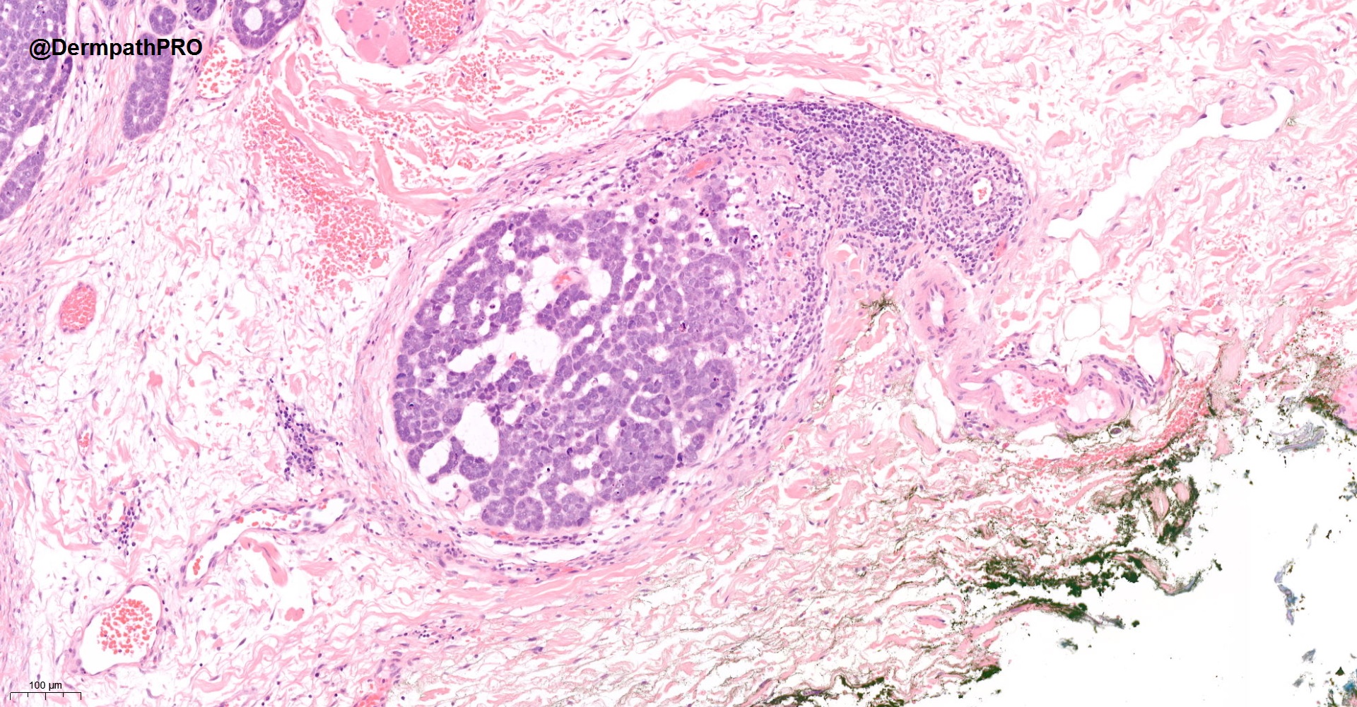
92M, excision right temple


  Report Record

User Feedback


Merkel Cell/polyomavirus-induced cutaneous carcinoma ( ?primary. Tumor embolism in last fig ). 

Share this comment


Link to comment
share_externally

Alex-Ventura-Leon

Posted

Yes, it has the architecture of a Neuroendocrine Carcinoma but let me add Sebaceous Carcinoma in the differential.  

Share this comment


Link to comment
share_externally

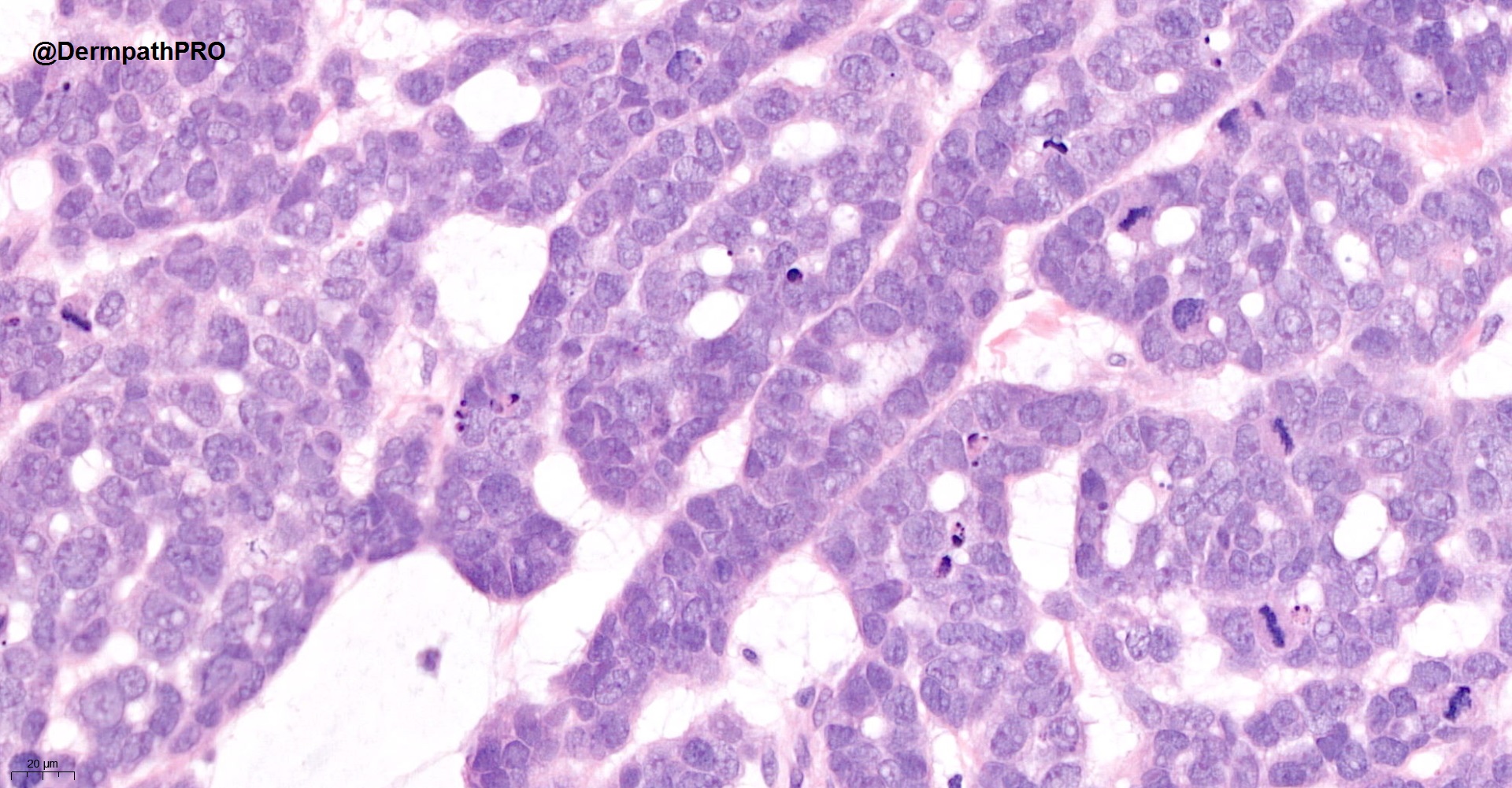
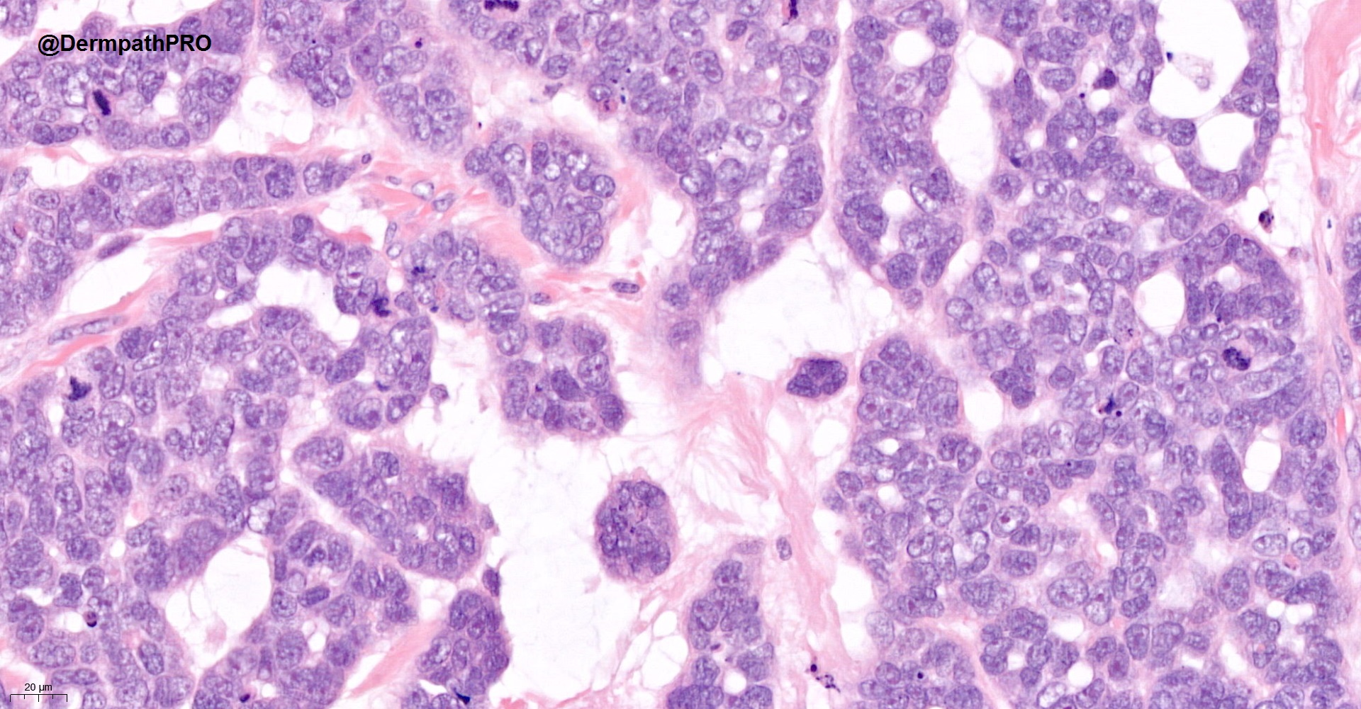
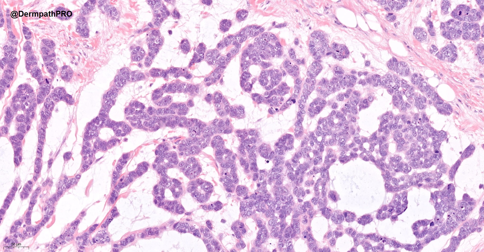
Nodular and infiltrative lesion in the dermis and subcutis, with tumour cells exhibiting high nuclear-to-cytoplasmic ratio with fine stippled chromatin, multiple small nucleoli and some degree of nuclear moulding. Mitoses/apoptosis frequent. Formation of acini, trabeculae and cords/ribbons. Lymphovascular permeation+.

Agree that we should consider a neuroendocrine tumour, in particular Merkel cell carcinoma of skin (CK20+ CAM5.2+ dot-like paranuclear staining; neuroendocrine marker+).

Have also thought of sebaceous carcinoma though with focal finely vacuolated cytoplasm but can it give rise to this architectural pattern?

Have encountered CK20- neuroendocrine carcinoma of skin, some call it CK20- Merkel cell carcinoma (less commonly associated with Merkel cell polyomavirus) but I am sometimes confused with the terminology. Any thoughts?

Share this comment


Link to comment
share_externally

Saleem Taibjee

Posted

Interim update: neuroendocrine markers (CD56, synaptophysin and chromogranin) are negative. Further thoughts?

Share this comment


Link to comment
share_externally

In that case we have a malignant basaloid tumour.

The white spaces in the image appear strange - probably represent mucin.

Seems that sebaceous carcinoma can show such architecture and should be excluded with adipophillin.

(Less likely with mucin pools)

Thought about a strange-looking BCC, but no conventional appearing areas found from the images. Could still try stains.

Other possibilities include other sweat-gland derived basaloid tumours...

Interested to know thoughts from others.

Share this comment


Link to comment
share_externally

? Primary Mucinous Carcinoma of the skin...or metastatic..

Share this comment


Link to comment
share_externally

Dr. Mona Abdel-Halim

Posted

I am favoring metastatic mucinous carcinoma

Share this comment


Link to comment
share_externally

Leila Ahmed

Posted

I would have called it endocrine mucin producing sweat gland carcinoma but neuroendocrine markers are negative! Although WHO book says it can be variably expressed! What about ER, PR, GCDFP15?

Share this comment


Link to comment
share_externally

Saleem Taibjee

Posted

Yes I too favoured this as mucinous carcinoma. It is an unusually ‘solid’ example, rather than the more classical examples of tumour islands within larger lakes of mucin.

This is a recent referral case, courtesy of Dr David Nicholas.

I must admit that my first low power impression was of basal cell carcinoma. But it is on closer scrutiny that one appreciates that the cytology is not quite right and somewhat ‘high grade’, there is a lack of peripheral palisading, and that the mucin is rather striking.

Of course, it is mandatory to consider metastasis in such cases, although primary cutaneous mucinous carcinoma also occurs (most commonly on the eyelid, but can be elsewhere).

This patient has a history of prostate cancer, with an elevated PSA of 50.

Many of the stains were negative. As I mentioned, neuroendocrine markers (CD56, synaptophysin, chromogranin) were negative. BerEP4 was negative, helpful in essentially ruling out BCC. But also negative were: CK20, CEA, TTF1, GCDFP15, CDX2, PSA.

Positive: p63, p53, p16, CK5/6 (patchy), AE1/AE3 (patchy), CK7 (focal), CAM5.2 (focal), EMA (weak patchy).

I have suggested to the referring pathologist to extend the panel to include prostatic acid phosphatase, as well as to review the histology of the original prostate cancer (from 2005).

BW
Saleem

Share this comment


Link to comment
share_externally

Thanks for sharing this case, Saleem. Quite an interesting one. Important to exclude metastasis from uncommon tumour subtypes for these cases. Wonder if the original prostate pathology shows some rare subtypes like basal cell carcinoma of prostate.

For interest, from experience of readers, do usual primary skin mucinous carcinomas have such basaloid appearance, or the usual adenocarcinoma with more cytoplasm?

Share this comment


Link to comment
share_externally

Saleem Taibjee

Posted

Hi Tyelaine. Yes, I think you have a point here. The cytology is somewhat high grade and basaloid compared to the rare examples of primary cutaneous mucinous carcinoma I have come across. I will try to feedback once I hear what the review of the original prostatic histology shows.

BW
Saleem

Share this comment


Link to comment
share_externally

Saleem Taibjee

Posted

The PSMA stain has now been received and shows some staining within the periphery only. But I think that further supports a cutaneous metastasis from the original prostatic carcinoma.

Share this comment


Link to comment
share_externally


Join the conversation

You can post now and register later. If you have an account, sign in now to post with your account.

Guest
Add a comment...

×   Pasted as rich text.   Paste as plain text instead

  Only 75 emoji are allowed.

×   Your link has been automatically embedded.   Display as a link instead

×   Your previous content has been restored.   Clear editor

×   You cannot paste images directly. Upload or insert images from URL.

Loading...

×
×
  • Create New...