Jump to content
In this section we have spot diagnoses posted on a daily basis since June 2010, now over 4000! You can review the archived cases and read the suggested diagnoses by users and the final comment by the contributors.
Case are uploaded each week day by 10 am UK time with the correct diagnosis will generally be posted at 8 pm UK time. Why not view the most recent spot diagnosis and proffer a diagnosis?

Case Number : Case 2749 - 19 January 2021 Posted By: Uma Sundram

Please read the clinical history and view the images by clicking on them before you proffer your diagnosis.
Submitted Date :
   (0 reviews)

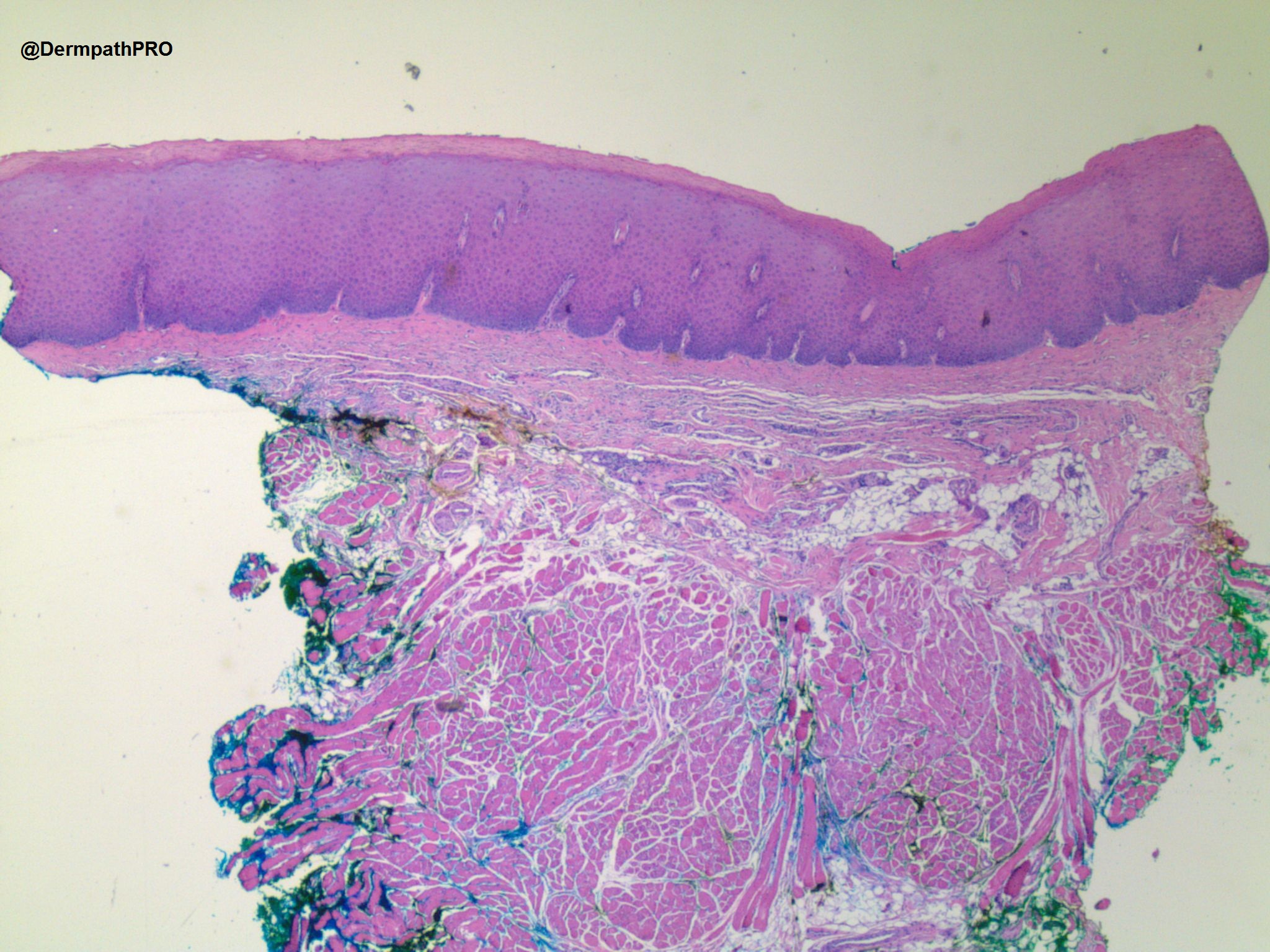
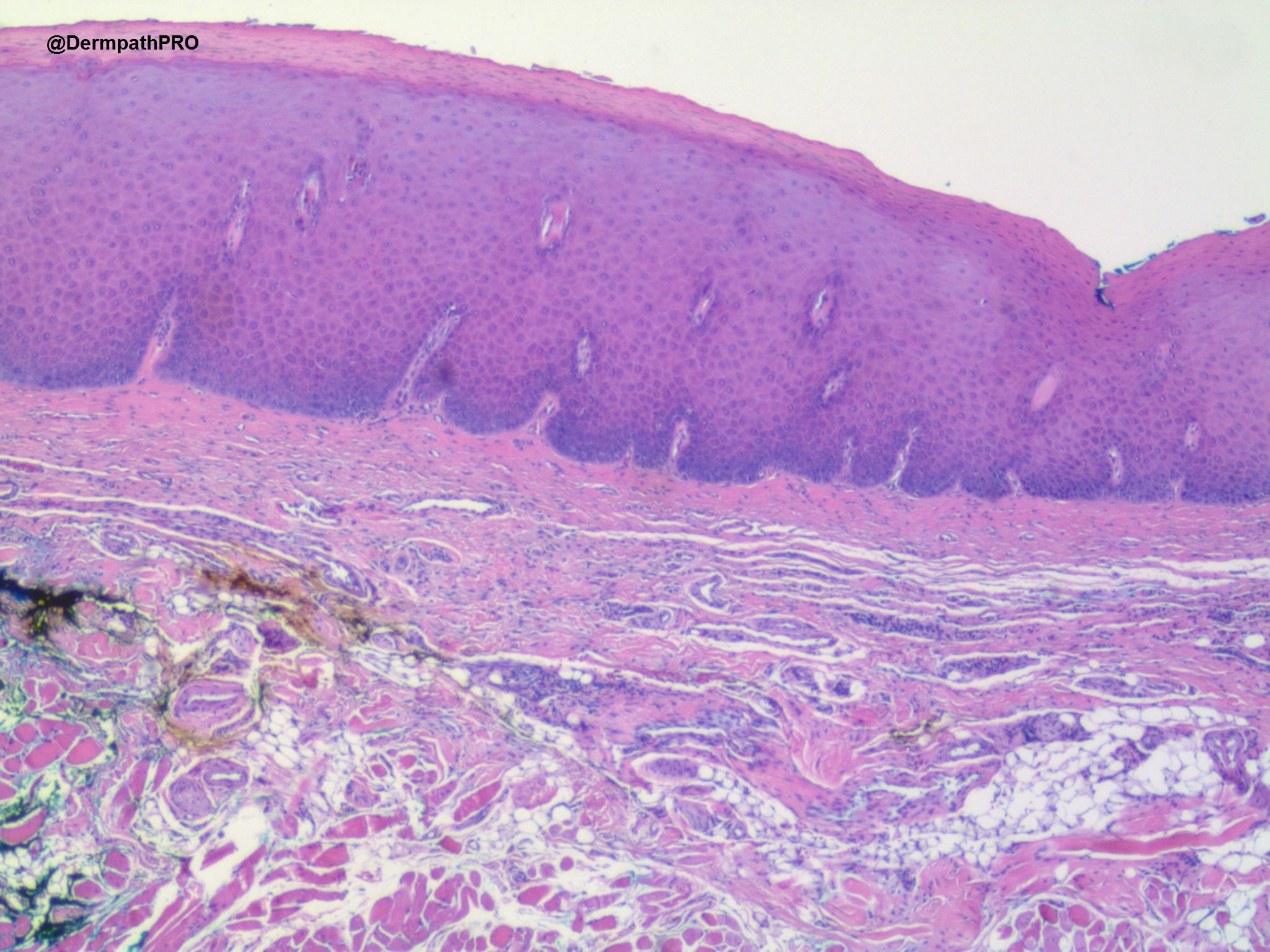
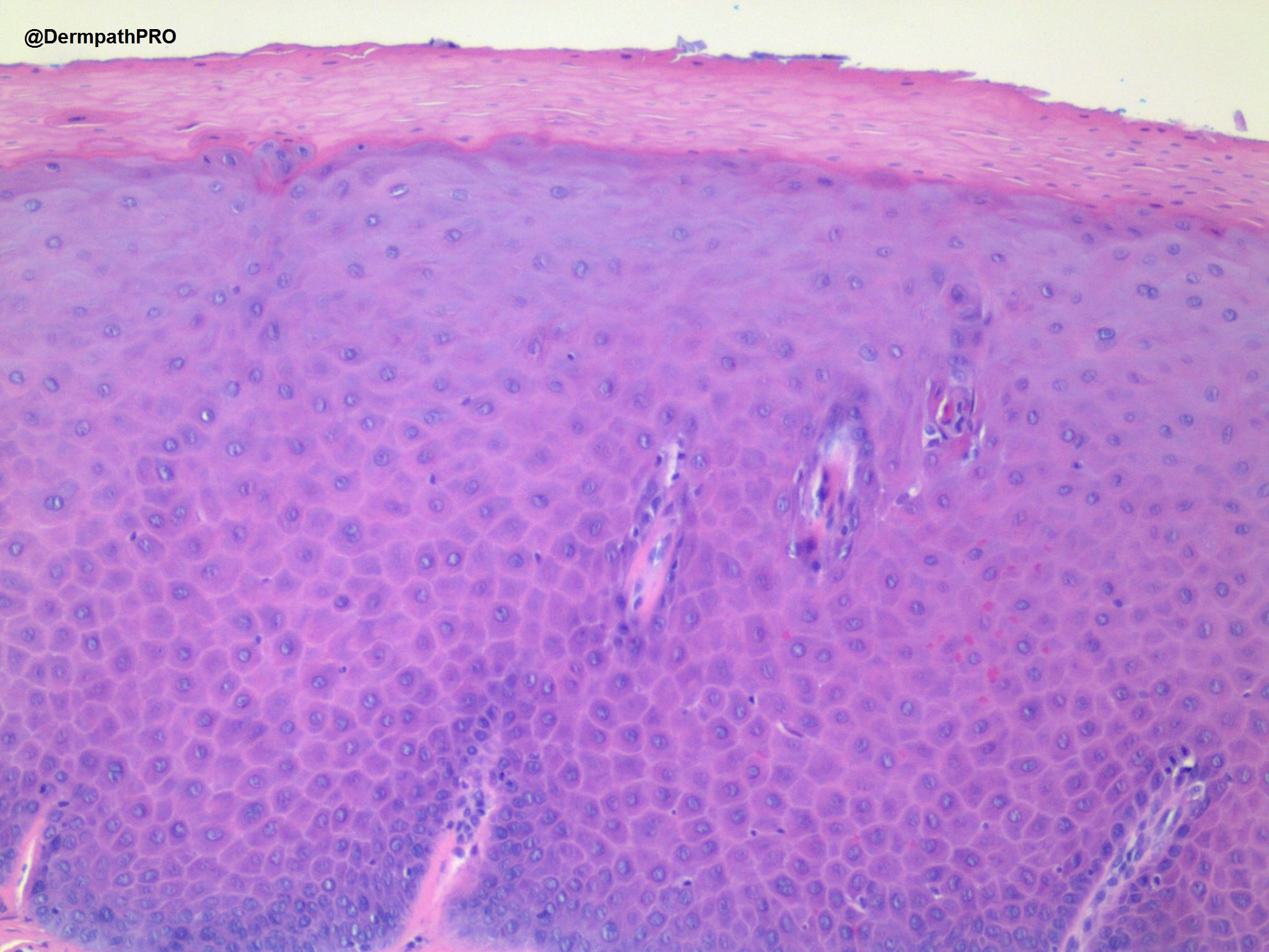
52 year old female with lesion on left lateral tongue.


  Report Record

User Feedback


Richard Logan

Posted

I was wondering about oral hairy leucoplakia, because there is a hint of pallor in the upper levels of the malpighian layer.  However, this pallor is nowhere near as obvious as that shown in two previous examples of that entity (Case 622 26th October 2012; Case 2438 6th November 2019), but that may just be a staining variation.

Failing that there is psoriasiform epidermal hyperplasia with loss of filiform papillae and fusion of adjacent rete ridges suggesting geographic tongue.

Clinical details vitally important here.

Share this comment


Link to comment
share_externally

Alex-Ventura-Leon

Posted

Oral lesions are difficult for me.

My interpretation: Leukoplakia whit mild dysplasia.

Share this comment


Link to comment
share_externally

Oral lesions are challenging!

Would agree with observations of psoriasiform epidermal hyperplasia with loss of filiform papillae. I am not sure about dysplasia. The epidermis just looks strange and a bit thickened. No spongiosis/spongiotic vesicles. I agree we need fungal stains too.

Geographic tongue seems possible. Looking forward to a clinical photo...

Share this comment


Link to comment
share_externally

Uma Sundram

Posted

So sorry, no clinical photo. Pt with white lesion on lateral tongue. Pas negative. Recent development after patient started chewing anti nicotine gum. This caused local irritant contact which became a white patch. 

Share this comment


Link to comment
share_externally

Uma Sundram

Posted

Dx, possible irritant contact Derm from chwing/holding nicotine gum in lateral tongue. Will update w follow up if I get it.

Share this comment


Link to comment
share_externally

Interesting clinicopathological correlation! Thanks for sharing this case.

Share this comment


Link to comment
share_externally


Join the conversation

You can post now and register later. If you have an account, sign in now to post with your account.

Guest
Add a comment...

×   Pasted as rich text.   Paste as plain text instead

  Only 75 emoji are allowed.

×   Your link has been automatically embedded.   Display as a link instead

×   Your previous content has been restored.   Clear editor

×   You cannot paste images directly. Upload or insert images from URL.

Loading...

×
×
  • Create New...