Jump to content
In this section we have spot diagnoses posted on a daily basis since June 2010, now over 4000! You can review the archived cases and read the suggested diagnoses by users and the final comment by the contributors.
Case are uploaded each week day by 10 am UK time with the correct diagnosis will generally be posted at 8 pm UK time. Why not view the most recent spot diagnosis and proffer a diagnosis?

Case Number : Case 2755 - 27 January 2021 Posted By: Saleem Taibjee

Please read the clinical history and view the images by clicking on them before you proffer your diagnosis.
Submitted Date :
   (0 reviews)

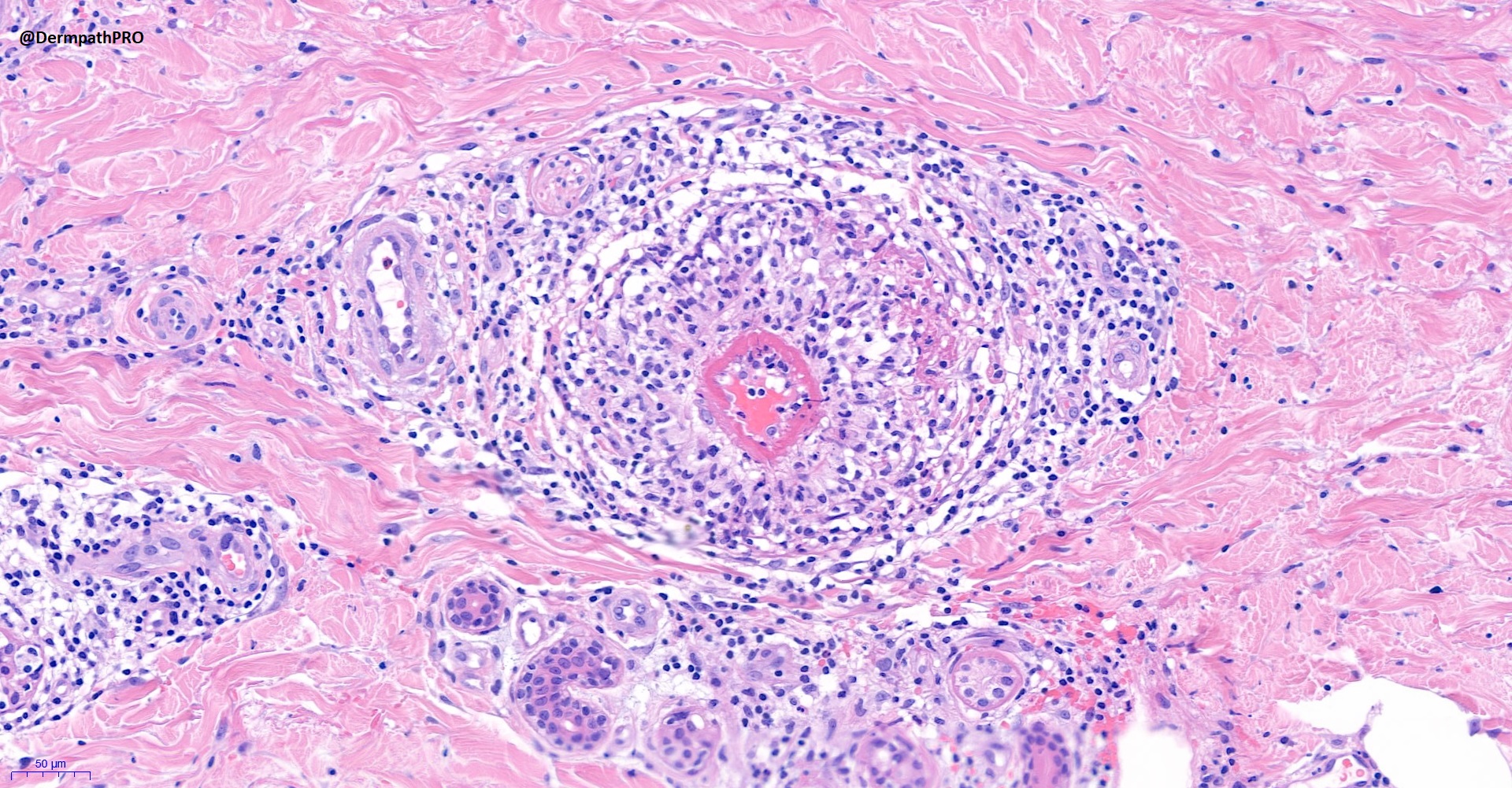
48F, widespread erythematous papules.

  • Thanks 1
  Report Record

User Feedback


Richard Logan

Posted

I'm struggling to come up with a unifying diagnosis for this histology. 

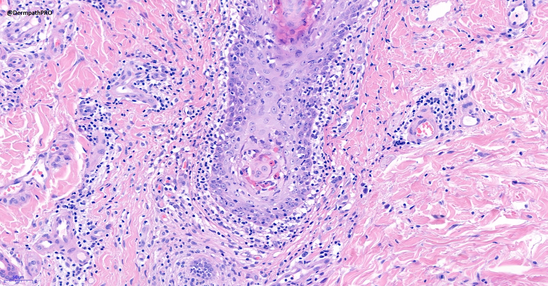
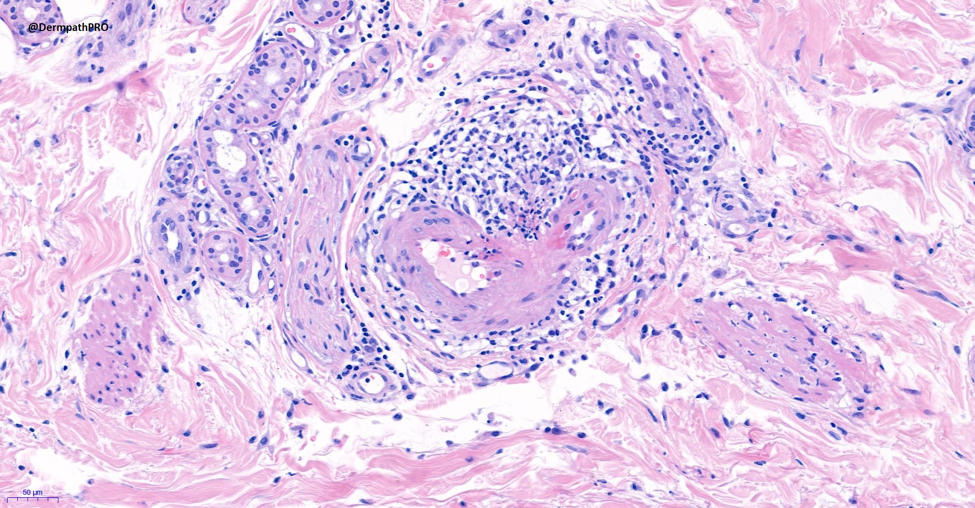
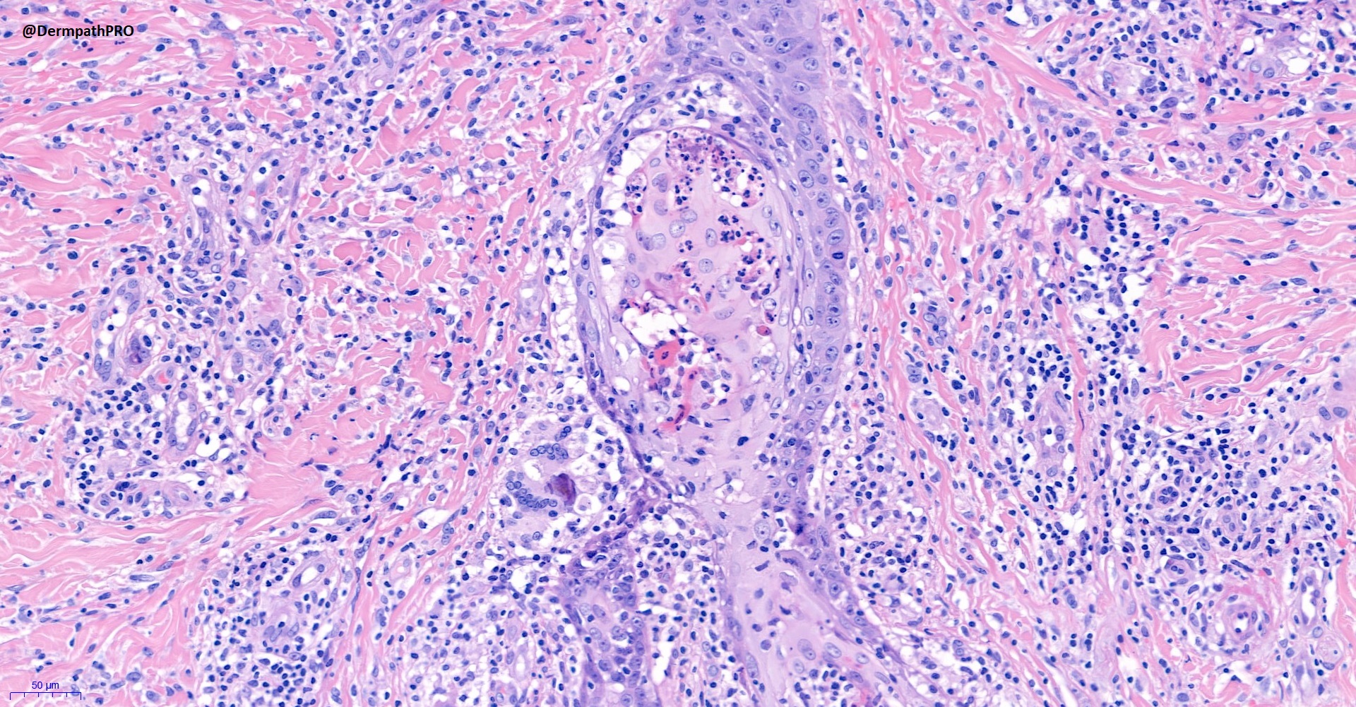
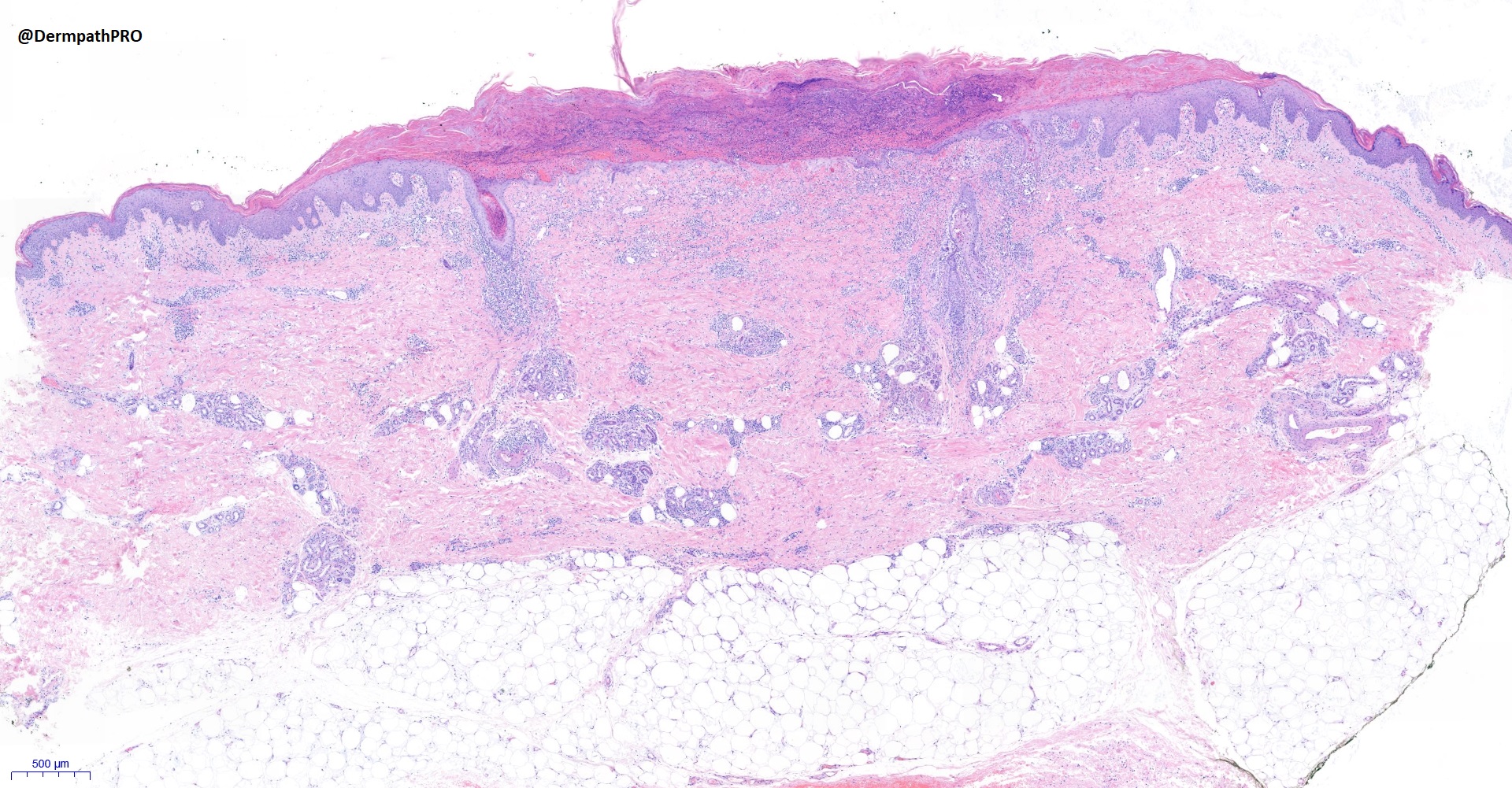
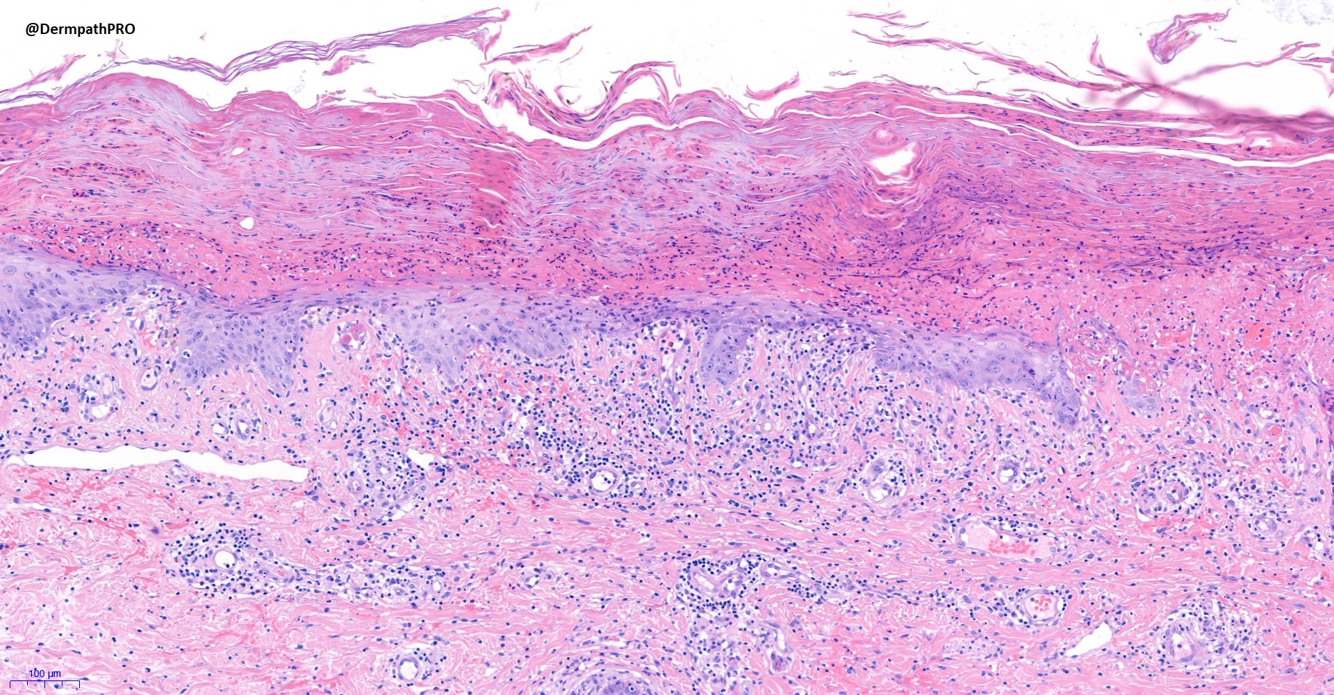
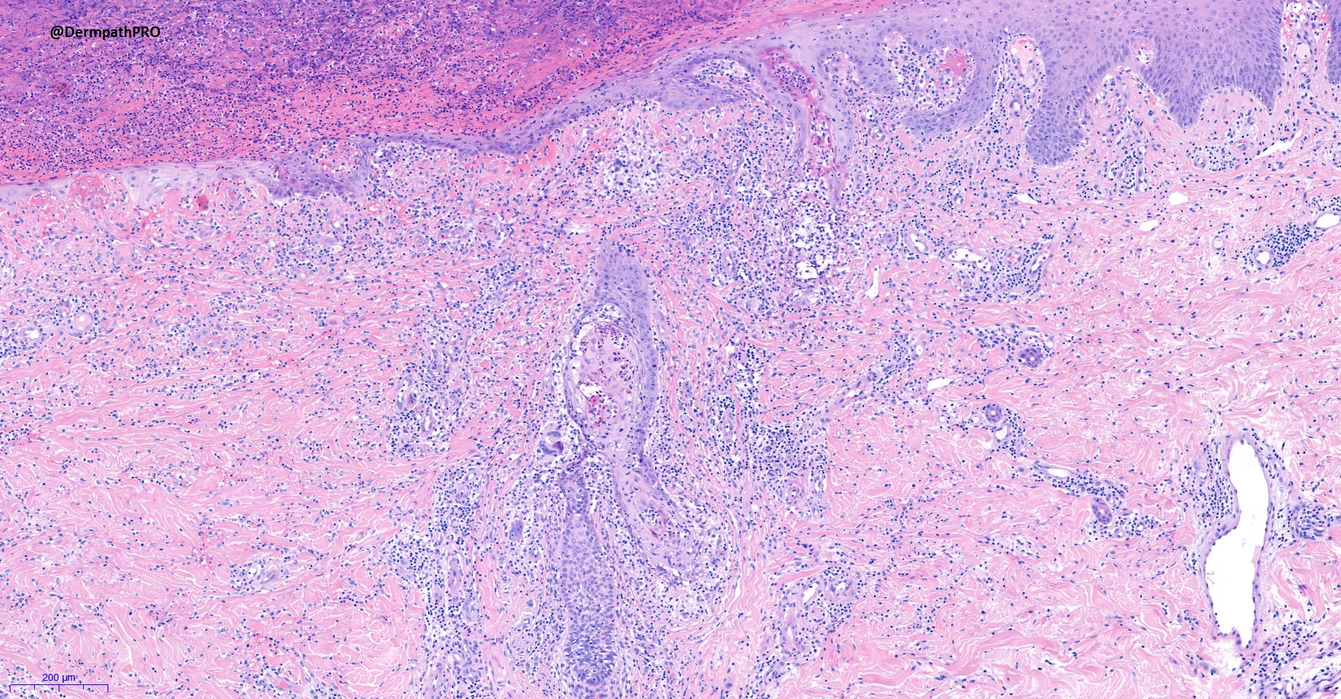
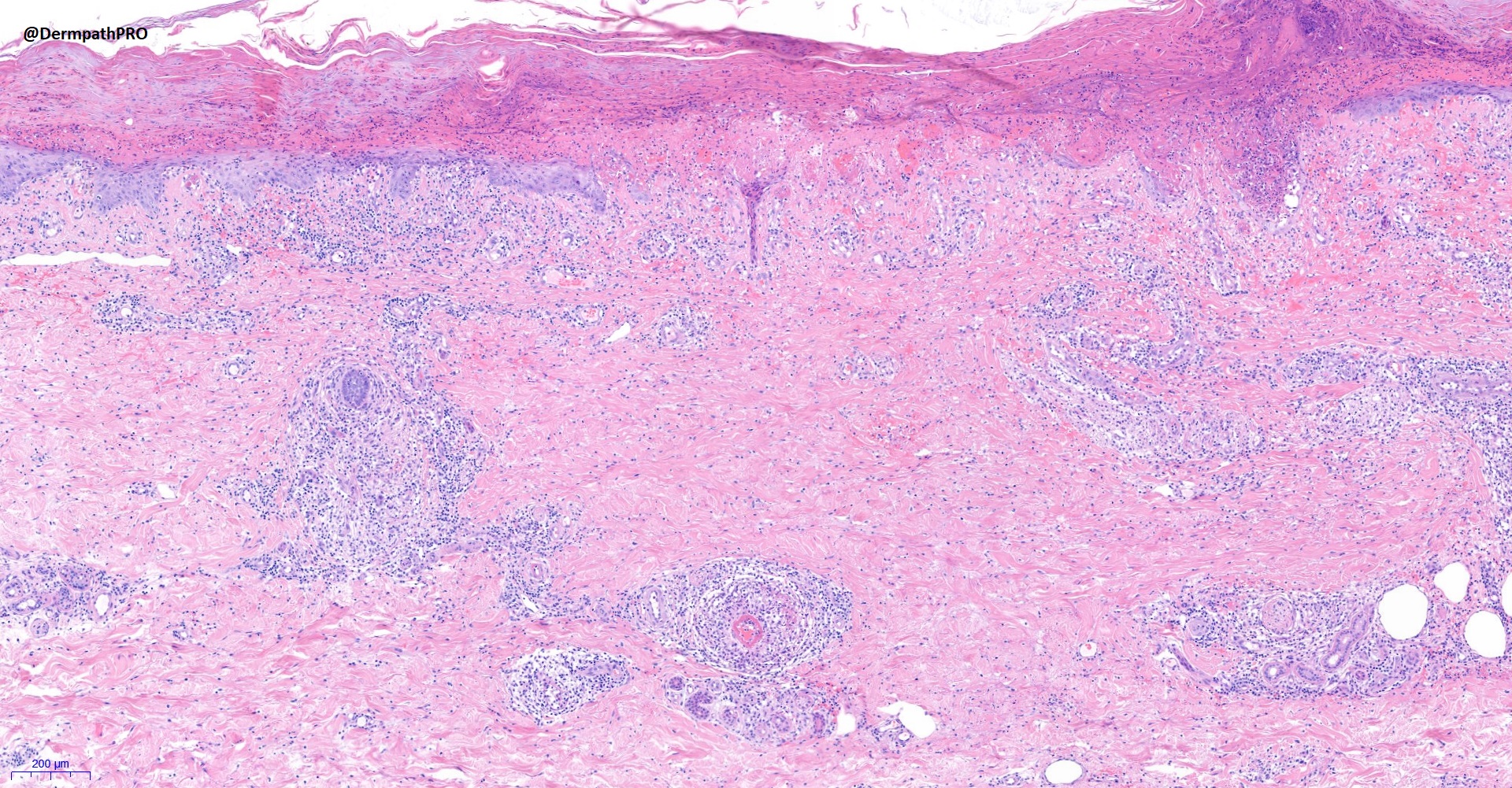
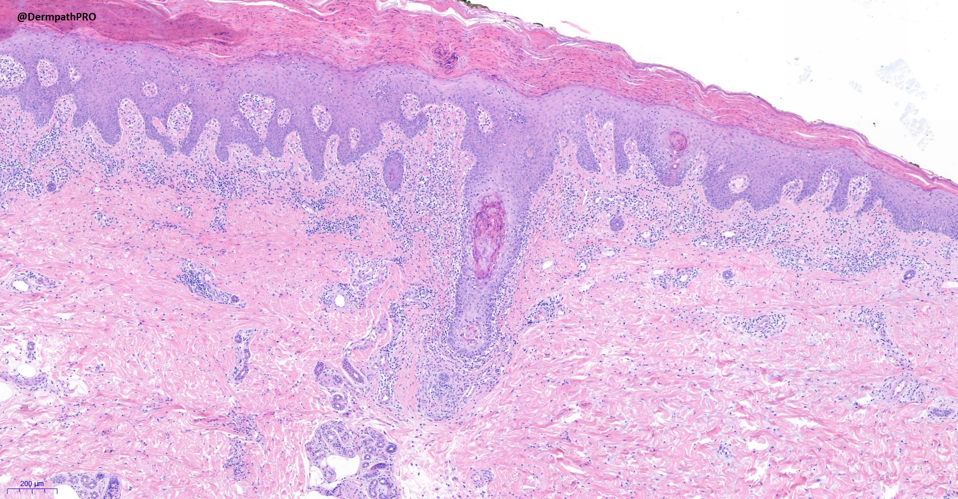
There is an area of epidermal ulceration surmounted by a compact scale crust.  There is a perivascular and periadnexal lymphocytic infiltrate which looks benign cytologically, but which suggest a diagnosis of lupus.  In places the infiltrate is almost granulomatous and there is a focus of thrombotic vasculopathy.  In places the follicles show apoptosis with a neutrophilic infiltrate.

Is this a patient with lupus and infective folliculitis, maybe fungal?

Share this comment


Link to comment
share_externally

Saleem Taibjee

Posted

Some of the histology images also seem to have been omitted. See below:

25475_10.0x.jpg

25475_20.0xe.jpg


Share this comment


Link to comment
share_externally

Saman Fatah

Posted

Pityriasis lichenoides/Lymphomatoid papulosis spectrum depending on further IHC and clinical course (though some cases can be challenging to confidently classify).

Share this comment


Link to comment
share_externally

Neutrophilic folliculitis and vasculitic changes. Suspected herpetic infection.

Share this comment


Link to comment
share_externally

Richard Logan

Posted

This looks like pityriasis lichenoides acuta clinically.  There doesn't seem to be sufficient cytological atypia to support lymphomatoid papulosis, and I still think the overall pattern is more lupus-like.  I agree that disseminated herpes is also a possbility.  Is the patient immunosuppressed?

Share this comment


Link to comment
share_externally

Saleem Taibjee

Posted

Here is the CD30 below, rather unexciting I'm afraid.

25475_5.0 CD30.jpg

25475_10.0x CD30.jpg


Share this comment


Link to comment
share_externally

Saman Fatah

Posted

Thanks Saleem for further immunos..

Last through of the dice, was there any involvement of palms and soles? the great mimicker (syphilis) can have a clinical resemblance to pityriasis lichenoides? 

Share this comment


Link to comment
share_externally

Saleem Taibjee

Posted

Thanks to everyone for the interesting dialogue on this challenging case.

I too initially found it difficult to reconcile the various histological features (ulceration centrally, adjacent epidermis showing parakeratotic foci with apoptotic keratinocytes and interface change including basal vacuolar degeneration, somewhat wedge-shaped infiltrate, foci of genuine vasculitis, and most curiously, the necrotic keratinocytes conspicuously involving hair follicles) to come up with a unifying diagnosis based on the initial clinical information provided.

However, once the clinician provided the clinical photos I was immediately drawn to a diagnosis of PLEVA, and after a further clinicopathological multidisciplinary meeting I understand that is the favoured diagnosis. The patient did not respond to initial treatment including erythromycin, but is now doing well on methotrexate.

All of the various differential diagnoses as mentioned above were considered. As you can see the CD30 stain is not suggestive of LyP. Various infections were excluded including syphilis (negative serology), HIV, toxoplasma, CMV, EBV, etc. IHC for Herpes simplex and zoster were negative. Vasculitis and connective tissue disease screening also negative.

As I mention, perhaps the most interesting aspect of the case is the follicular apoptosis/necrotic keratinocytes. After an extensive review of the literature and textbooks, I have found virtually no mention of this in PLEVA, so I think this is an important 'new' observation. We intend to review past cases and examine deeper levels to ascertain if this is more commonly observed than previously appreciated. Please do let me know if you come across any such cases, which could be included for a possible publication (saleemtaibjee@gmail.com).

I wish to acknowledge Malcolm Rustin (clinician), Alistair Robson and Florence Deroide in this interesting case.

Share this comment


Link to comment
share_externally

Richard Logan

Posted

Fascinating Saleem.  I think it has long been suspected that pityriasis lichenoides may be a reaction pattern to a viral infection.  Perhaps we haven't yet found the right virus?

Share this comment


Link to comment
share_externally


Join the conversation

You can post now and register later. If you have an account, sign in now to post with your account.

Guest
Add a comment...

×   Pasted as rich text.   Paste as plain text instead

  Only 75 emoji are allowed.

×   Your link has been automatically embedded.   Display as a link instead

×   Your previous content has been restored.   Clear editor

×   You cannot paste images directly. Upload or insert images from URL.

Loading...

×
×
  • Create New...