Jump to content
In this section we have spot diagnoses posted on a daily basis since June 2010, now over 4000! You can review the archived cases and read the suggested diagnoses by users and the final comment by the contributors.
Case are uploaded each week day by 10 am UK time with the correct diagnosis will generally be posted at 8 pm UK time. Why not view the most recent spot diagnosis and proffer a diagnosis?

Case Number : Case 2757 - 29 January 2021 Posted By: Dr. Richard Carr

Please read the clinical history and view the images by clicking on them before you proffer your diagnosis.
Submitted Date :
   (0 reviews)

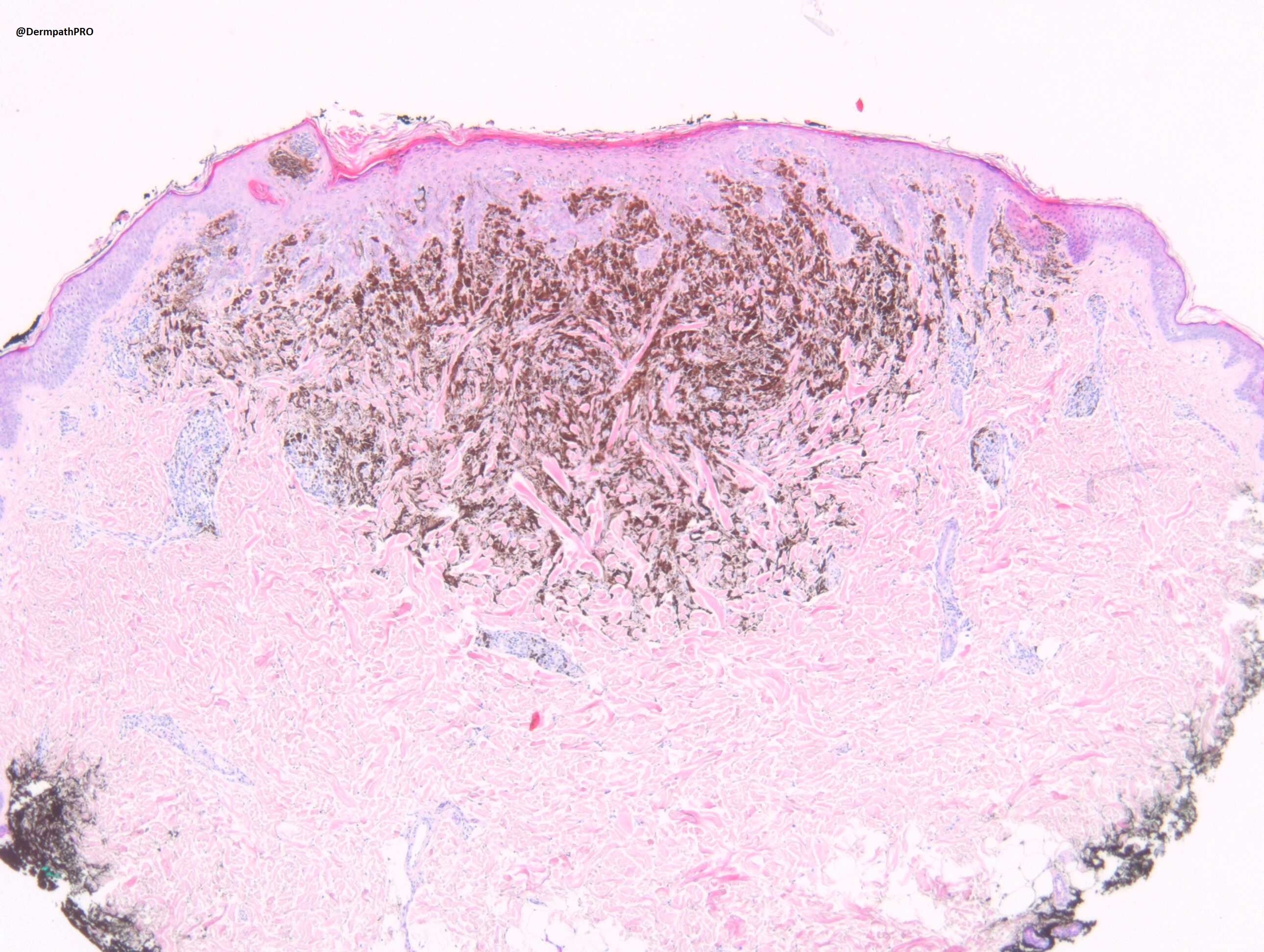
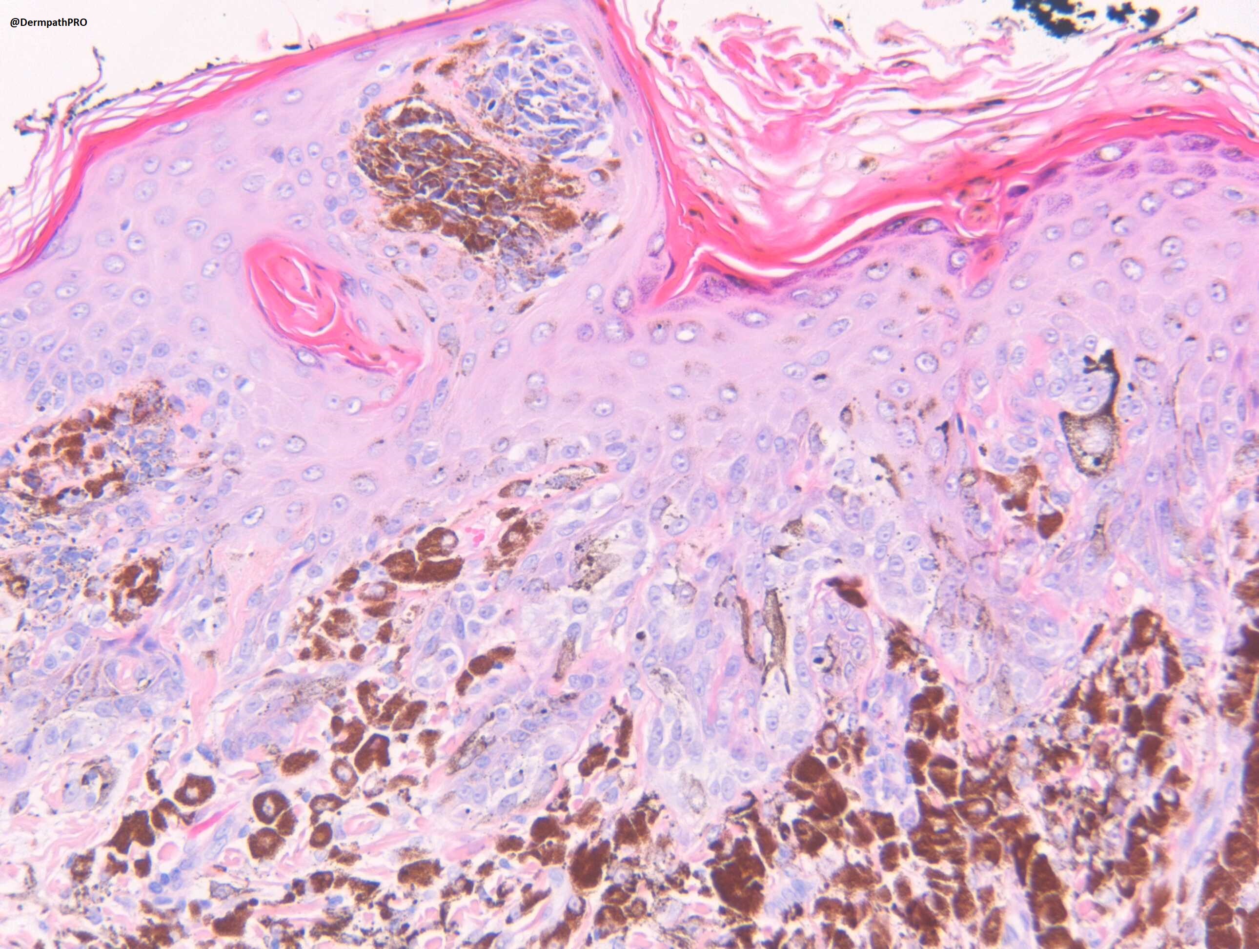
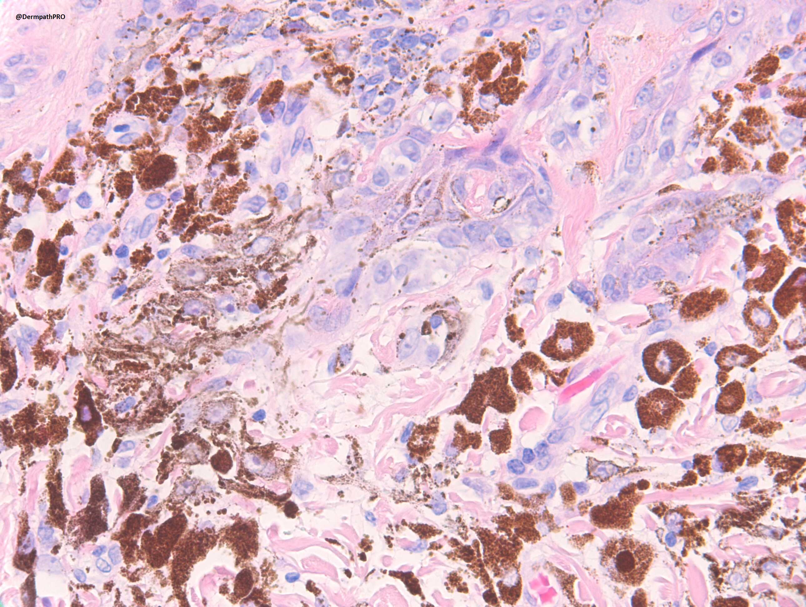
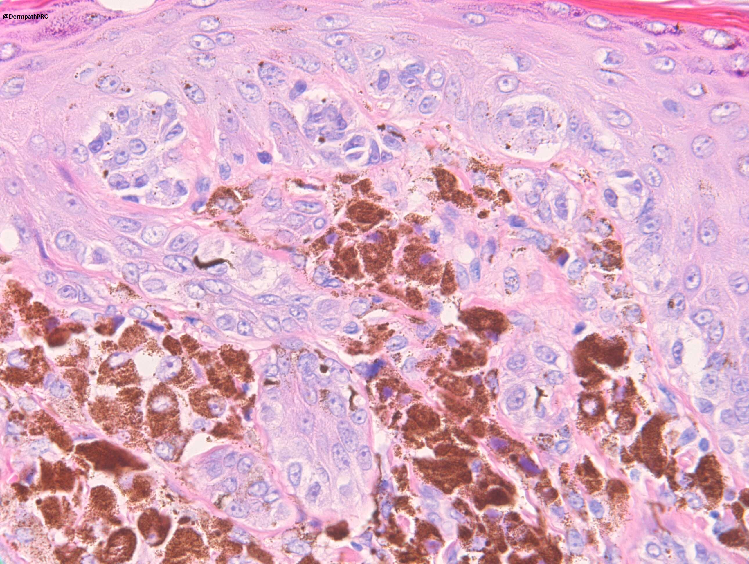
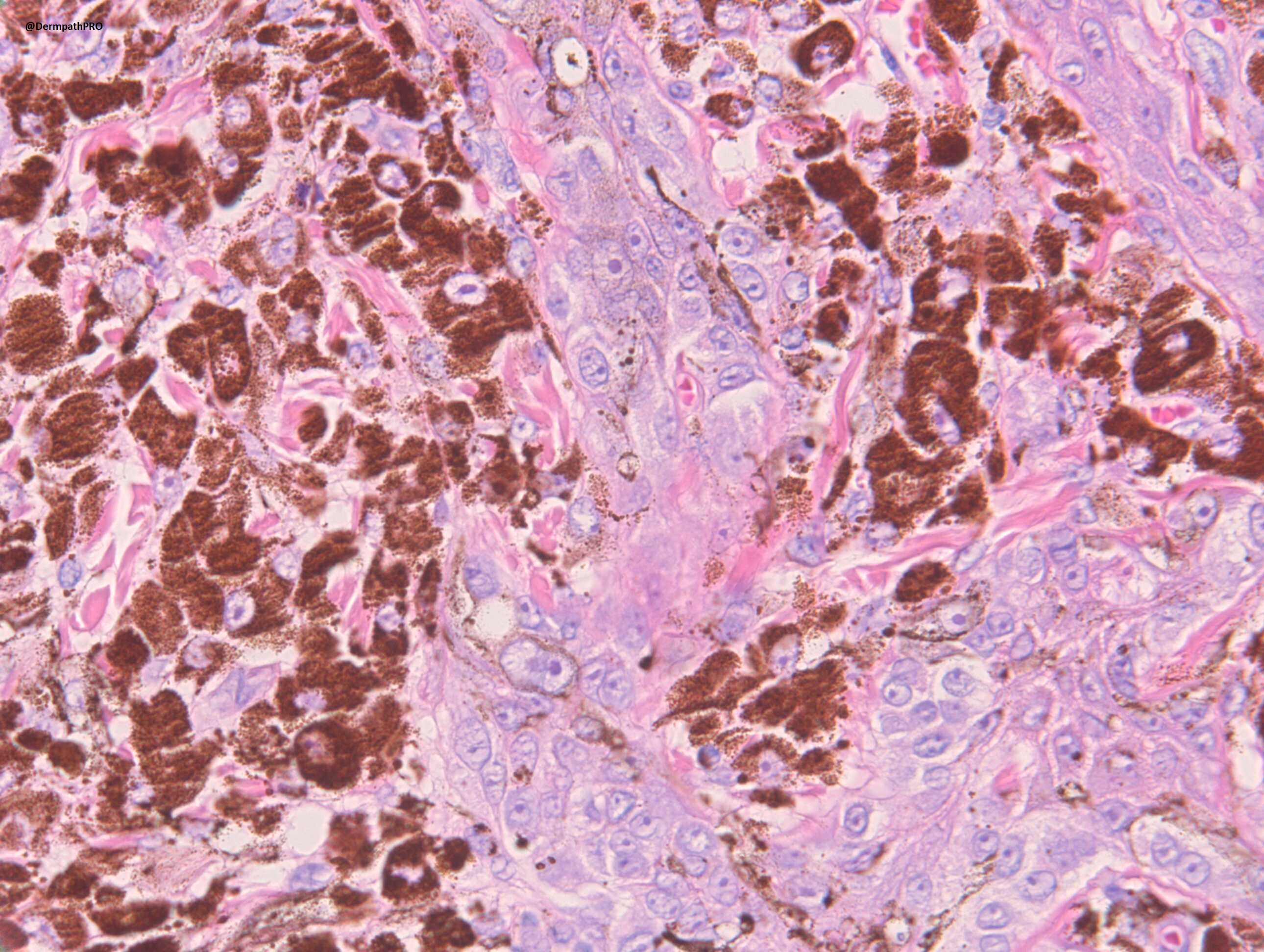
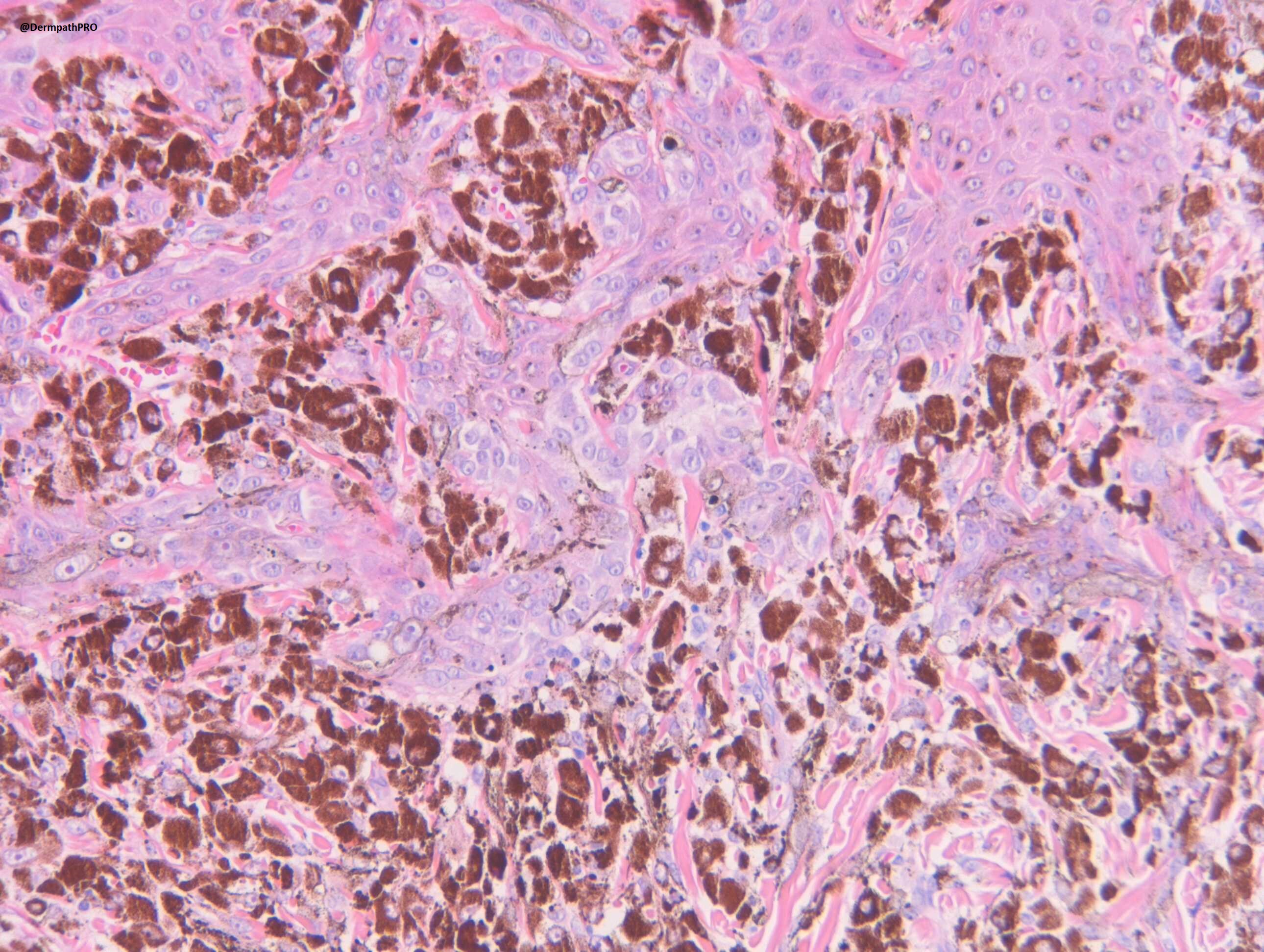
M10. Site not stated. Exclude melanoma.


  Report Record

User Feedback


Carmen Lisievici

Posted

I would take into consideration a pigmented epithelioid melanocytoma, mostly due to the presence of epidermal hyperplasia and prominent nucleoli.

Share this comment


Link to comment
share_externally

Totally agree: PEM. It could show a PRKRA1 related cytoplasmic protein loss.  

Share this comment


Link to comment
share_externally

Meenakshi Batrani

Posted

I would consider what some call as BLITZ (combination of Blue nevyus+spitz nevus) or some other call such lesions as superficial DPN. PEMC also a possibility. Tough to differentiate between these lesions. 

Share this comment


Link to comment
share_externally

Dr. Richard Carr

Posted

Agree with the favoured diagnosis of PEM. Clues here include a slightly more naevocellular junctional nest (PEM are often combined with naevocellular naevi that are BRAF positive - although this case was BRAF negative). They can be near impossible to distinguish from epithelioid variant of blue naevus but blue naevi are usually S100 weak or negative (compared with strong MelanA) and this case was S100 positive. DPN is another challenging differential but they usually show cytoplasmic & nuclear beta-catenin. We don't have PRKRA1 available but I think given the lesion is fairly typical for PEM and lacked worrying histological features I was happy to sign it out as a PEM. One should probably ensure complete removal and enquire as to the possibility of associated Carney's. Regarding BLITZ this is not a good term "blue" and "Spitz" are mutually exclusive based on driver mutations. So you can have a blue naevus-like Spitz or a Spitz-like blue neavus but BLITZ is not a thing. It should be noted that or two PEMs in one of the larger papers are re-classified as blue naevi based on their driver mutations. Also it is possible to have a blue naevus driver mutation with a PRKRA1 abnormality! i.e. a "blue PEM"

Share this comment


Link to comment
share_externally


Join the conversation

You can post now and register later. If you have an account, sign in now to post with your account.

Guest
Add a comment...

×   Pasted as rich text.   Paste as plain text instead

  Only 75 emoji are allowed.

×   Your link has been automatically embedded.   Display as a link instead

×   Your previous content has been restored.   Clear editor

×   You cannot paste images directly. Upload or insert images from URL.

Loading...

×
×
  • Create New...