Jump to content
In this section we have spot diagnoses posted on a daily basis since June 2010, now over 4000! You can review the archived cases and read the suggested diagnoses by users and the final comment by the contributors.
Case are uploaded each week day by 10 am UK time with the correct diagnosis will generally be posted at 8 pm UK time. Why not view the most recent spot diagnosis and proffer a diagnosis?

Case Number : Case 2866 - 01 July 2021 Posted By: Saleem Taibjee

Please read the clinical history and view the images by clicking on them before you proffer your diagnosis.
Submitted Date :
   (0 reviews)

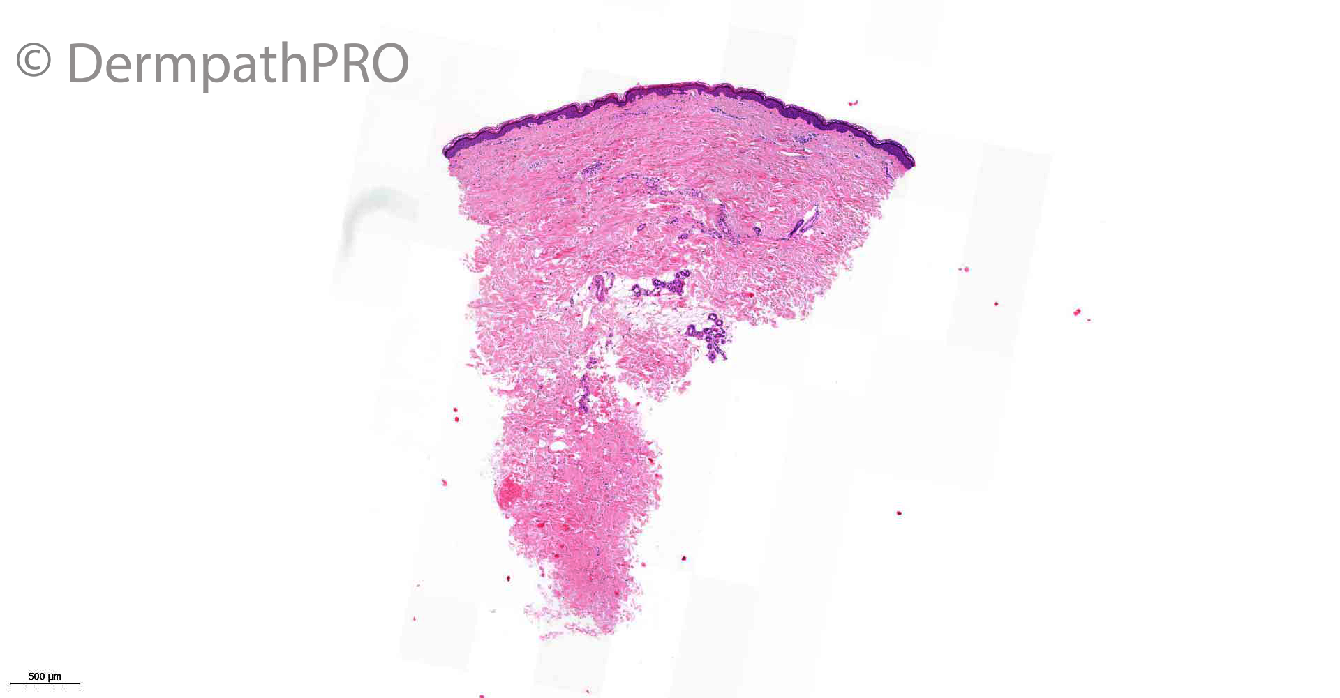
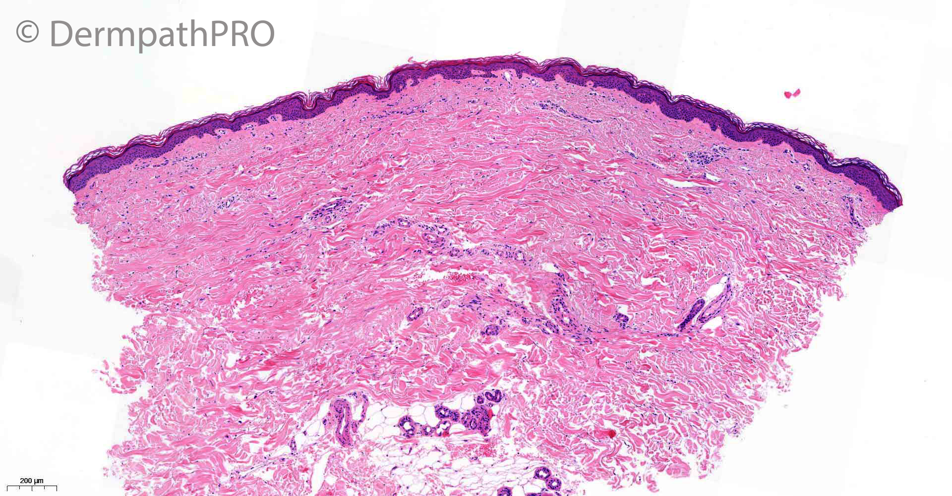
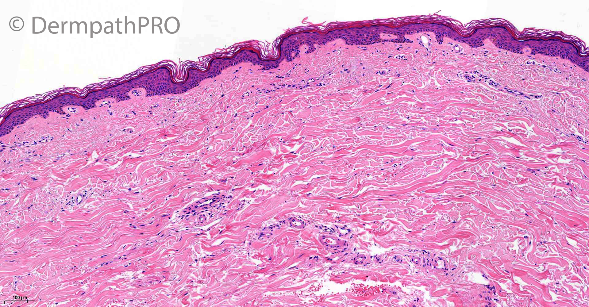
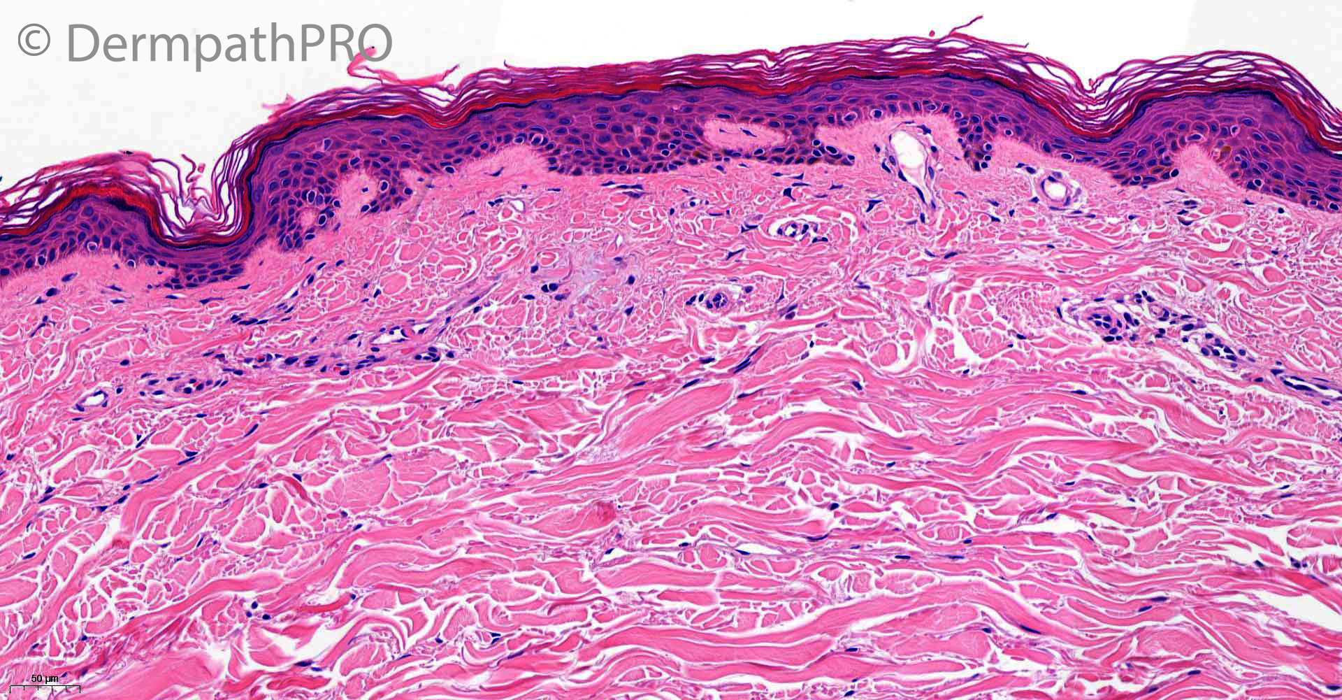
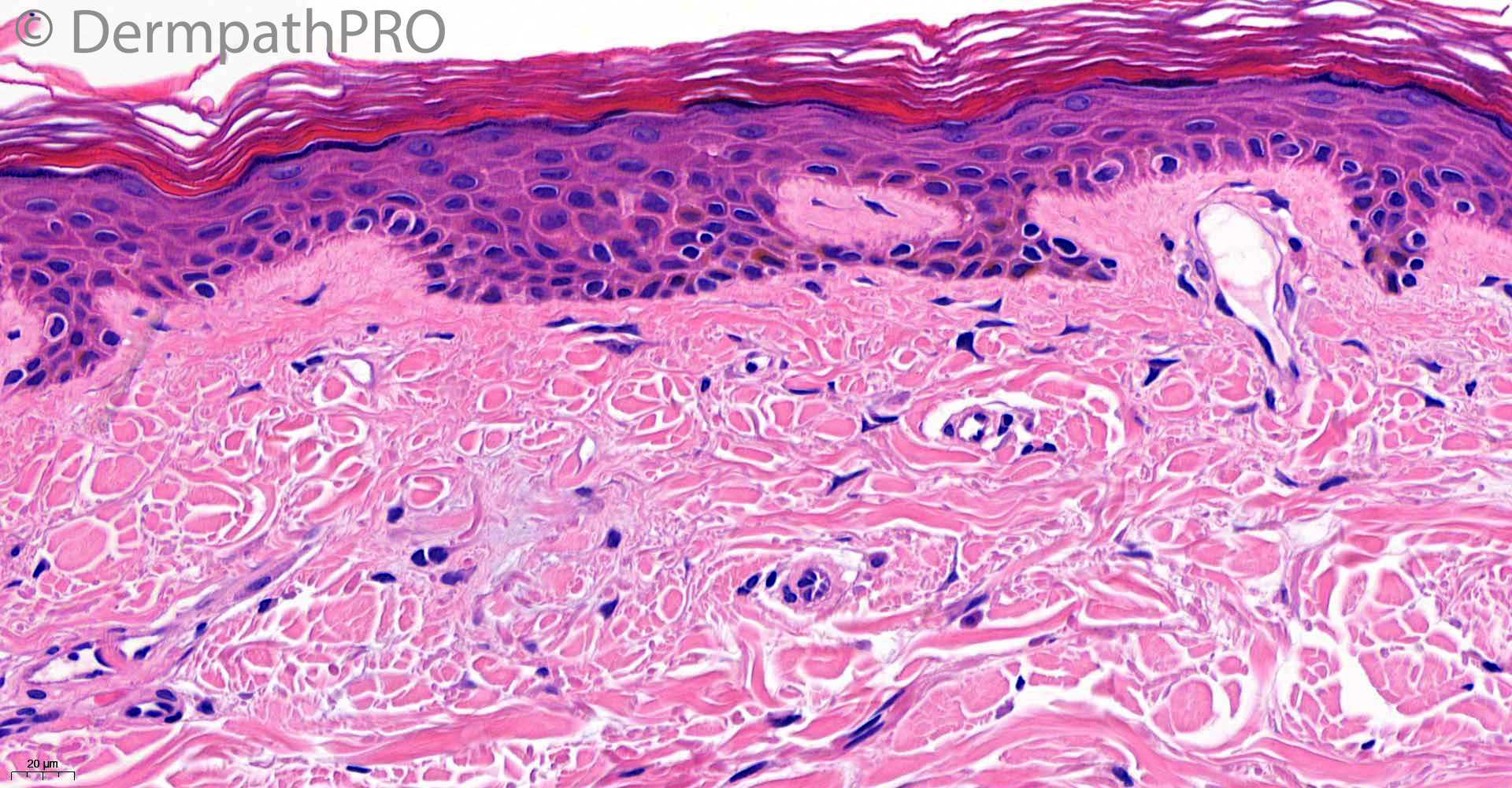
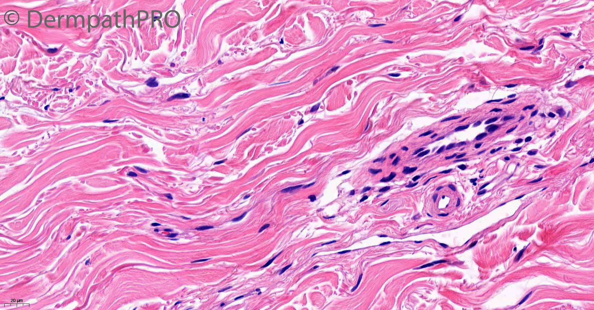
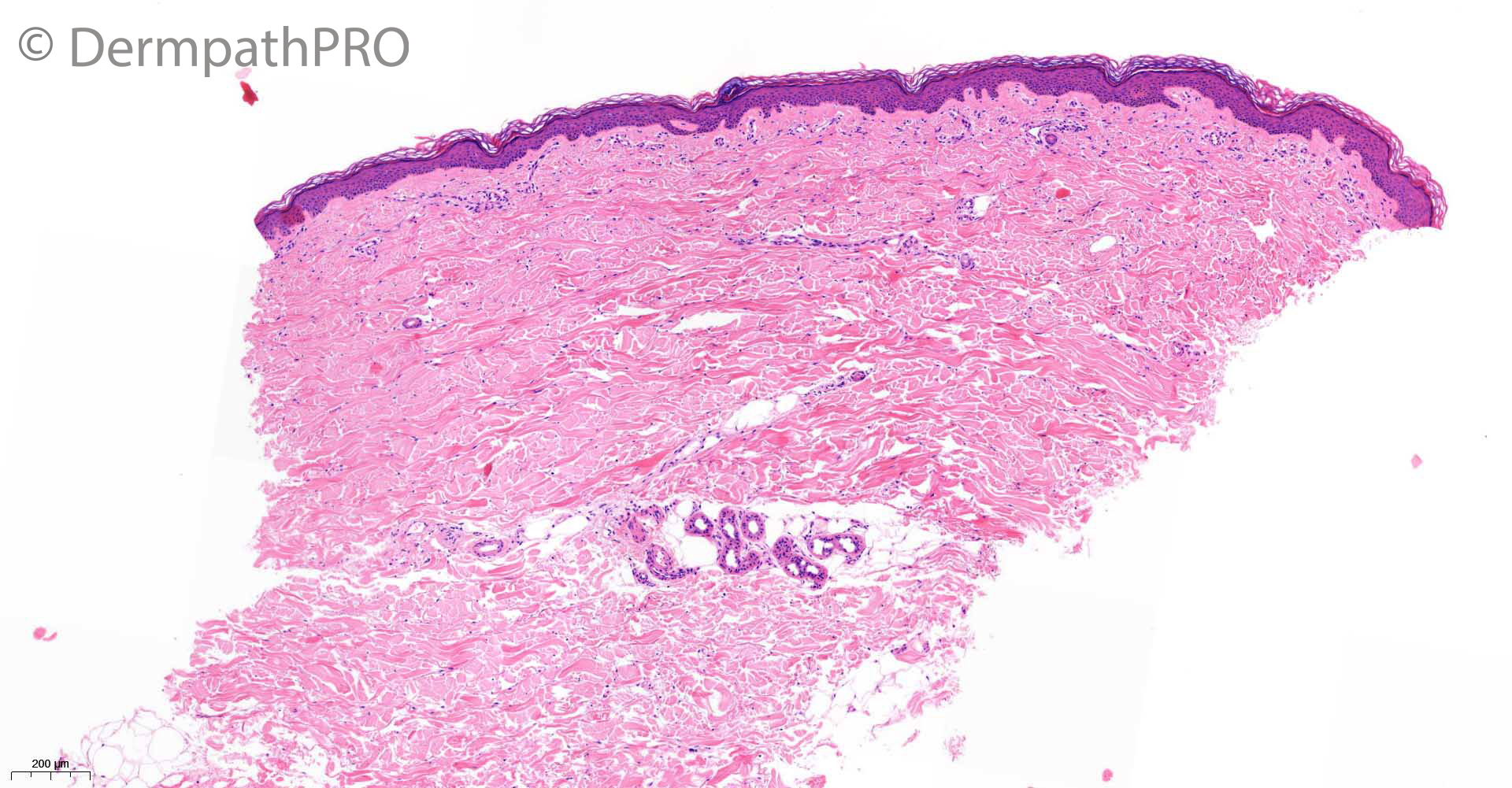
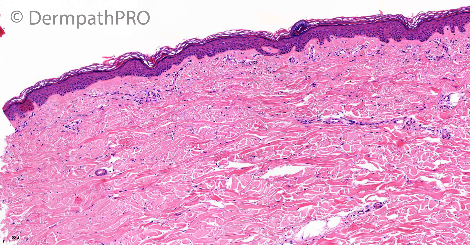
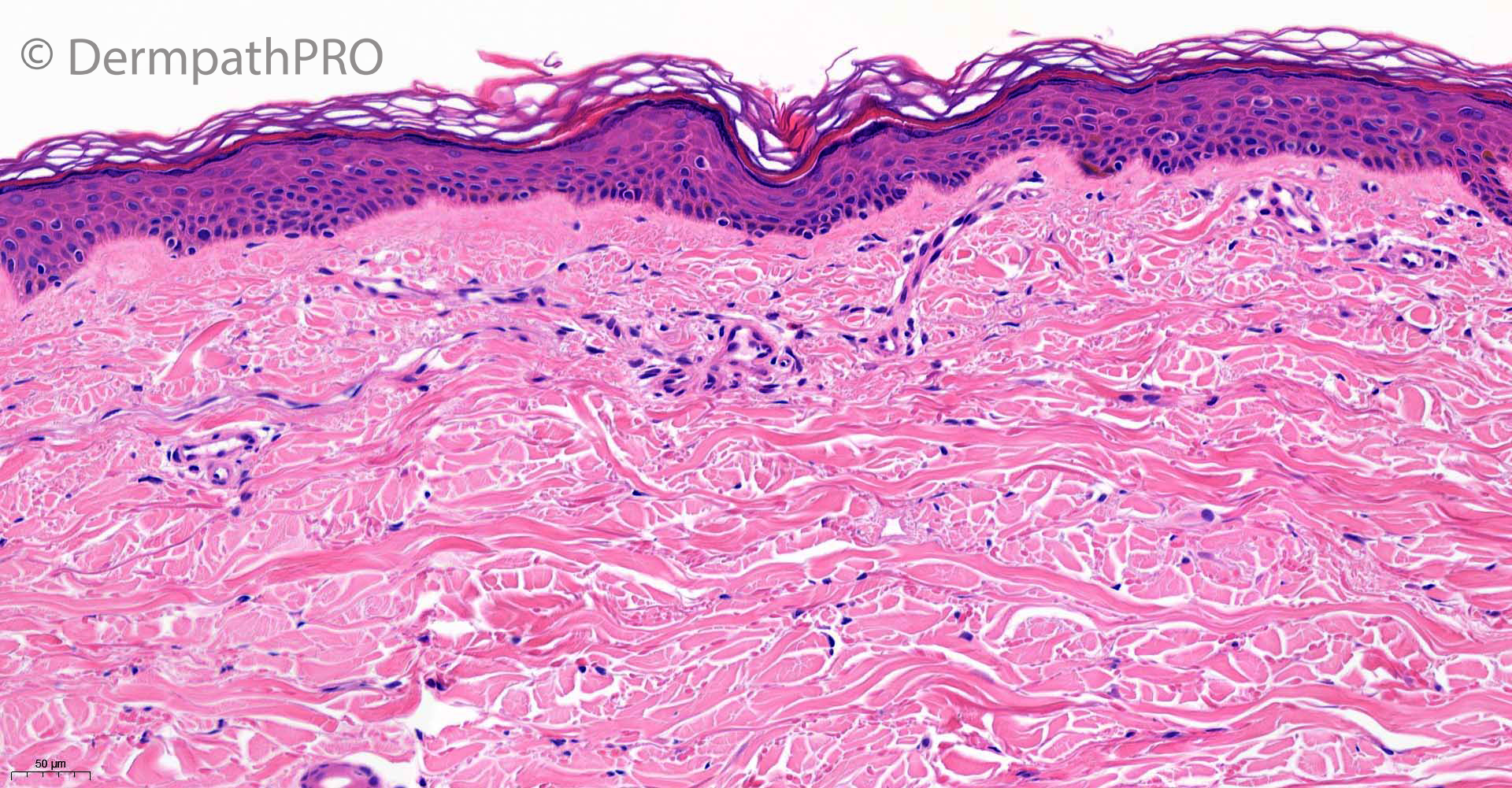
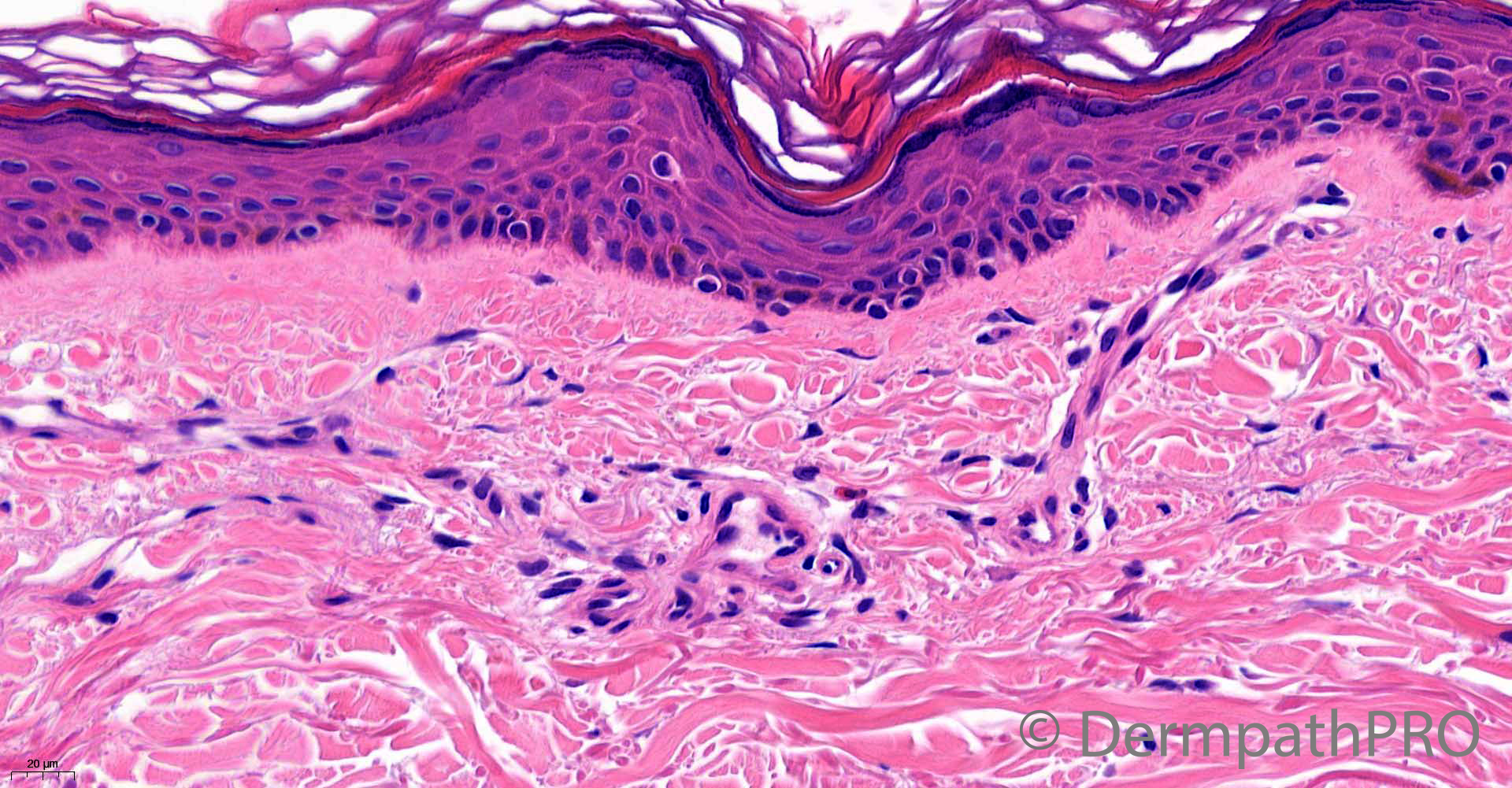
64F, punch biopsies x 2, left thigh. Widespread red/brown macules on both thighs.


  • Like 1
  Report Record

User Feedback


Krishnakumar subramanian

Posted

epidermis looks normal. there is one or two superficial dermal perivascular eosinophil and lymphocyte

? drug rash [ no basal cell damage]


Share this comment


Link to comment
share_externally

Eman El-Nabarawy

Posted

Patch stage kaposi sarcoma. Nice promontory!

Share this comment


Link to comment
share_externally

daniellindsay

Posted

Finding this difficult. There are some extravasated erythrocytes, made me consider a pigmented purpuric dermatosis, but no haemosiderin deposition! Would do levels, subtle changes in what we have here. 

Share this comment


Link to comment
share_externally

Lymphocytic vasculitis, tight cuffing of blood vessels with lymphocytes, some extravasated RBCs, May be a sort of pigmented purpuric dermatosis

Share this comment


Link to comment
share_externally

Eman El-Nabarawy

Posted (edited)

? Telangiectasia macularis eruptiva perstans. 

Edited by Eman El-Nabarawy

Share this comment


Link to comment
share_externally

vincenzo

Posted

7 hours ago, Eman El-Nabarawy said:

? Telangiectasia macularis eruptiva perstans. 

Very difficult. But I would agree with Eman. 

Share this comment


Link to comment
share_externally

Alex-Ventura-Leon

Posted

After the clue of "invisible dermatoses" agree with mastocytosis (TEMP)

Share this comment


Link to comment
share_externally

Krishnakumar subramanian

Posted

Thanks a lot sir for discussing this important topic

Share this comment


Link to comment
share_externally

Saleem Taibjee

Posted

Yes, this is a recent case of mastocytosis. But I have to give all the credit to the clinician who had suggested mastocytosis. This prompted the original pathologist to request CD117 (shown below). The case was passed to me to report in the absence of the original pathologist since the report was required urgently with the patient due back to clinic. I was quite surprised when I saw the CD117, and I am certain I would have missed the diagnosis otherwise. I can see that some of you have got the diagnosis, perhaps slightly aided my clue of 'invisible diagnosis'. Certainly it would have been invisible to me but for the CD117 already requested. Many of the mast cells are rather spindly and easily missed on the H&E. The clinician did not suggest a subtype of mastocytosis, and I would probably not attempt to subtype based on the histology. I don't think the vessels are particularly telangiectatic, and would probably favour urticaria pigmentosa (macular-papular cutaneous mastocytosis), but as I say. this aspect needs clinical correlation. Of course, such a patient also needs a work up for potential systemic involvement.

BW, Saleem

22552_20.0x CD117 2nd biopsy.jpg

22552_40.0x CD117 2nd biopsy.jpg

Share this comment


Link to comment
share_externally

Saman Fatah

Posted

Thanks for sharing this great case.


From clinical prospective maculopapular cutaneous mastocytosis in increasing used that encompass both UP/TMEP and much easier for “lumpers”. TMEP is not a common clinical pattern of presentation and even in such cases one can still see tan brown macules and barely raised papules of UP if you look hard enough for them. 
 

Saleem, the reference you kindly provided looks like a possible book chapter in PDF rather than an article. Would you kindly be able to provide further details of that source please (if feasible)?

Share this comment


Link to comment
share_externally

Saleem Taibjee

Posted

Hi Saman. Unfortunately I don't know the source of the list, it came up directly from Google.

Here is another article which looks useful https://www.diagnostichistopathology.co.uk/article/S1756-2317(18)30107-5/fulltext but I don't have access to the pdf. I think I will ask our hospital library to request this article. I have heard Dr Tomasini previously lecture on this subject.

BW
Saleem

Share this comment


Link to comment
share_externally

Saman Fatah

Posted

Thanks very much Saleem, will source the other article too. 

Share this comment


Link to comment
share_externally

Saleem Taibjee

Posted

I have the pdf of the article. If you want to give me your email address, I can email it to you.

Share this comment


Link to comment
share_externally

Saman Fatah

Posted

Very noble of you Saleem, I sent a separate message which I hope you will be able to access in the top right corner next to the bell once you signed in.

Share this comment


Link to comment
share_externally

Very interesting discussion about mastocytosis as an "invinsive dermatosis". I think there has been quite some arguments about "how much" is "too much" as for the exact numbers of mast cells count. what criteria you guys use to define it is too much and need be consider as cutanoeus mastocytosis? and then when you think it is too much, how do you define the increased mast cells are reactive like those seen in inflmmatory dermatosis, or neoplastic as in mastocytosis? love to hear your input. Saleem.

On 06/07/2021 at 12:12, Saleem Taibjee said:

Yes, this is a recent case of mastocytosis. But I have to give all the credit to the clinician who had suggested mastocytosis. This prompted the original pathologist to request CD117 (shown below). The case was passed to me to report in the absence of the original pathologist since the report was required urgently with the patient due back to clinic. I was quite surprised when I saw the CD117, and I am certain I would have missed the diagnosis otherwise. I can see that some of you have got the diagnosis, perhaps slightly aided my clue of 'invisible diagnosis'. Certainly it would have been invisible to me but for the CD117 already requested. Many of the mast cells are rather spindly and easily missed on the H&E. The clinician did not suggest a subtype of mastocytosis, and I would probably not attempt to subtype based on the histology. I don't think the vessels are particularly telangiectatic, and would probably favour urticaria pigmentosa (macular-papular cutaneous mastocytosis), but as I say. this aspect needs clinical correlation. Of course, such a patient also needs a work up for potential systemic involvement.

BW, Saleem

22552_20.0x CD117 2nd biopsy.jpg

22552_40.0x CD117 2nd biopsy.jpg


Share this comment


Link to comment
share_externally

Saleem Taibjee

Posted

Hi Henry

Honest truth is that I don't have good criteria for how many mast cells are too many i.e. for when it is mastocytosis, and this is an area which also vexes me.

I don't know whether this has been systematically studied?

BW

Saleem

Share this comment


Link to comment
share_externally

9 hours ago, Saleem Taibjee said:

Hi Henry

Honest truth is that I don't have good criteria for how many mast cells are too many i.e. for when it is mastocytosis, and this is an area which also vexes me.

I don't know whether this has been systematically studied?

BW

Saleem

Thank you Saleem. here are some from the Steven Billings book. but still not clear as to what about cases with  12 mast cell/HPF? 图像

图像


Share this comment


Link to comment
share_externally

In the following book, For TMEP, the criteria is

on special stains:

>=10 masts/HPF/perivacular: Dx

6-9/HPF: Suspicious

3-5/HPF: Typical Clinical Features for Dx

图像

Then, there are papers stating this:  https://pubmed.ncbi.nlm.nih.gov/19719830/   ( All these materials are courtesy from Twitter discussions about TMEP)

图像


Share this comment


Link to comment
share_externally

Saleem Taibjee

Posted

It's a confusing area. I found this table from a recent paper Kirsten N et al, JEADV 2017, but noting that the numbers are /mm2 rather than /HPF. One of the authors' message is that mast cell number count can vary significantly from one section to the next in the same biopsy. There did not seem to be significant differences in staining between IHC for CD117 and tryptase. The mean values in TMEP are really quite low, very much seeming to emphasise that the mast cell numbers can be at the upper end of 'normal'. The diagnosis seems to depend very much on Clinicopathological correlation, which does feel a bit unsatisfactory from an objective aspect, and one also wonders the effect of anatomical site. This is starting to show some parallels with examples of paucicellular lentigo maligna, where again absolute numbers of cells can be unreliable.

image.png

2016 Jan;107(1):12-7.


Overexpression of p16(INK4a) in Mastocytosis (Urticarial Pigmentosa)

 
  • PMID: 27333655

Abstract

The expression of p16(INK4a) has been reported to induce cell-cycle arrest and cellular senescence. The p16(INK4a) expression has never been examined in human mast cells and mastocytosis. We immunohistologically examined the expression of p16(INK4a) and tryptase in 5 normal human skin and 4 mastocytosis. In normal mast cells, only 5.9 ± 3.4 (mean ± standard deviation) % of tryptase-positive mast cells coexpressed p16(INK4a). However, significantly higher percentage (86.0 ± 14.1%) of tryptase-positive tumor cells was immunoreactive to p16(INK4a) in all of 4 mastocytosis. The p16(INK4a) overexpression may induce the senescence of neoplastic mast cells to undergo spontaneous regression of mastocytosis.

Share this comment


Link to comment
share_externally

Saleem Taibjee

Posted

Going forwards, ideally we need a marker of neoplastic vs reactive mast cells, if such will ever exist, assuming that the mast cells in mastocytosis are clonally expanded/neoplastic? I don't get the impression that histology morphology of the mast cells allows us to distinguish cutaneous mastocytosis from normal.

The paper above on p16 looks interesting. I think I might try it out!

Share this comment


Link to comment
share_externally


Join the conversation

You can post now and register later. If you have an account, sign in now to post with your account.

Guest
Add a comment...

×   Pasted as rich text.   Paste as plain text instead

  Only 75 emoji are allowed.

×   Your link has been automatically embedded.   Display as a link instead

×   Your previous content has been restored.   Clear editor

×   You cannot paste images directly. Upload or insert images from URL.

Loading...

×
×
  • Create New...