-
 1
1
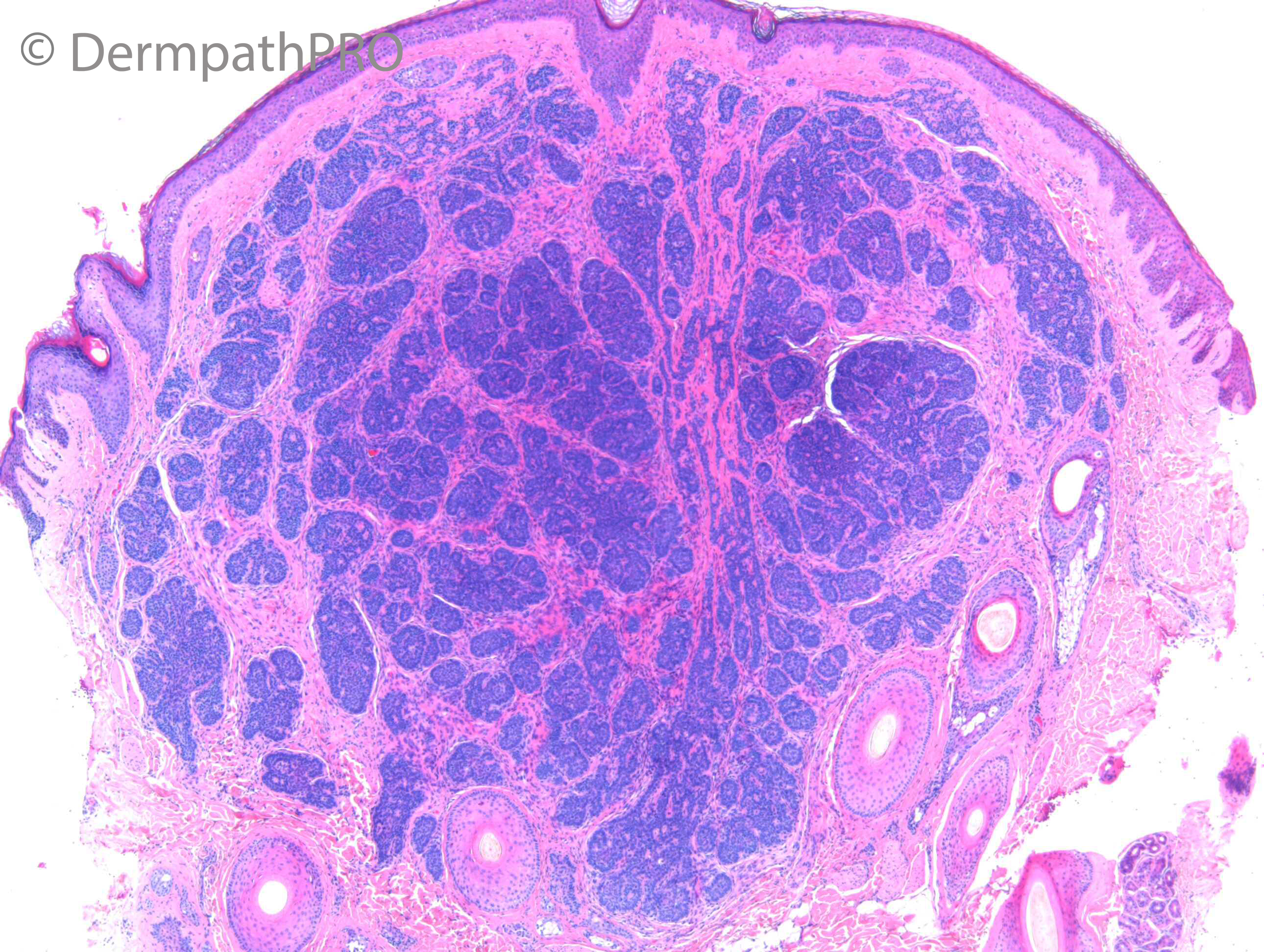
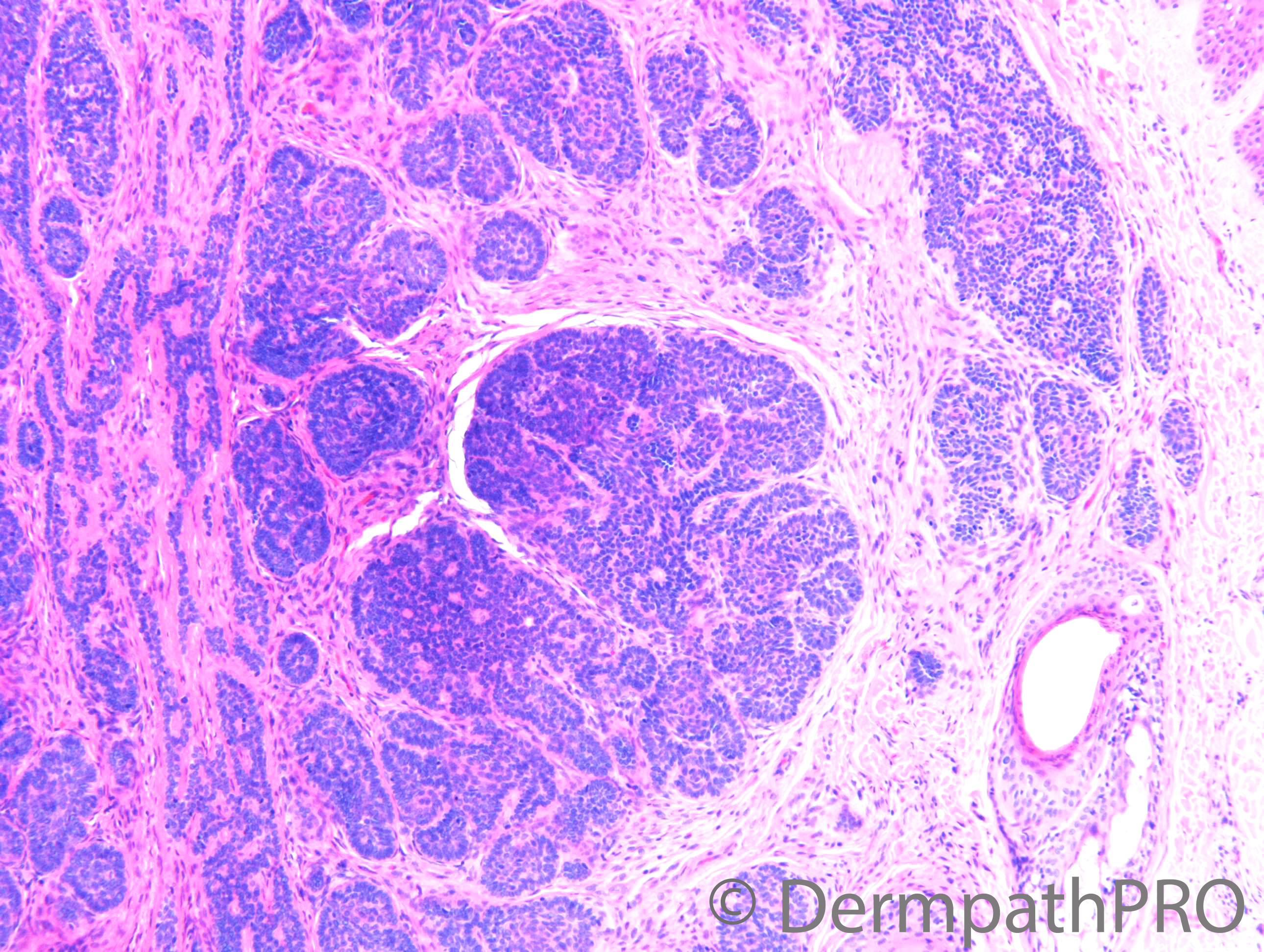
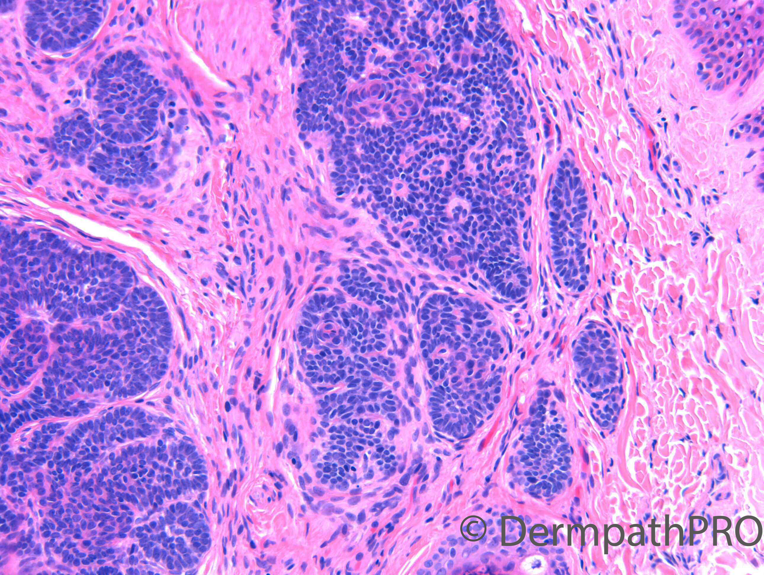
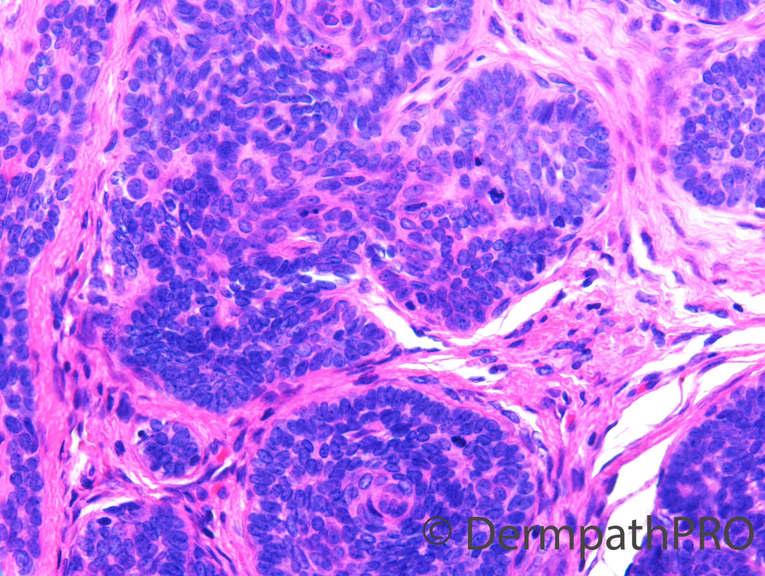
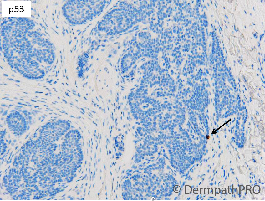
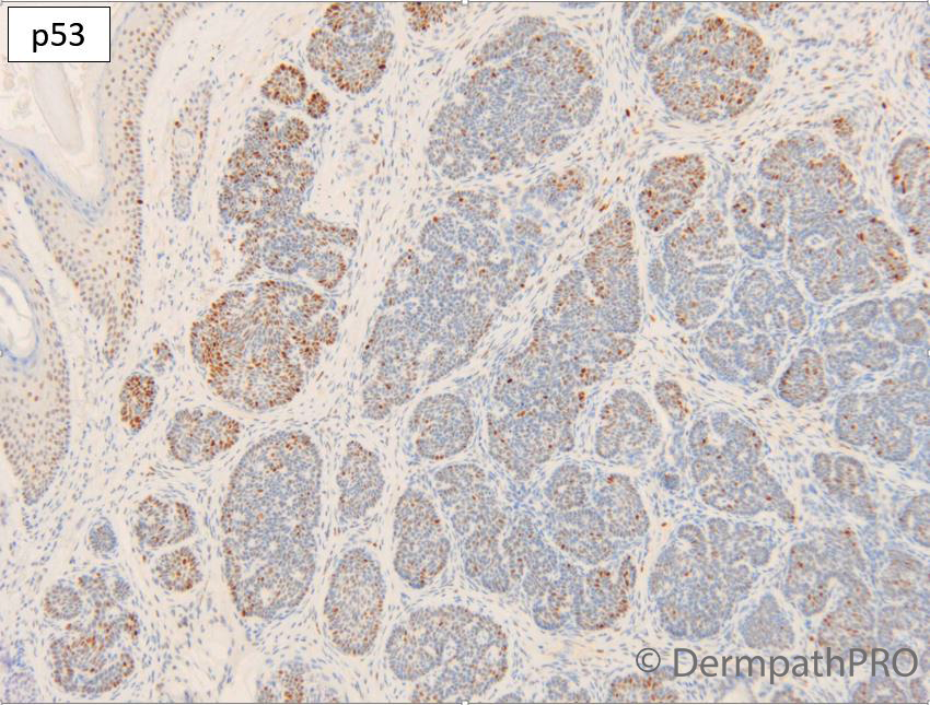
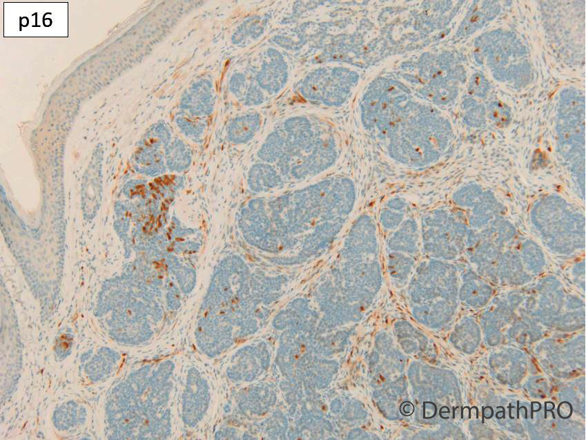
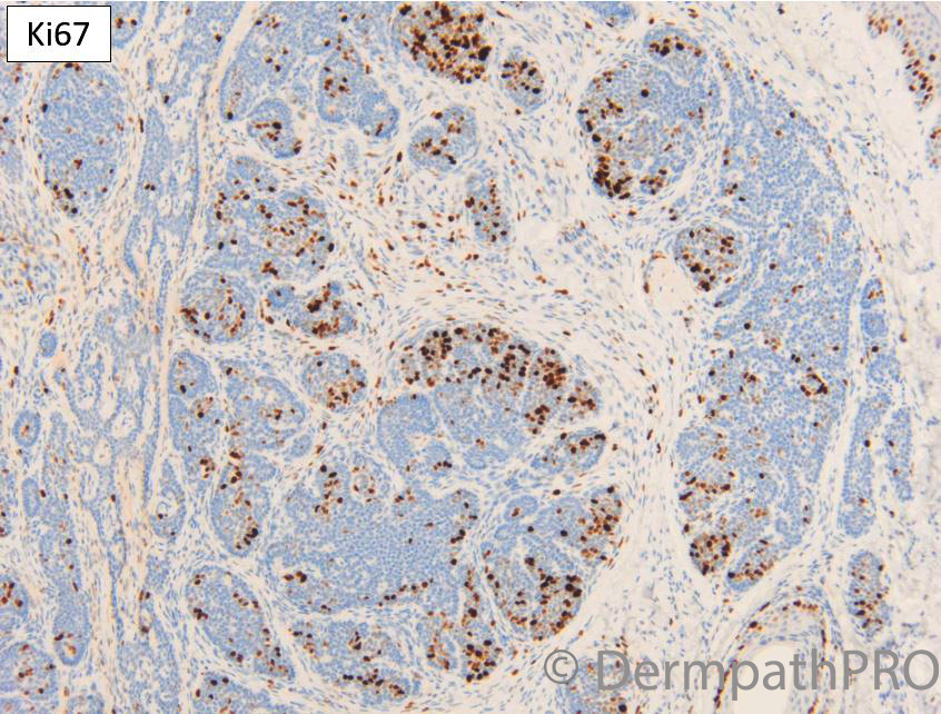
Diagnostic Pearls : Case 2867 - 02 July 2021
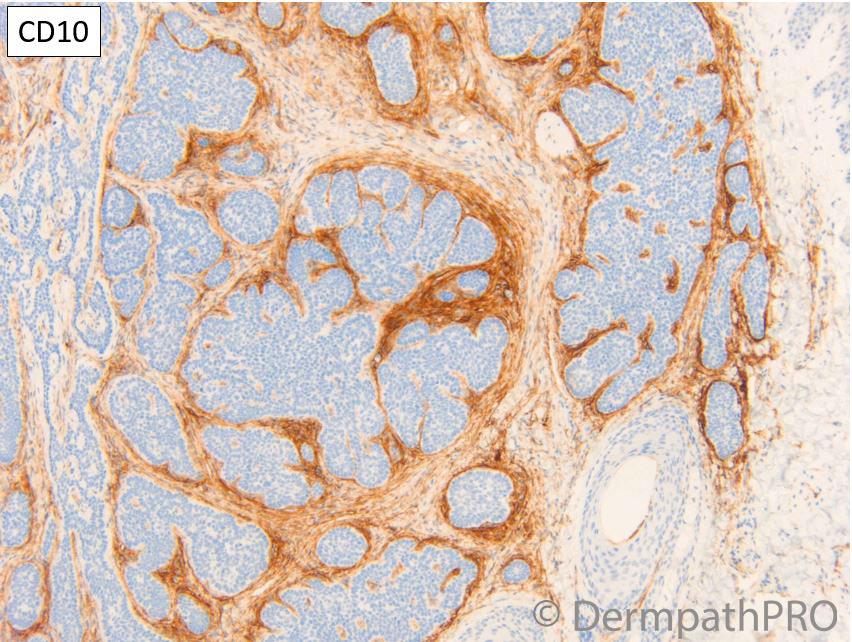
F20. Scalp cyst.
Dr. Richard Carr
Posted 30/06/21
Posted 30/06/21
 1
1
F20. Scalp cyst.
Join the conversation
You can post now and register later. If you have an account, sign in now to post with your account.