Jump to content
In this section we have spot diagnoses posted on a daily basis since June 2010, now over 4000! You can review the archived cases and read the suggested diagnoses by users and the final comment by the contributors.
Case are uploaded each week day by 10 am UK time with the correct diagnosis will generally be posted at 8 pm UK time. Why not view the most recent spot diagnosis and proffer a diagnosis?

Case Number : Case 2869 - 06 July 2021 Posted By: Iskander H. Chaudhry

Please read the clinical history and view the images by clicking on them before you proffer your diagnosis.
Submitted Date :
   (0 reviews)

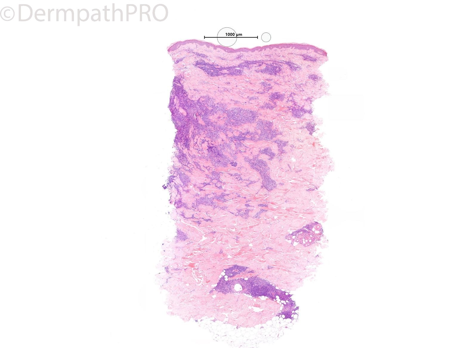
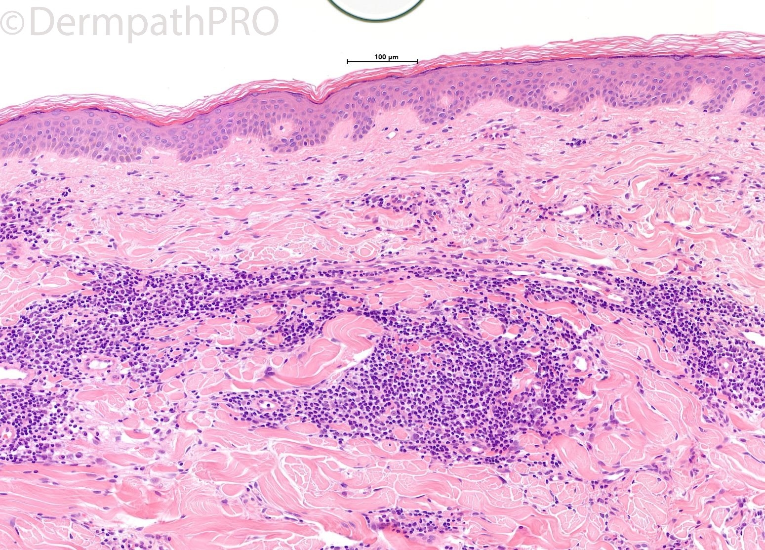
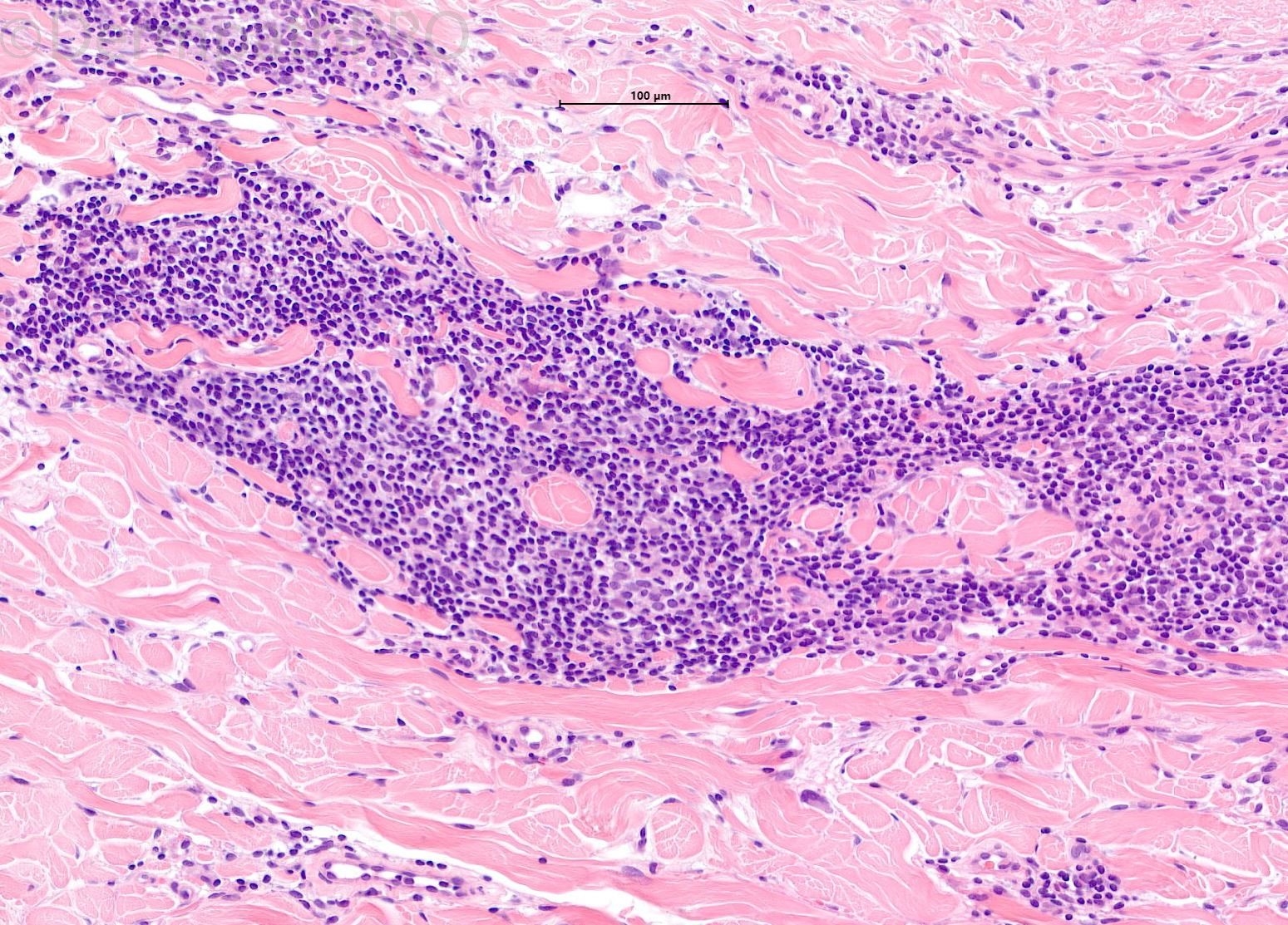
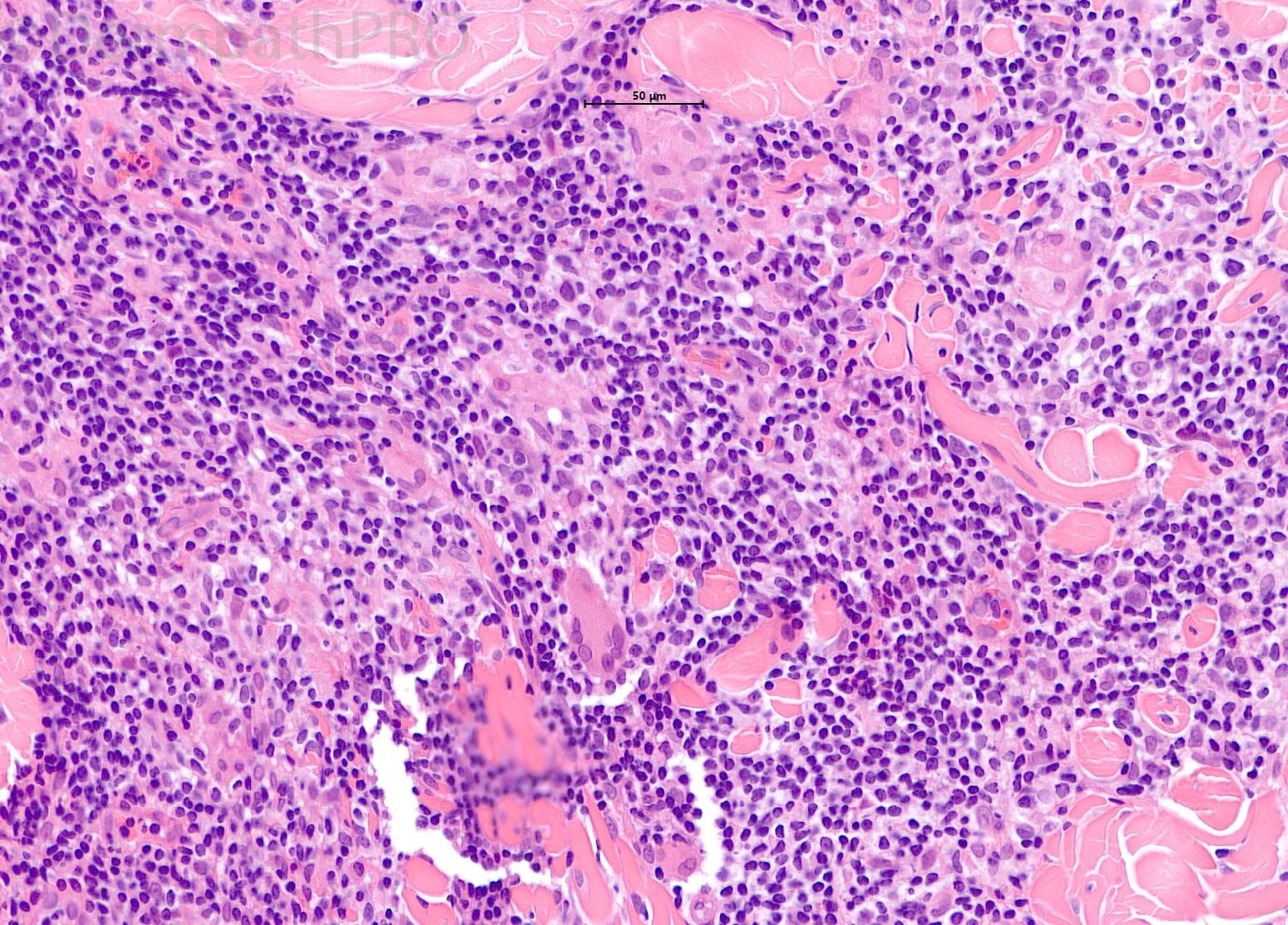
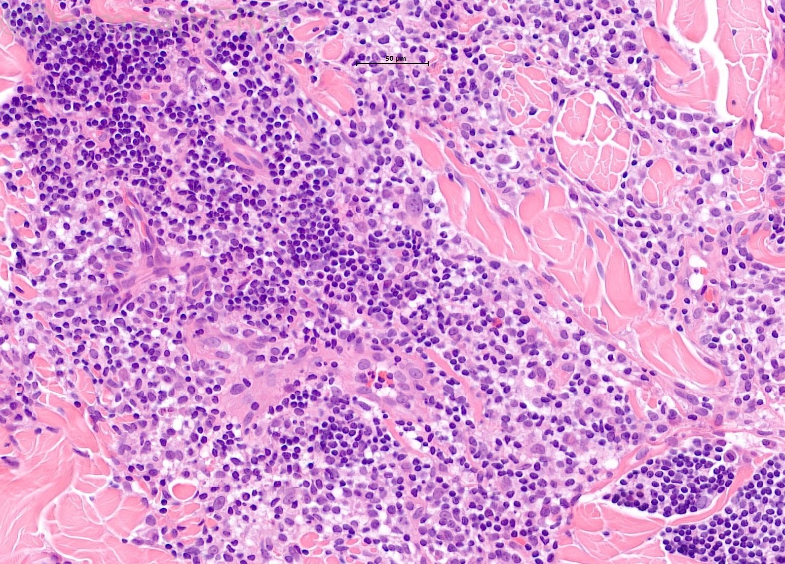
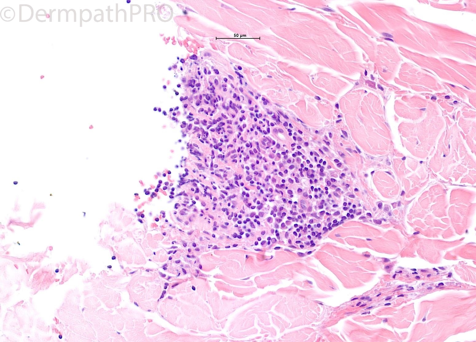
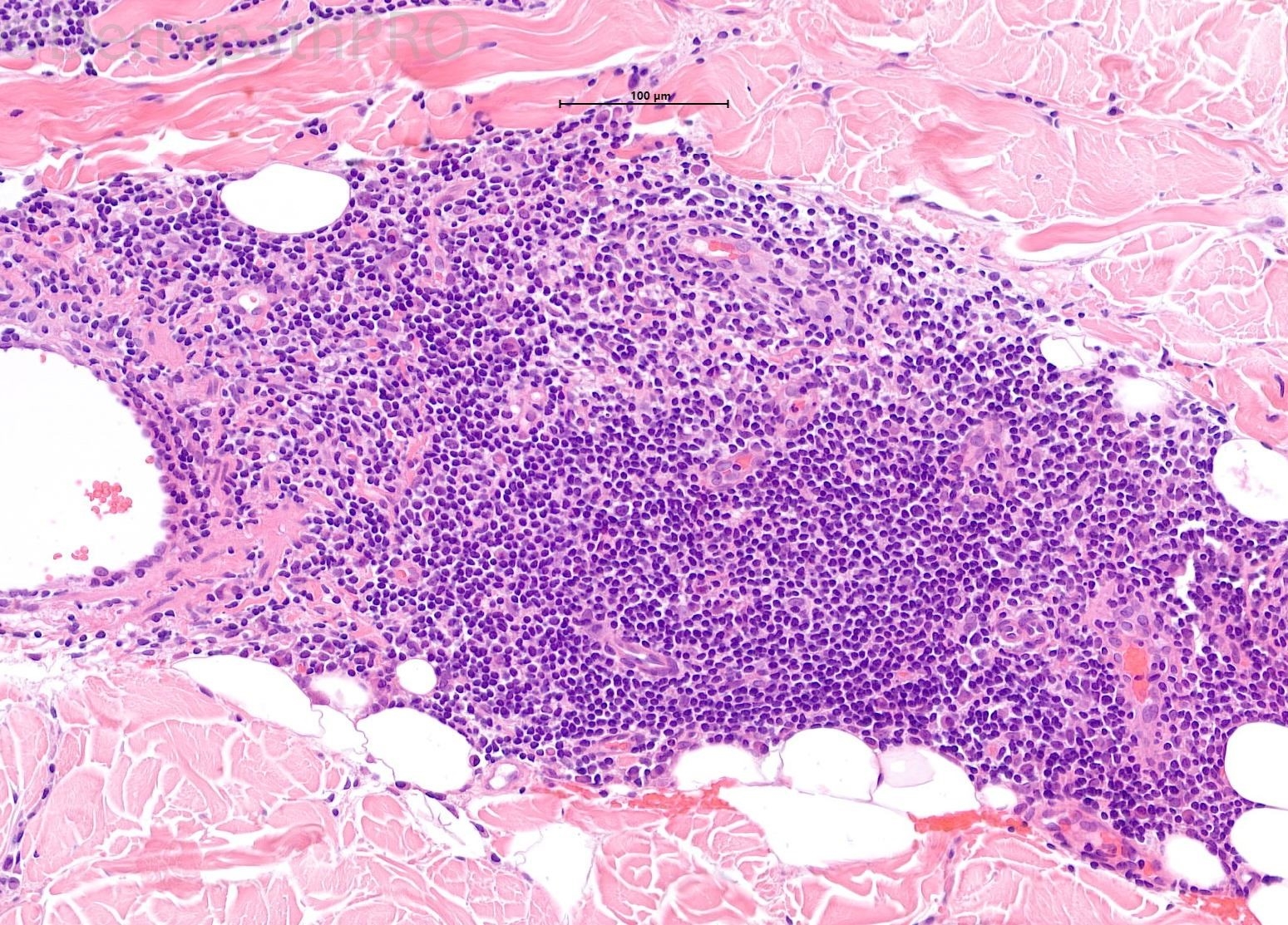
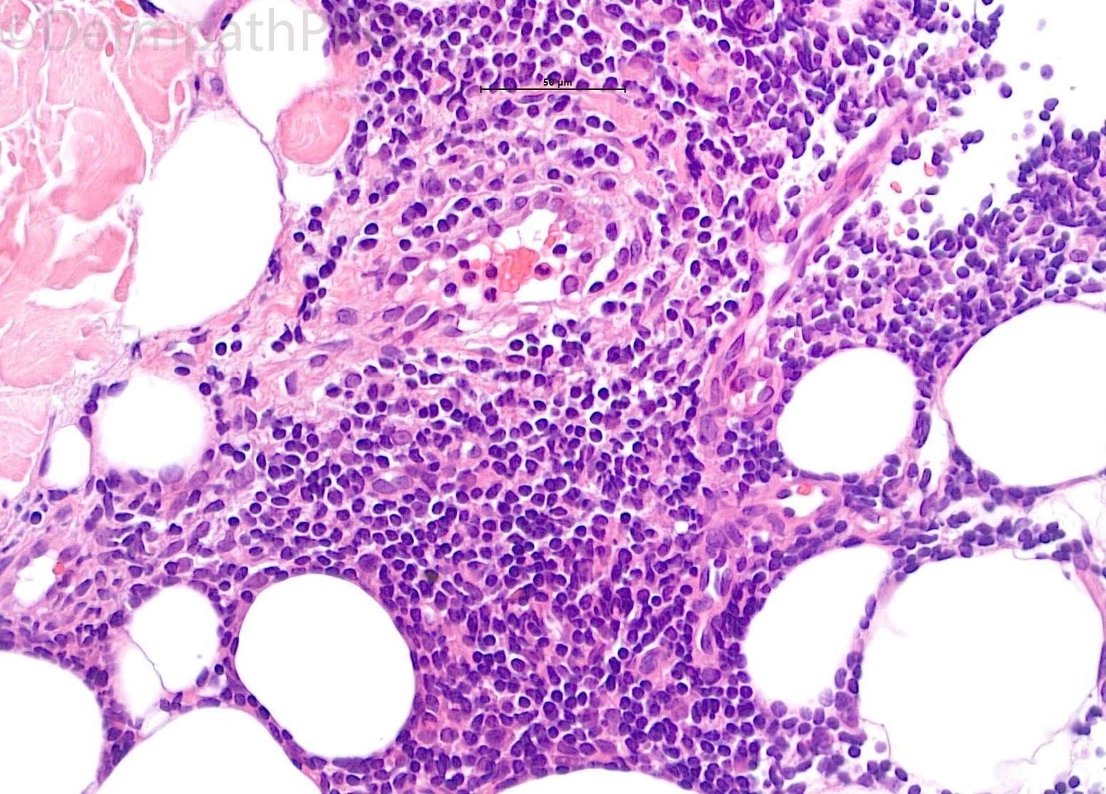
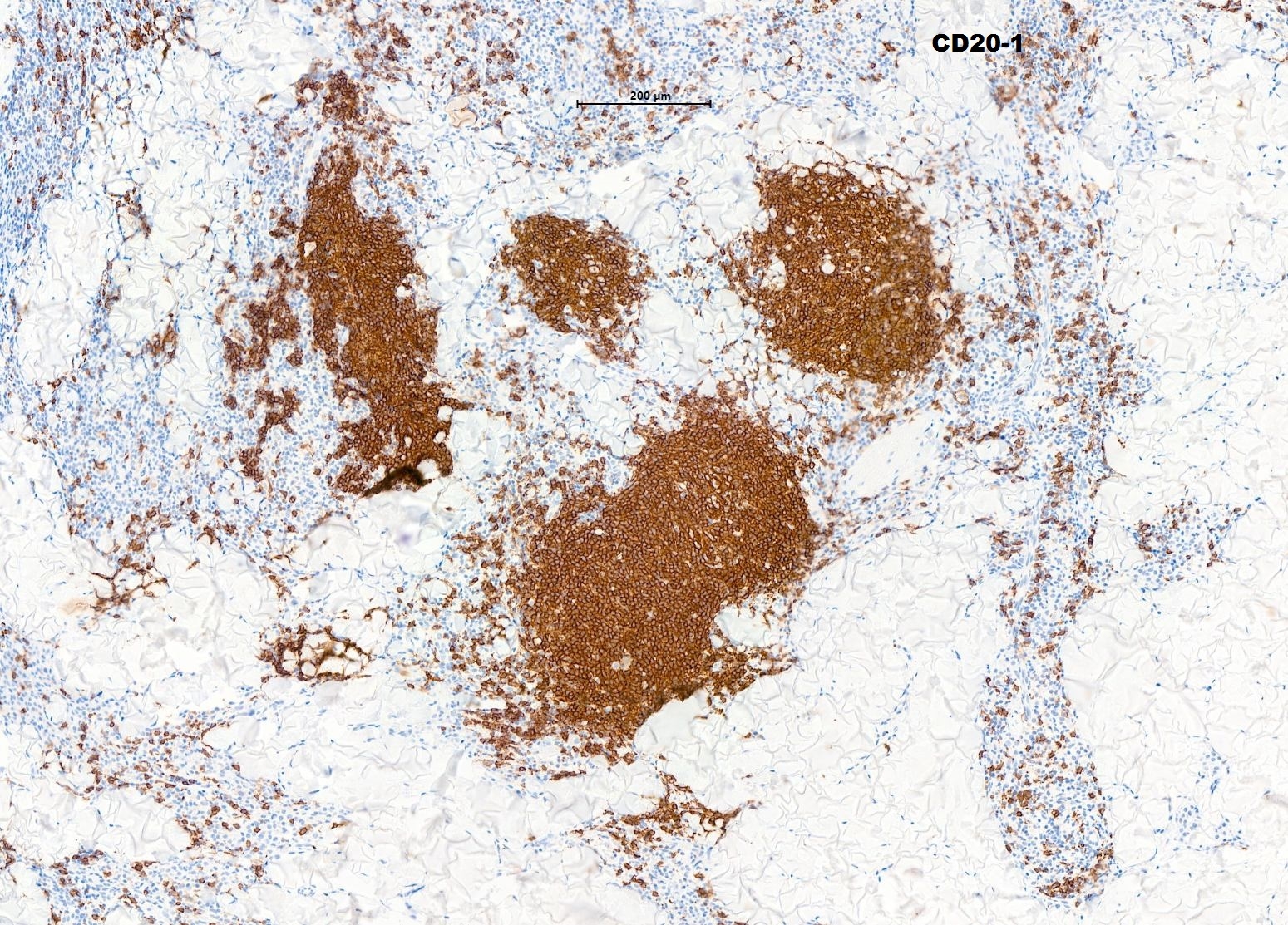
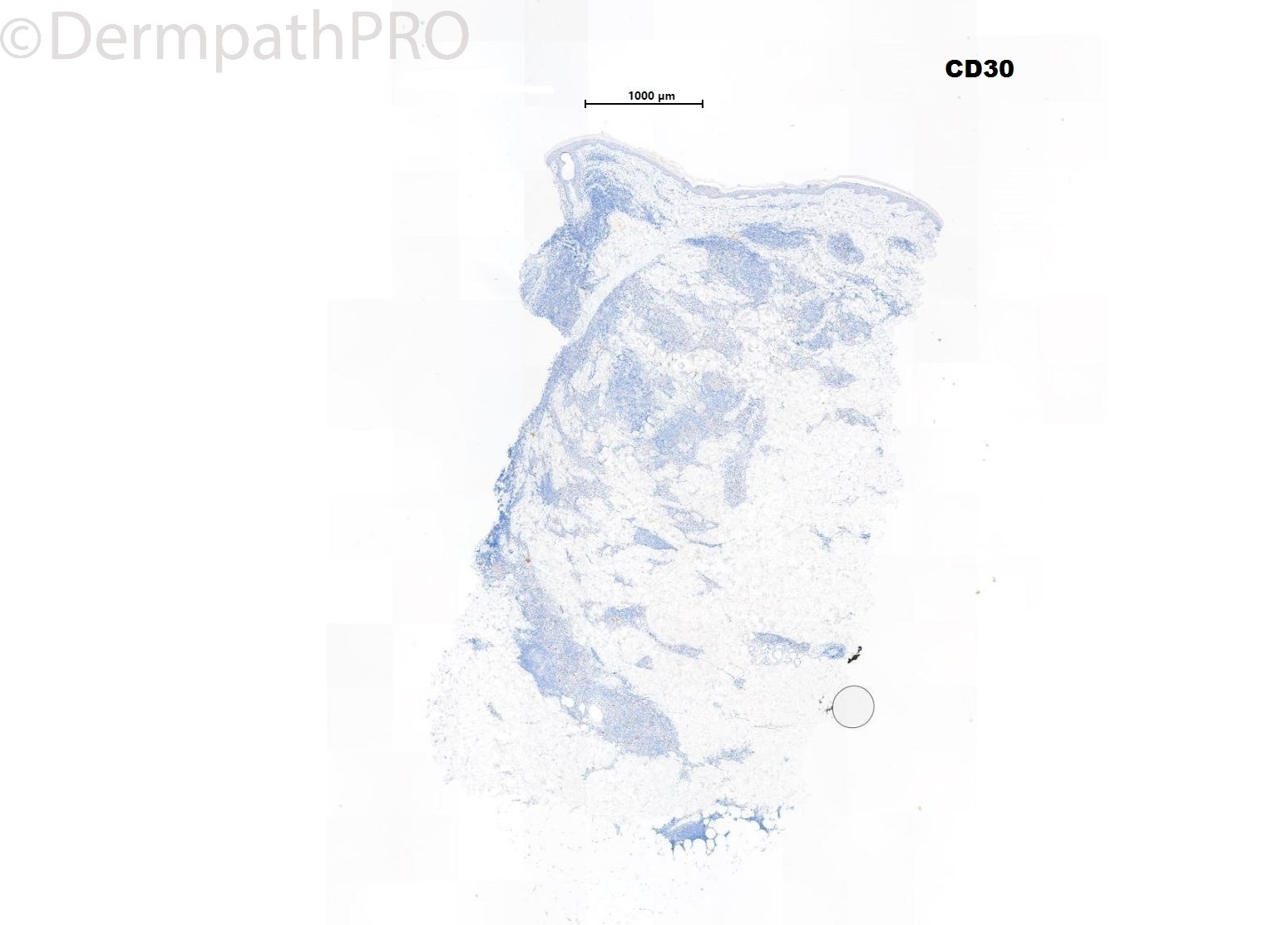
60 Male ,Punch biopsy left mid back. Granulomatous nodule? Persistent bite reaction ? Lymphoma


  Report Record

User Feedback


Richard Logan

Posted

My first thought looking at the low-power pattern was that this was Jessner's lymphocytic infiltrate.  However, the cell population is rather too heterogeneous and includes quite a few plasmacytoid cells.  I wonder if this is blastic plasmacytoid dendritic cell neoplasm.

Share this comment


Link to comment
share_externally

Saman Fatah

Posted

?PC CD4+ SM T-cell LPD vs PCMZL depending on further IHC/clonality studies. It is alway useful to consider syphilis serology in such cases as it is a great mimicker though the site is unusual especially if solitary lesion. 

Share this comment


Link to comment
share_externally

Eman El-Nabarawy

Posted

Pseudolymphoma vs Marginal zone lymphoma top my list for further immunohistochemical studies..

Share this comment


Link to comment
share_externally

Krishnakumar subramanian

Posted

granulomatous inflammation with dense lymphoplasmacytic cells with eosinophils

Any stains for infections such as PAS and GMS needed

Share this comment


Link to comment
share_externally

daniellindsay

Posted

I would also consider granulomatous slack skin.

Share this comment


Link to comment
share_externally

Admin_Dermpath

Posted

Dr Chaudhry Diagnosis:


Special stains were all negative and this was not really concerned by the clinician.

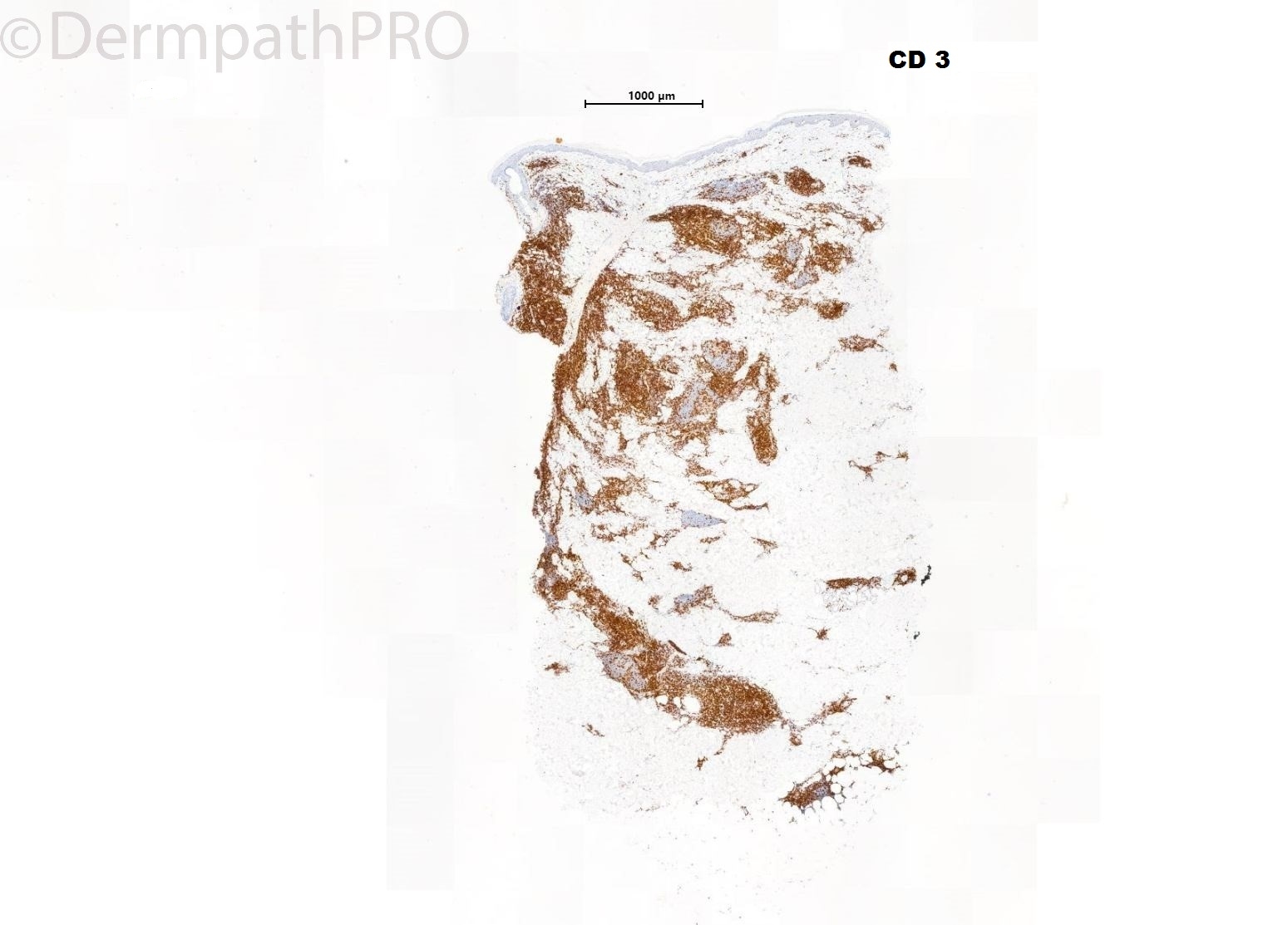
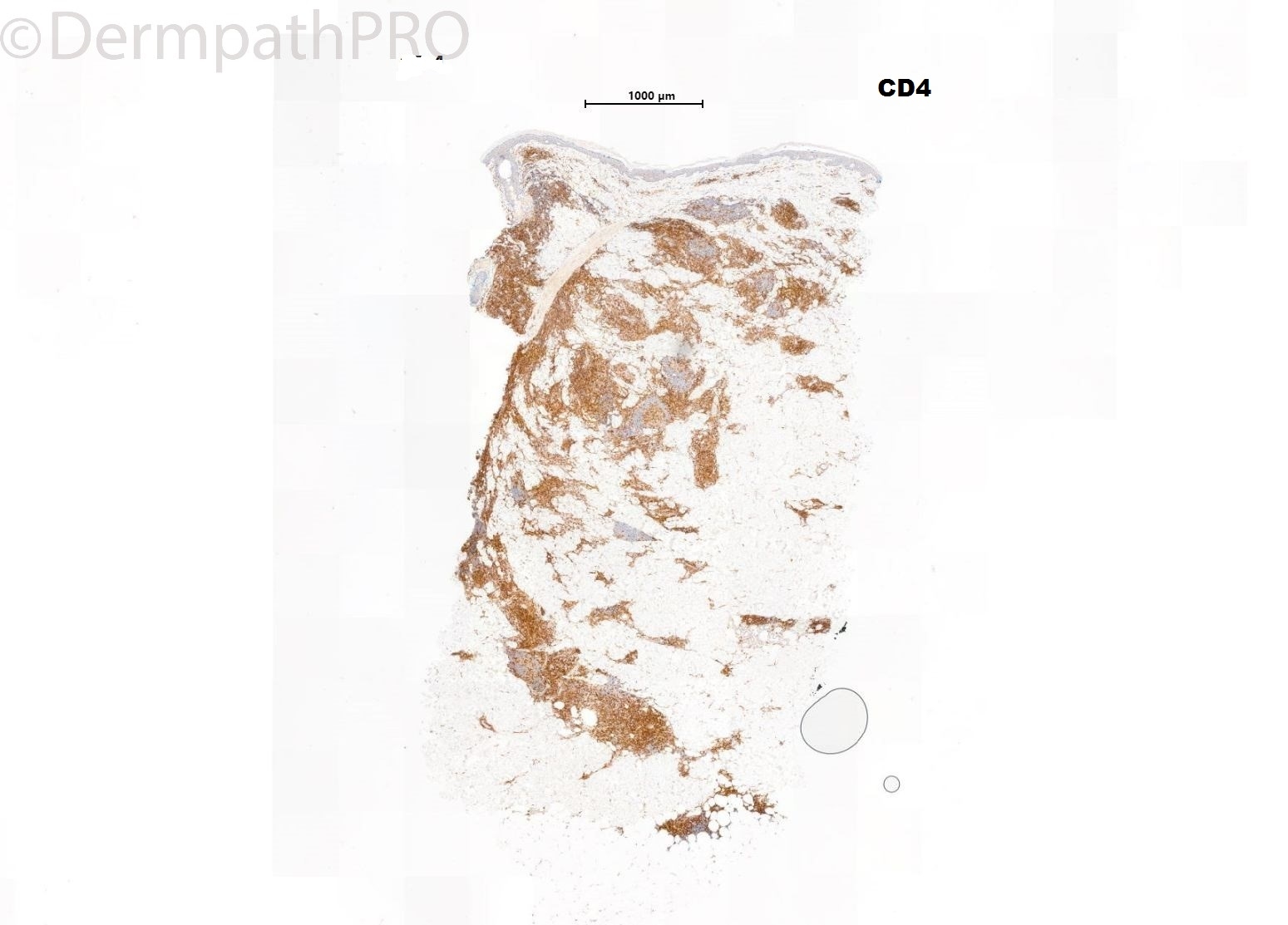
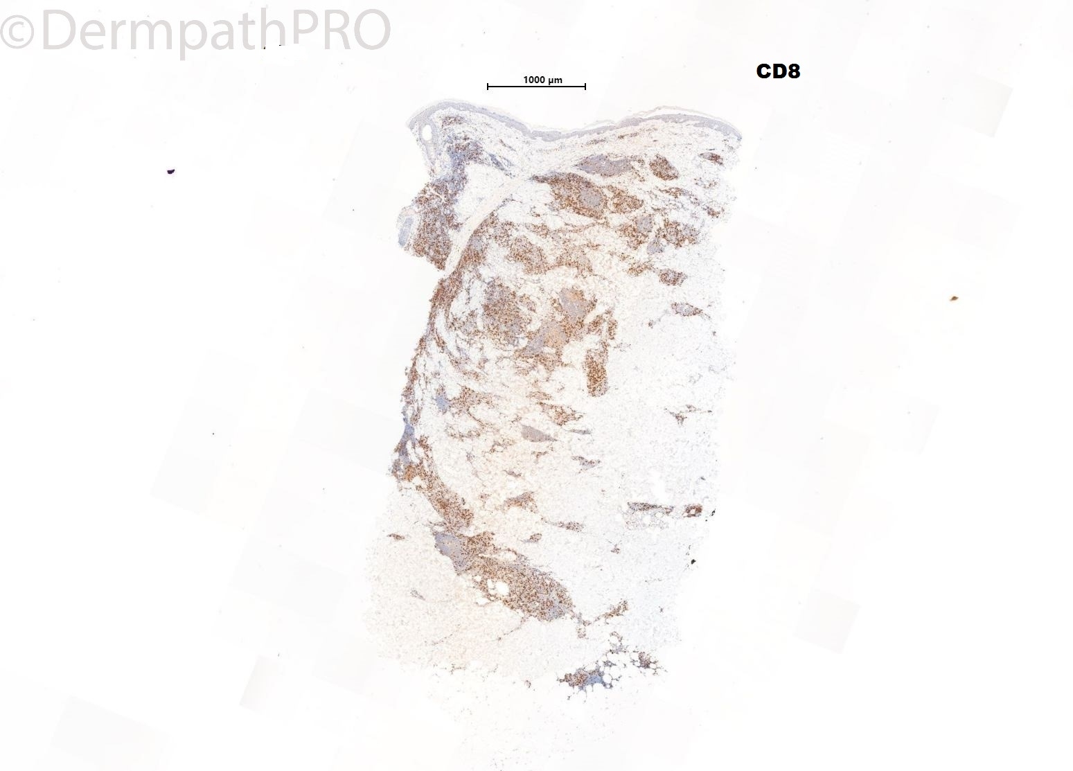
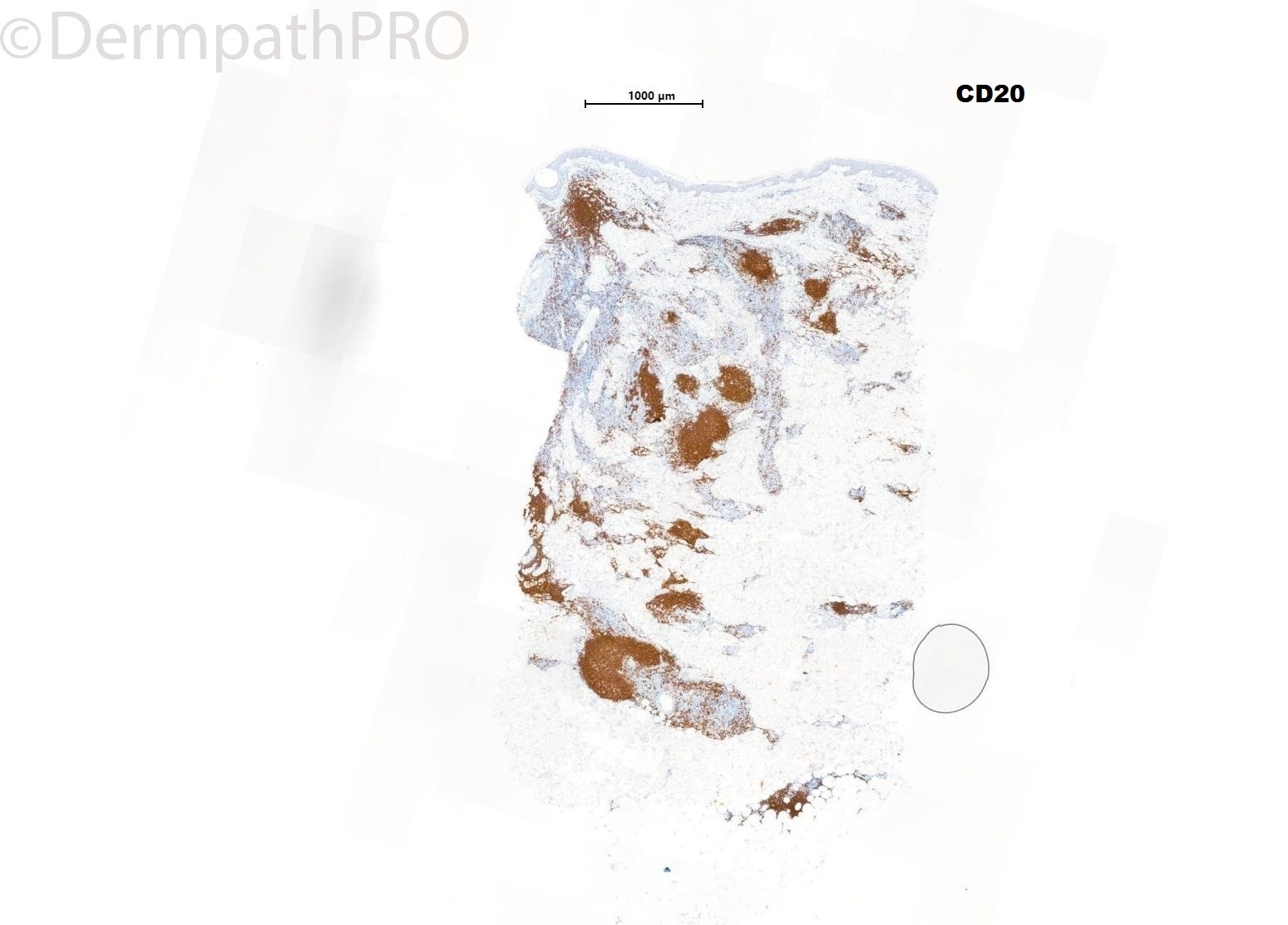
As the biopsy was to determine whether it was a Lymphoma or pseudo-lymphoma, based upon the morphology and immunohistochemistry I have come to the conclusion that the features are those of pseudo-lymphoma which requires clinical correlation. 

After speaking to the clinician, it was brought to light that there was a history of insect bite and we queried whether it could be an insect bite reaction.

Share this comment


Link to comment
share_externally

vincenzo

Posted

K/Lambda? Enough plasma cells in 6st picture. I’m not thinking about GSS-like MF because of the odd site(mid back); and not about BPDCN, because the BPDCN is a double negative ( CD3-/CD20-) and CD4+ neoplasm.  And morphologically this a small cell infiltrate, with some plasma cells and some monocytoid clear cell lymphocyte. Pseudolymphoma versus PCMZL. I don’t understand the last EmEo picture...it seems another case ( sarcoidosis? )

Share this comment


Link to comment
share_externally

Dr. Richard Carr

Posted

That is a great observation Vincenzo. Always consider a specimen mix-up when things don't add up!!

Kappa and lambda should confirm reactive cutaneous lymphoid hyperplasia and I often limit my markers in suspected lymphocytoma cutis, especially if the history is short. 

Without history inflammatory morphoea would be a consideration.

Share this comment


Link to comment
share_externally

18 hours ago, vincenzo said:

K/Lambda? Enough plasma cells in 6st picture. I’m not thinking about GSS-like MF because of the odd site(mid back); and not about BPDCN, because the BPDCN is a double negative ( CD3-/CD20-) and CD4+ neoplasm.  And morphologically this a small cell infiltrate, with some plasma cells and some monocytoid clear cell lymphocyte. Pseudolymphoma versus PCMZL. I don’t understand the last EmEo picture...it seems another case ( sarcoidosis? )

I agree with the image that looks like sarcoid, made me confused as well

Share this comment


Link to comment
share_externally

Admin_Dermpath

Posted

Apologies for the image (Sarcoidosis). That was an error and belongs to another case, wrong image have been deleted.

Really appreciates you for highlighting this issue. Thank you!

Share this comment


Link to comment
share_externally

the histomorphology and IHCs pattern fit perfect with T cell rich pseudolymphoma, which I find could be quite difficult to differentiate from CD4+SMLPD. but the latter rarely have germinal centers. so more pseudolymphoma to me.

Share this comment


Link to comment
share_externally


Join the conversation

You can post now and register later. If you have an account, sign in now to post with your account.

Guest
Add a comment...

×   Pasted as rich text.   Paste as plain text instead

  Only 75 emoji are allowed.

×   Your link has been automatically embedded.   Display as a link instead

×   Your previous content has been restored.   Clear editor

×   You cannot paste images directly. Upload or insert images from URL.

Loading...

×
×
  • Create New...