Jump to content
In this section we have spot diagnoses posted on a daily basis since June 2010, now over 4000! You can review the archived cases and read the suggested diagnoses by users and the final comment by the contributors.
Case are uploaded each week day by 10 am UK time with the correct diagnosis will generally be posted at 8 pm UK time. Why not view the most recent spot diagnosis and proffer a diagnosis?

Case Number : Case 2876 - 15 July 2021 Posted By: Saleem Taibjee

Please read the clinical history and view the images by clicking on them before you proffer your diagnosis.
Submitted Date :
   (0 reviews)

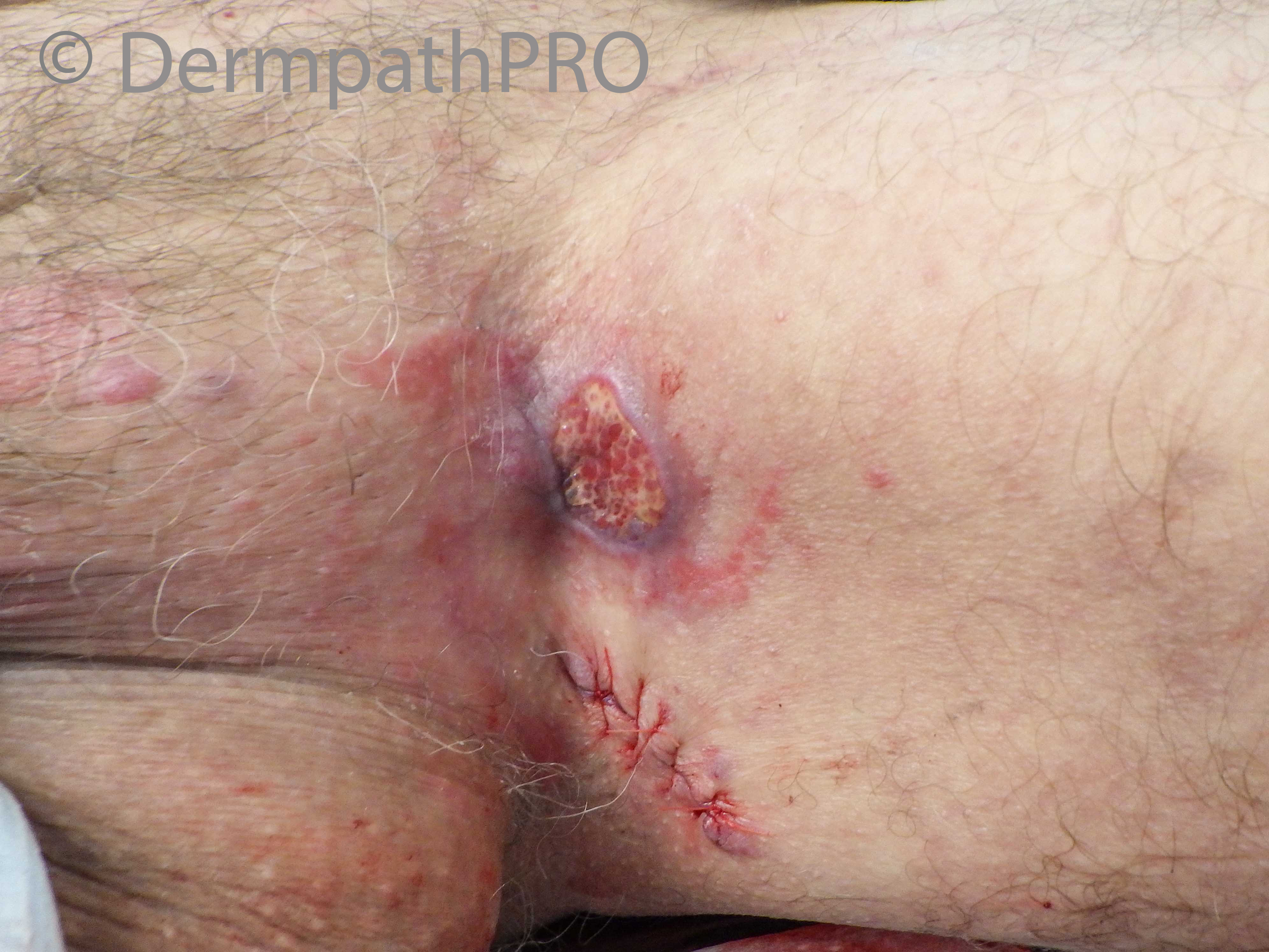
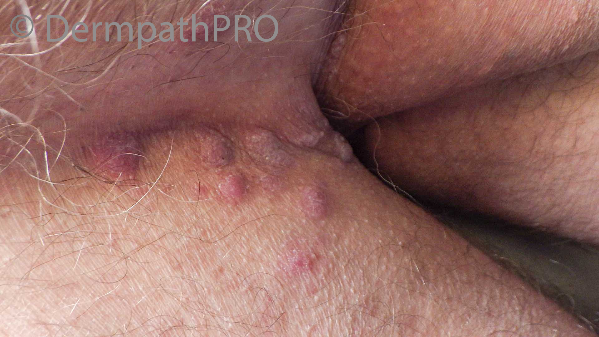
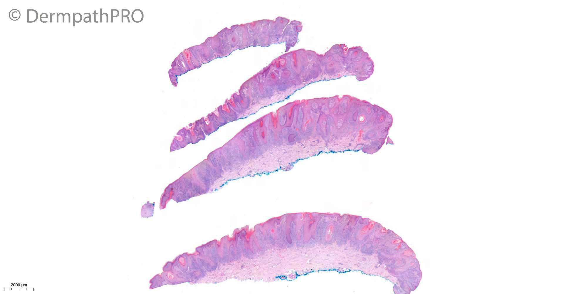
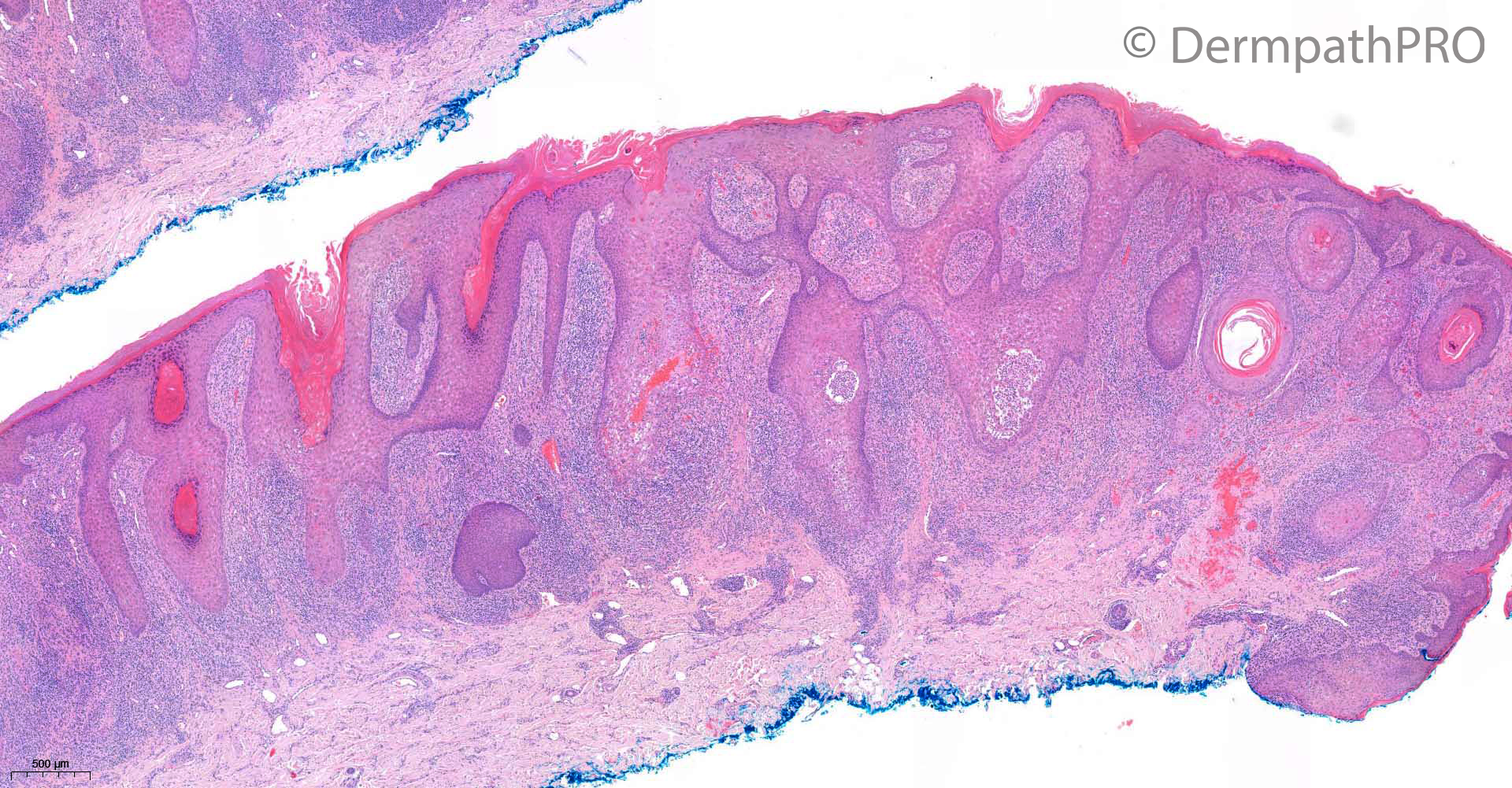
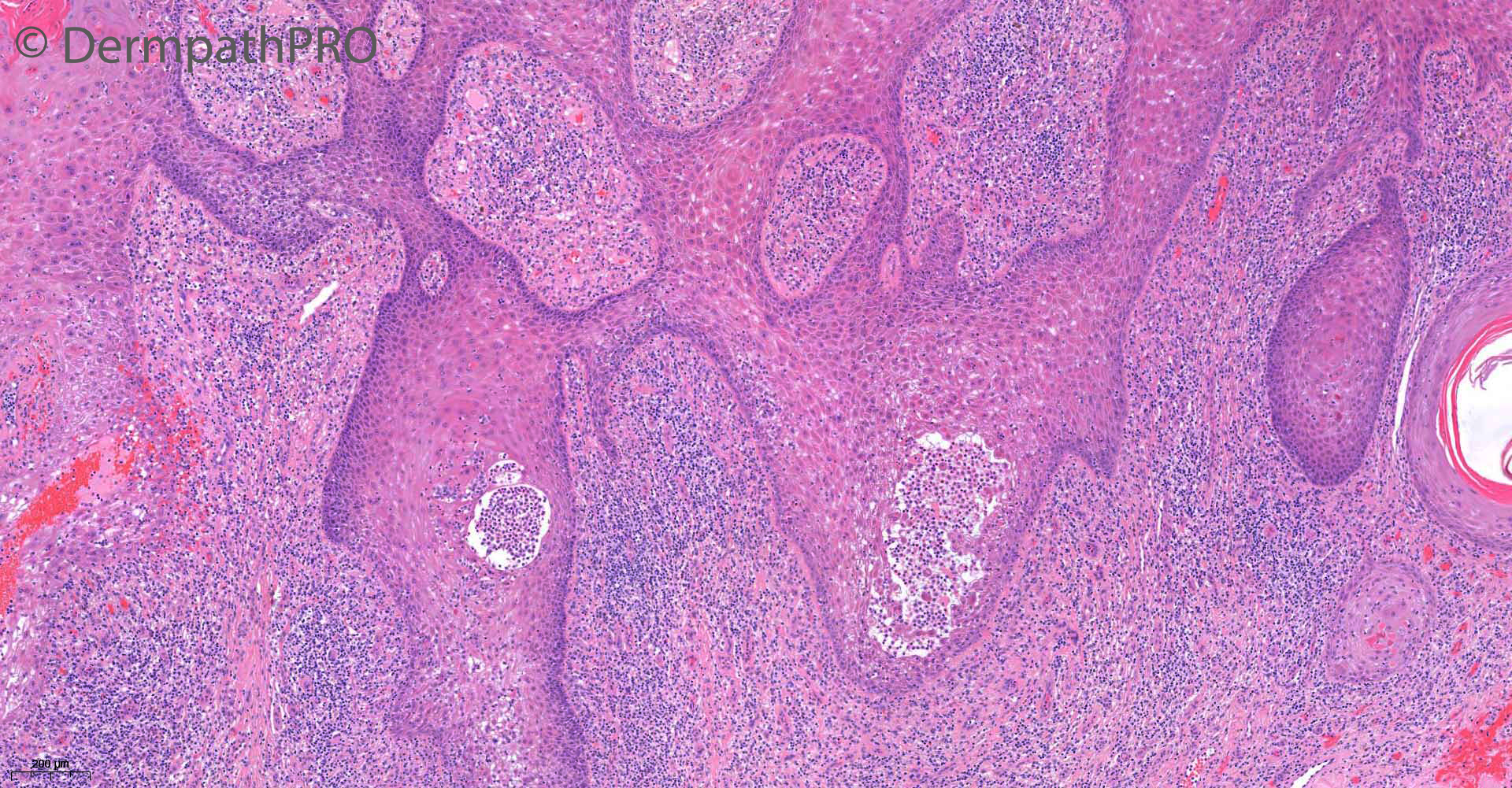
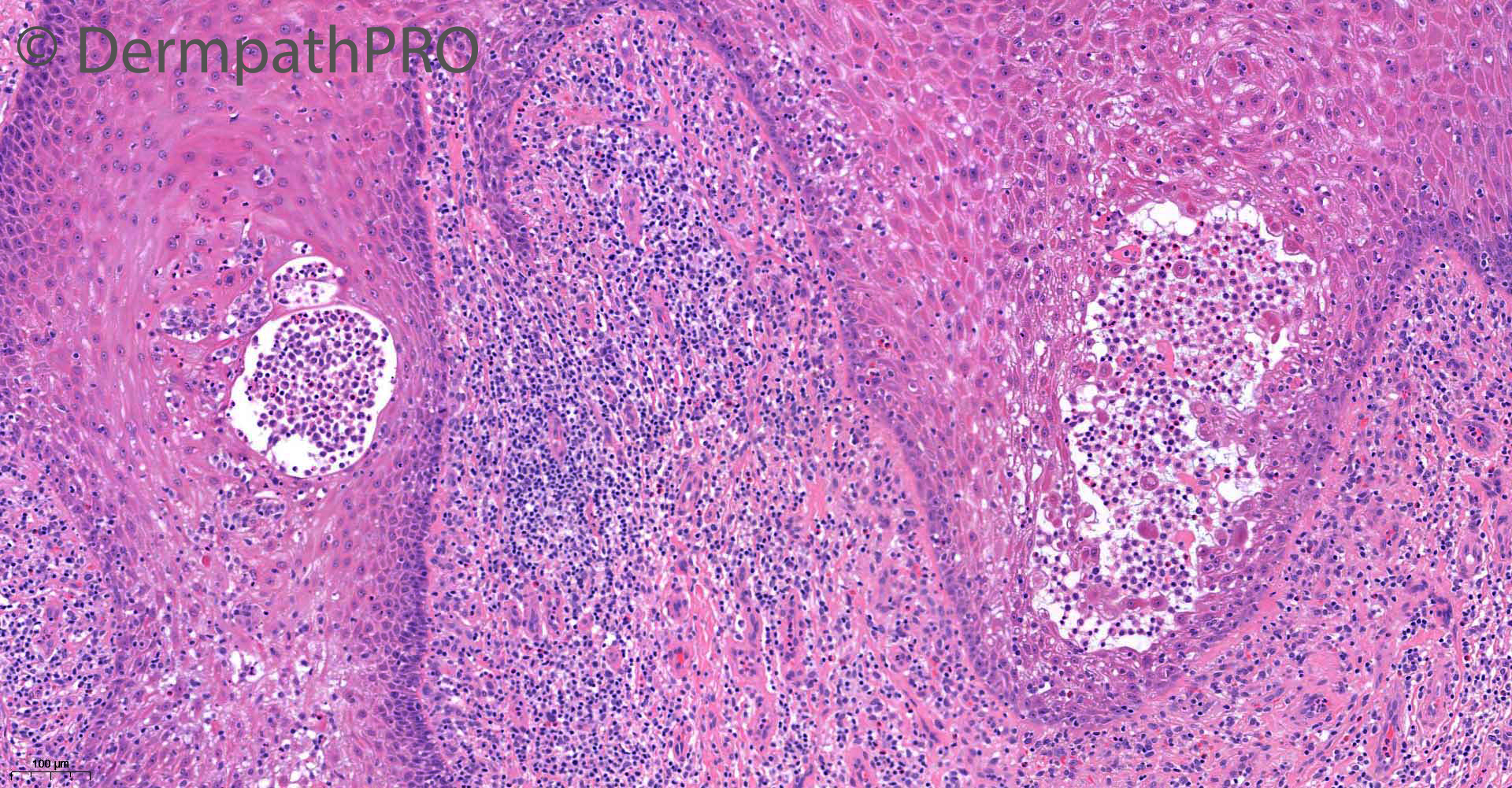
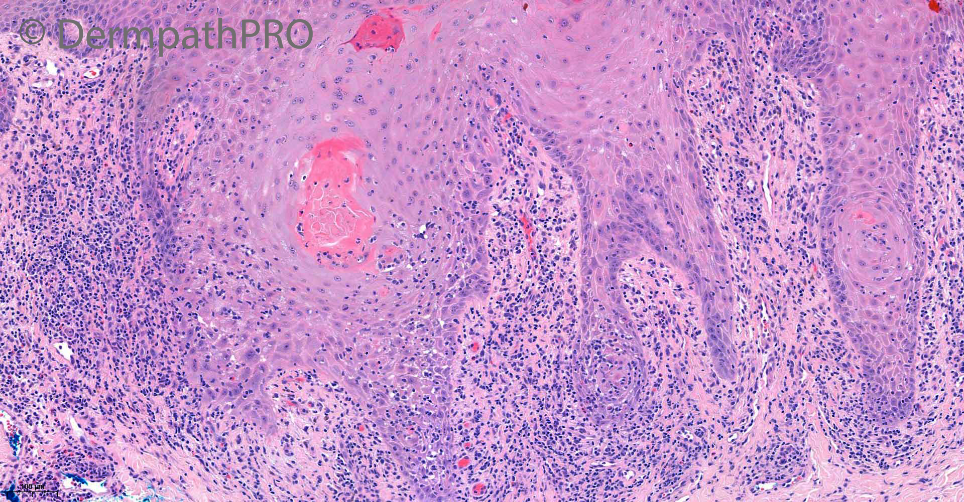
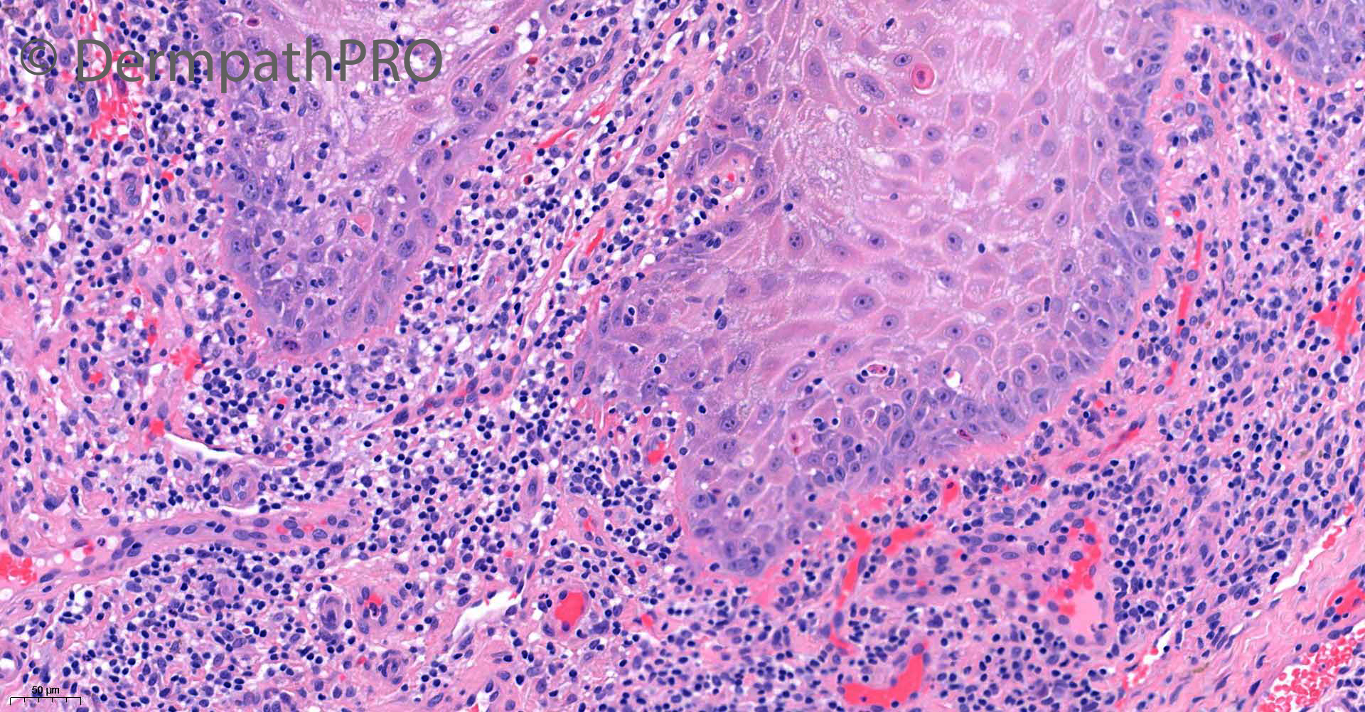
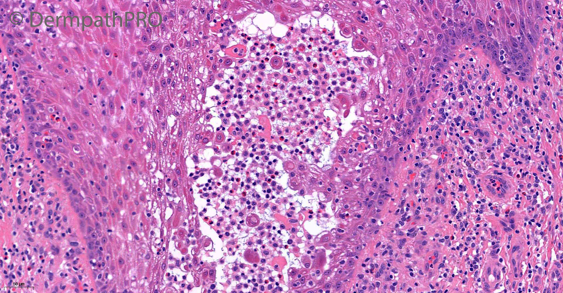
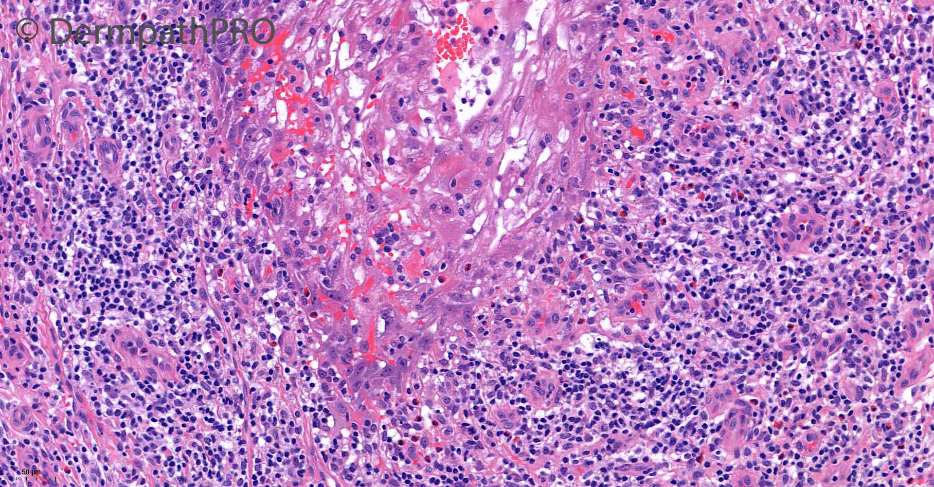
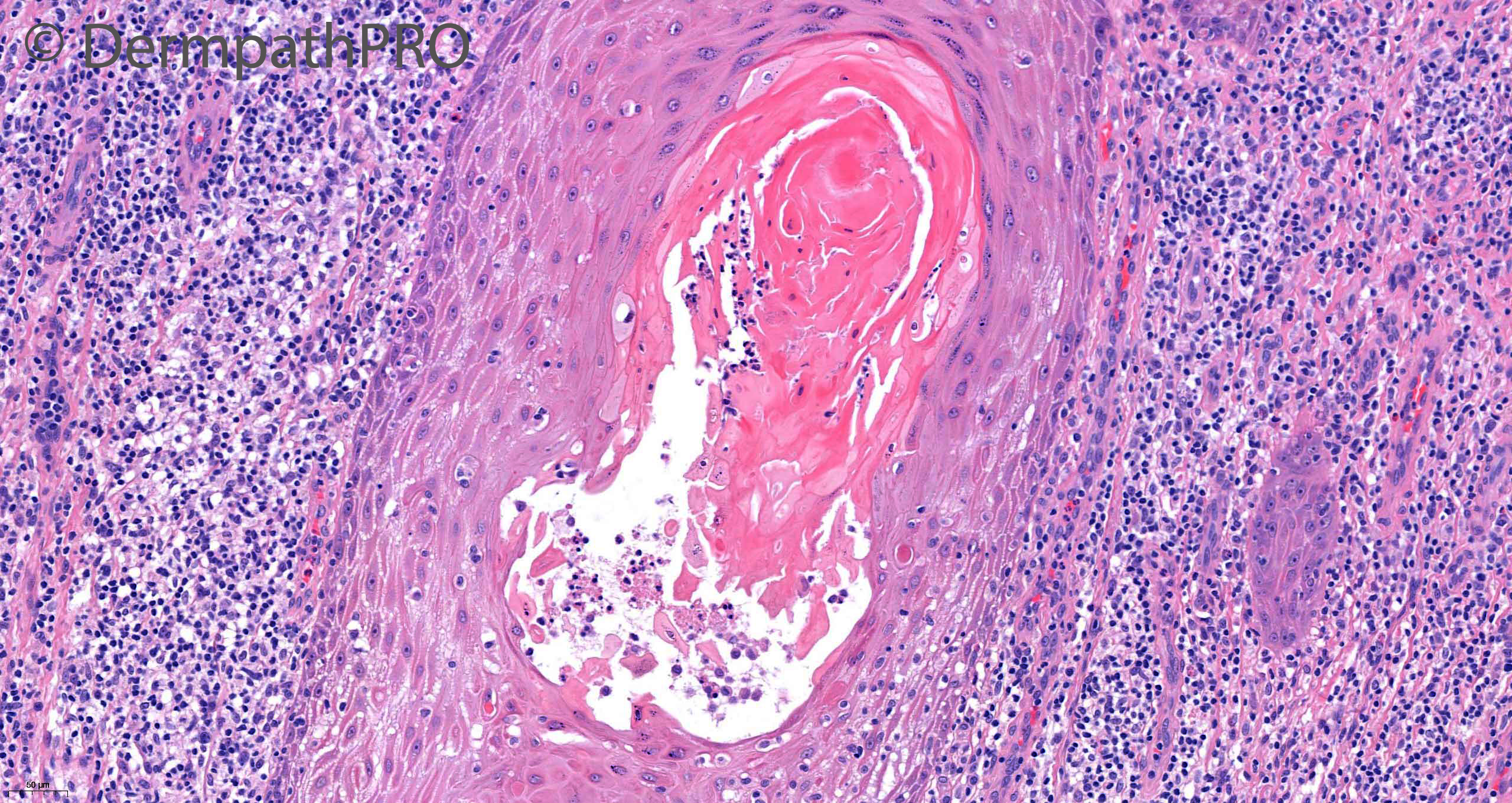
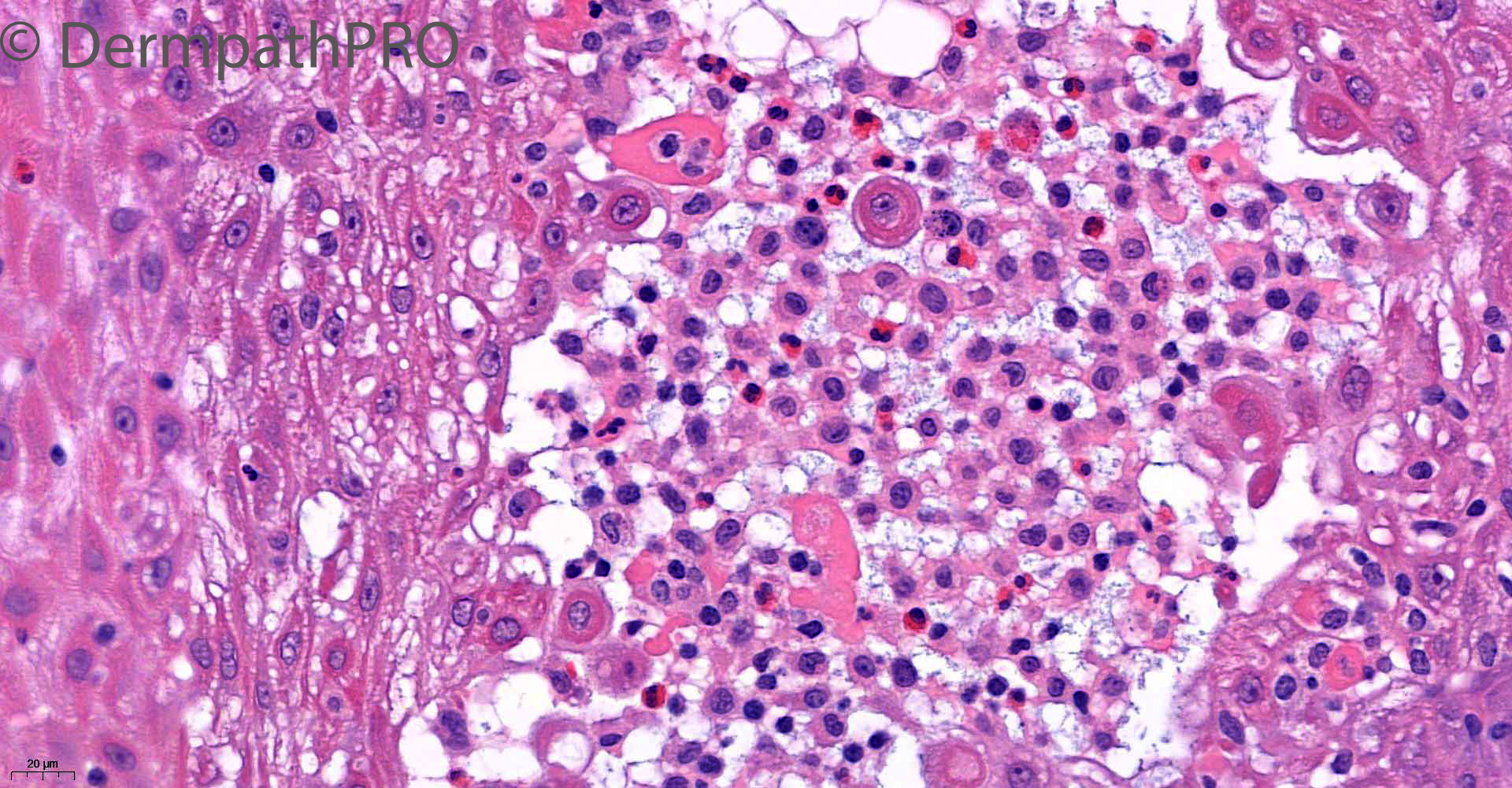
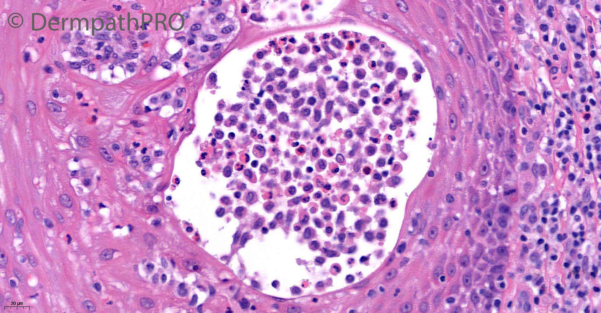
79M biopsy left groin ?malignancy ?lymphoma


  Report Record

User Feedback


Richard Logan

Posted

Nice example of pemphigus vegetans both clinically and histologically.

Share this comment


Link to comment
share_externally

Eman El-Nabarawy

Posted

My first perception is pemphigus vegetans too..

Share this comment


Link to comment
share_externally

8 hours ago, Richard Logan said:

Nice example of pemphigus vegetans both clinically and histologically.


7 hours ago, Eman El-Nabarawy said:

My first perception is pemphigus vegetans too..

Totally agree

Share this comment


Link to comment
share_externally

Saman Fatah

Posted

Pemphigus vegetans is rare and the cases I have seen were mostly exudative friable eroded vegetative plaques compared to a rather lichenified nodules especially on clinical image 2.

The histology pattern reminded me with a spectacular case of Dr Carr posted on 8th November 2019 (2440) of halogenoderma secondary to bromide and flourine.

Once infective cause excluded, I will cautiously suggest Halogenoderma acknowledging that it is also rare and hopefully Richard’s wait for the 3rd case will discontinue!

Looking forward to hear final verdict from Saleem. Great quality photo/photomicrographs and thanks for Dermpathpro team/editors for continuing to educate us. 

Share this comment


Link to comment
share_externally

Krishnakumar subramanian

Posted

i am bit concerned with site of occurrence and some histopath points

This flexural site, is also where histiocytic disorders appear. also i see numerous eosinophils and some histiocytic cells. I will get DIF done to confirm pemphigus, if negative do  markers for  CD 68, s100 and Cd1a

Share this comment


Link to comment
share_externally

Saleem Taibjee

Posted

Interim comment: Direct immunofluorescence was negative.

Share this comment


Link to comment
share_externally

Krishnakumar subramanian

Posted

so we must exclude histiocytic disorders with IHC panels

Share this comment


Link to comment
share_externally

Saleem Taibjee

Posted

Very well done to Saman!

This is a recent fascinating case. I wish to thank Alistair Robson also for his second opinion.

Like others here, my first reaction to the histology was pemphigus vegetans given the epidermal hyperplasia and microabscesses, some containing eosinophils, and even a hint of acantholysis (I had to hunt hard for this) as shown above in the penultimate image.

But as I mentioned, direct immunofluoresence was negative, and the clinical picture wasn't quite right for this.

I asked Alistair to have a look because the clinician was quite suspicious of lymphoma, and indeed the lymphocytic infiltrate is quite dense in this case. But like myself, he too favoured a reactive/inflammatory process.

The pattern of pseudoepitheliomatous hyperplasia with microabscesses, some containing eosinophils, as well as lichenoid element, did make me specifically mention Halogenoderma in the differential diagnosis in my original report.

I was able to review the patient's medication list on the digital patient record, and it transpired that the patient was taking a long-term inhaler called Trimbow which contains glycopyrronium bromide. I think it is fair to say that there was a little scepticism that this was the cause of the problem. However, the patient did eventually have a trial of stopping this. The nodules promptly completely settled within just a few days of stopping the medication, and there has been no recurrence on Dermatology review.

Hence this combination of PEH, microabcesses (including eosinophils) and lichenoid histology seems to be a recognisable pattern for Halogenoderma.

Of course, I need to thank Richard Carr for alerting me to this rare diagnosis. I would certainly not have recognised this possibility histologically, or pursued asking for the patient's inhaler to be discontinued, without Richard's previous instructive case which he had personally shared with me some time ago.

I can see that Krishnakumar has concerns of LCH or other form of histiocytosis. I agree that the abscesses do probably contain Langerhans cells also. But there is no evidence of a significant dermal infiltrate of Langerhans cells, and of course the resolution on stopping the inhaler is further reassuring. Hence I have not requested any histiocytic markers.

BW
Saleem

Share this comment


Link to comment
share_externally

Richard Logan

Posted

Another salutory case Saleem. 

I wonder what explains the anatomical distribution of the lesions?  Clearly there is systemic absorption of the bromide, which perhaps is concentrated in the apocrine sweat glands in this area.  Of course inhaled steroids can lead to cutaneous atrophy but that is more generalised than this example.

Share this comment


Link to comment
share_externally

Another spectacular and educative case. Thanks, Saleem. 

Share this comment


Link to comment
share_externally


Join the conversation

You can post now and register later. If you have an account, sign in now to post with your account.

Guest
Add a comment...

×   Pasted as rich text.   Paste as plain text instead

  Only 75 emoji are allowed.

×   Your link has been automatically embedded.   Display as a link instead

×   Your previous content has been restored.   Clear editor

×   You cannot paste images directly. Upload or insert images from URL.

Loading...

×
×
  • Create New...