Jump to content
In this section we have spot diagnoses posted on a daily basis since June 2010, now over 4000! You can review the archived cases and read the suggested diagnoses by users and the final comment by the contributors.
Case are uploaded each week day by 10 am UK time with the correct diagnosis will generally be posted at 8 pm UK time. Why not view the most recent spot diagnosis and proffer a diagnosis?

Case Number : Case 2877 - 16 July 2021 Posted By: Dr. Richard Carr

Please read the clinical history and view the images by clicking on them before you proffer your diagnosis.
Submitted Date :
   (0 reviews)

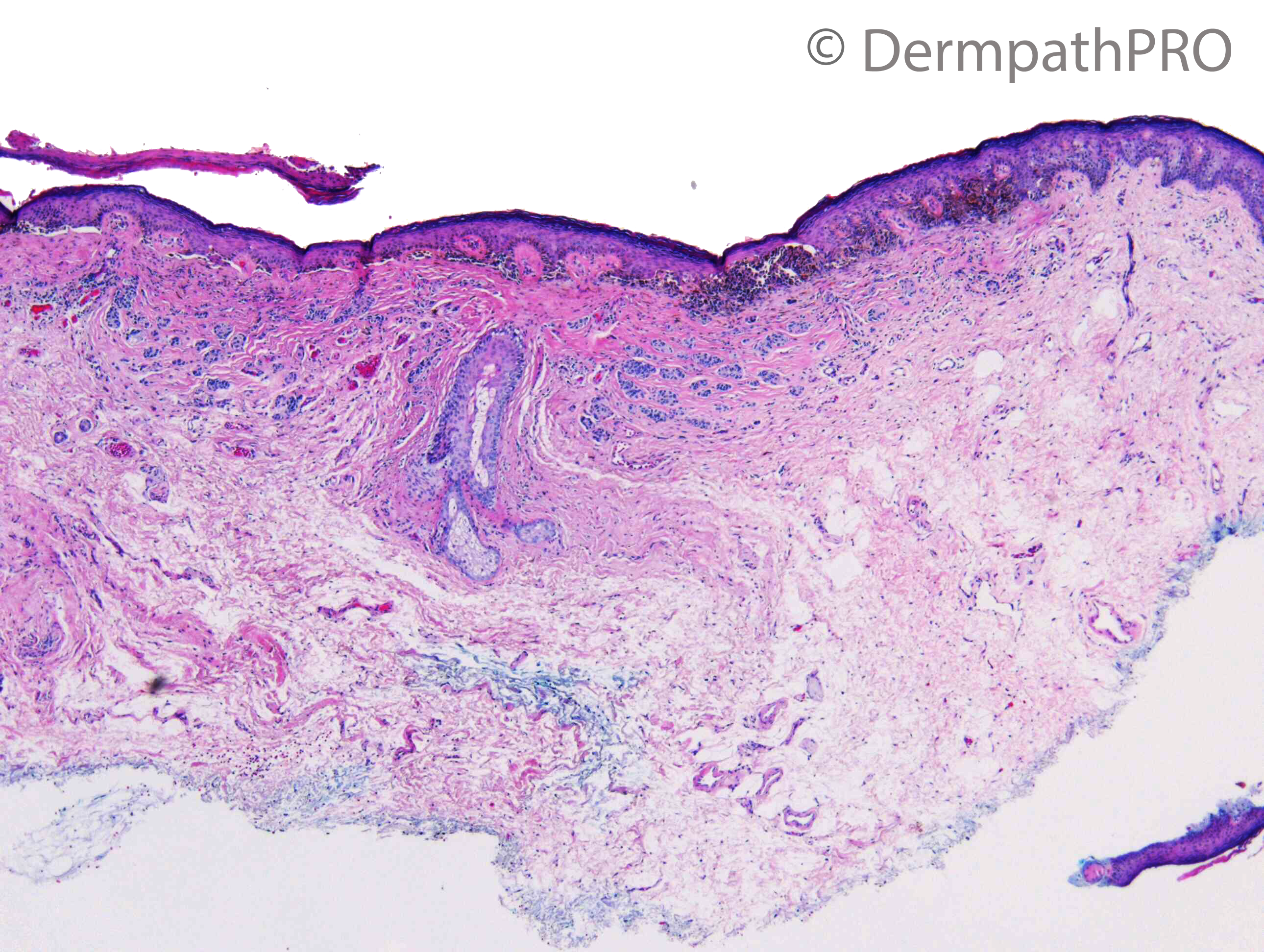
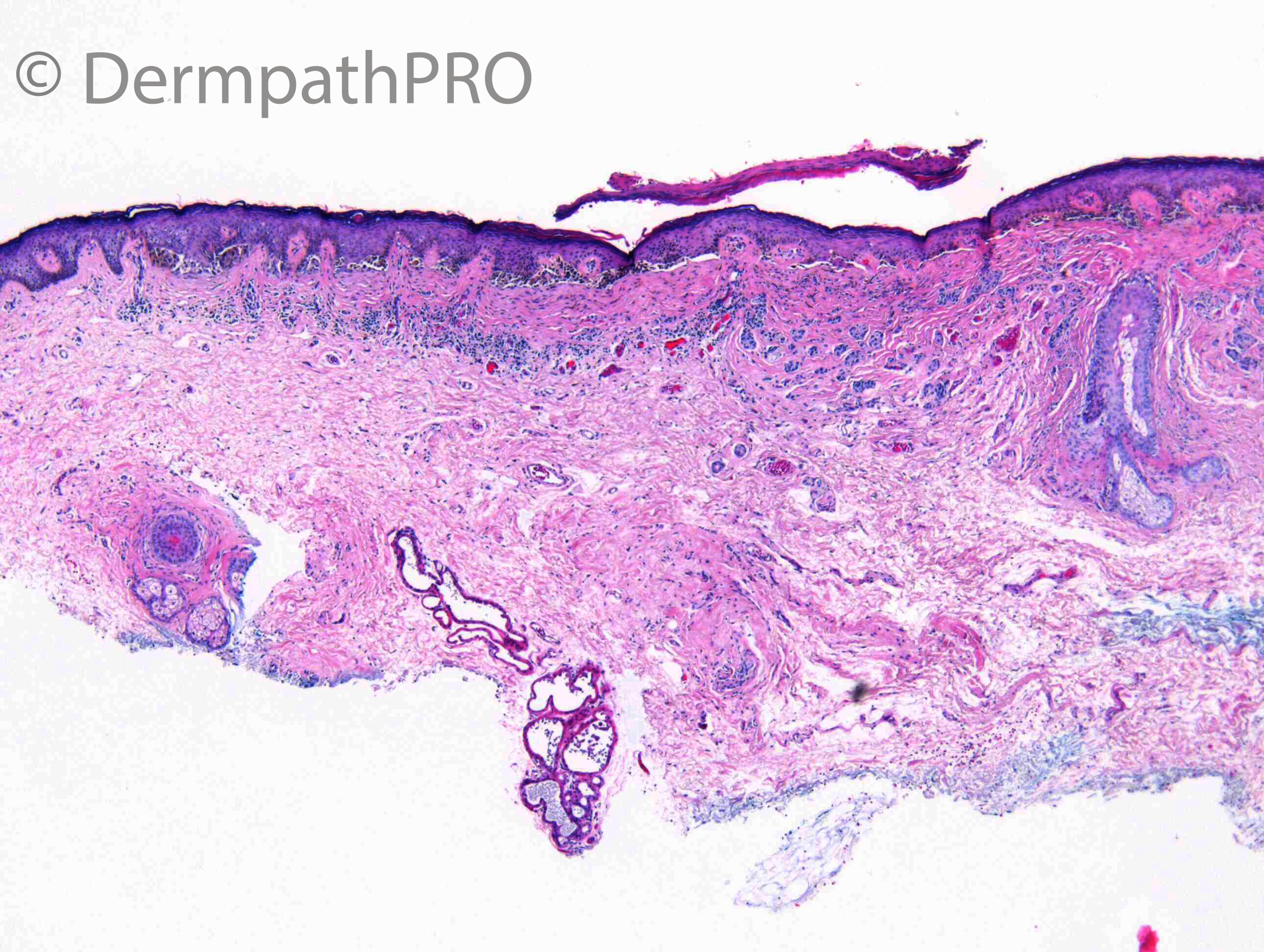
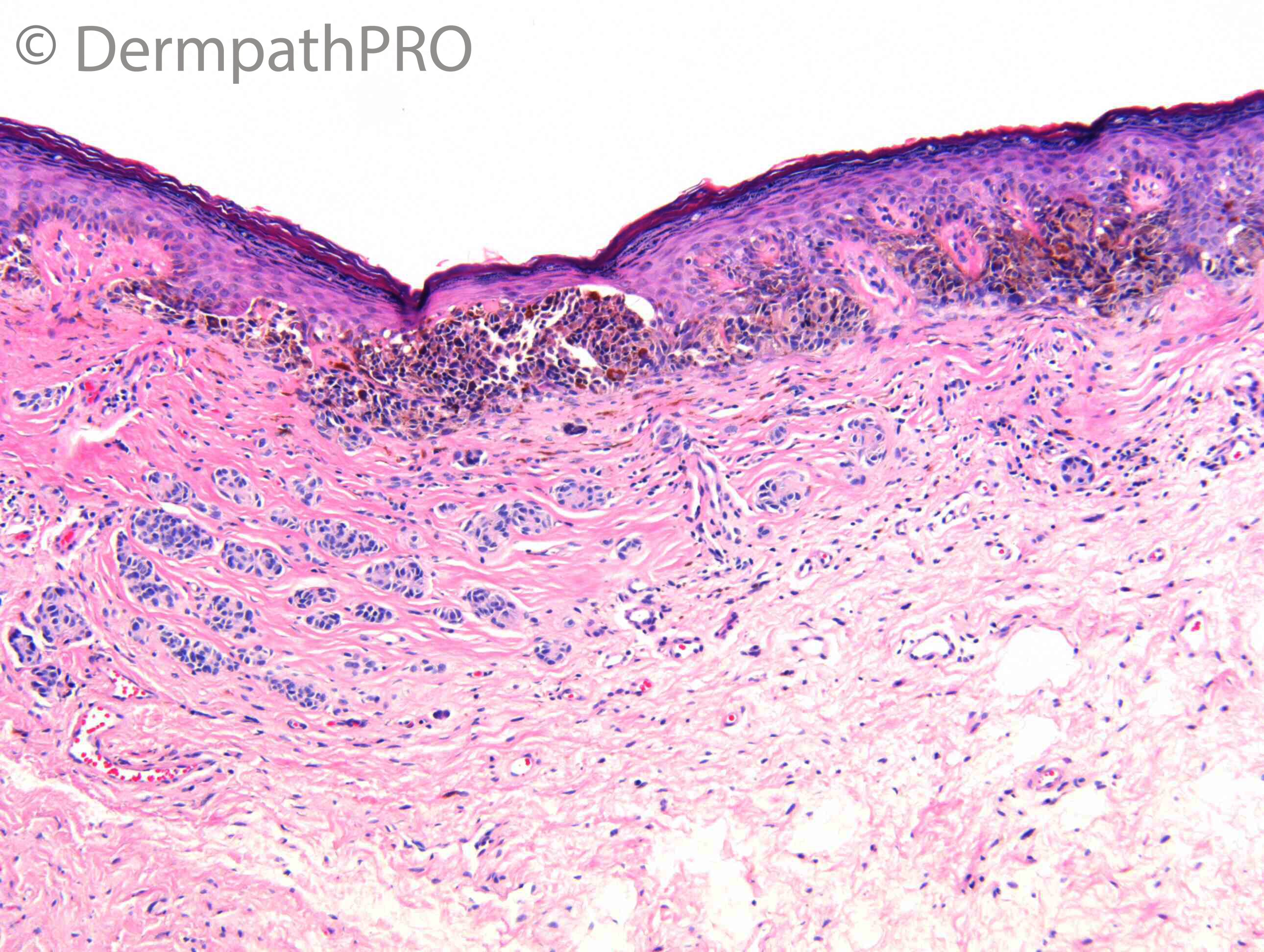
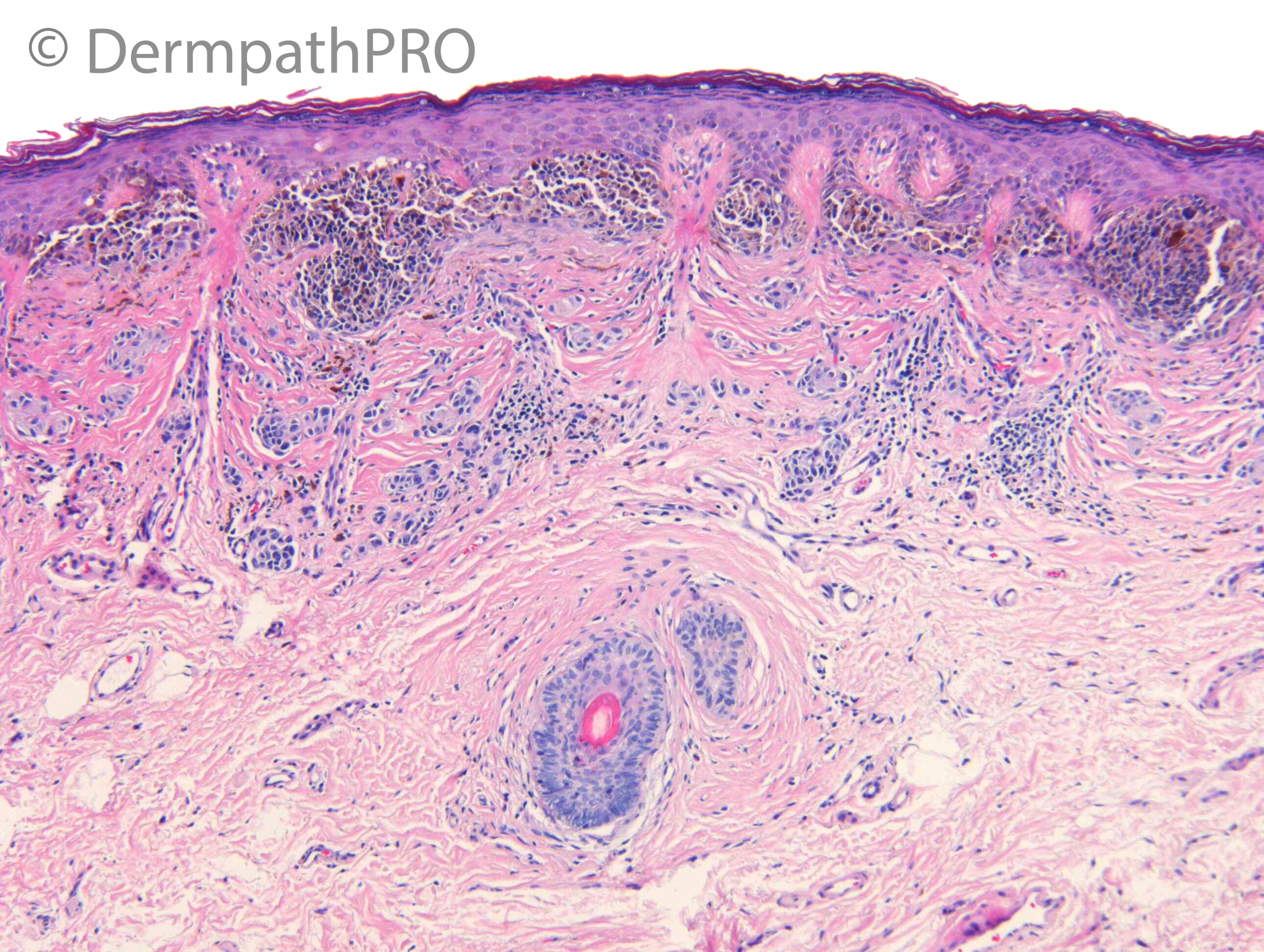
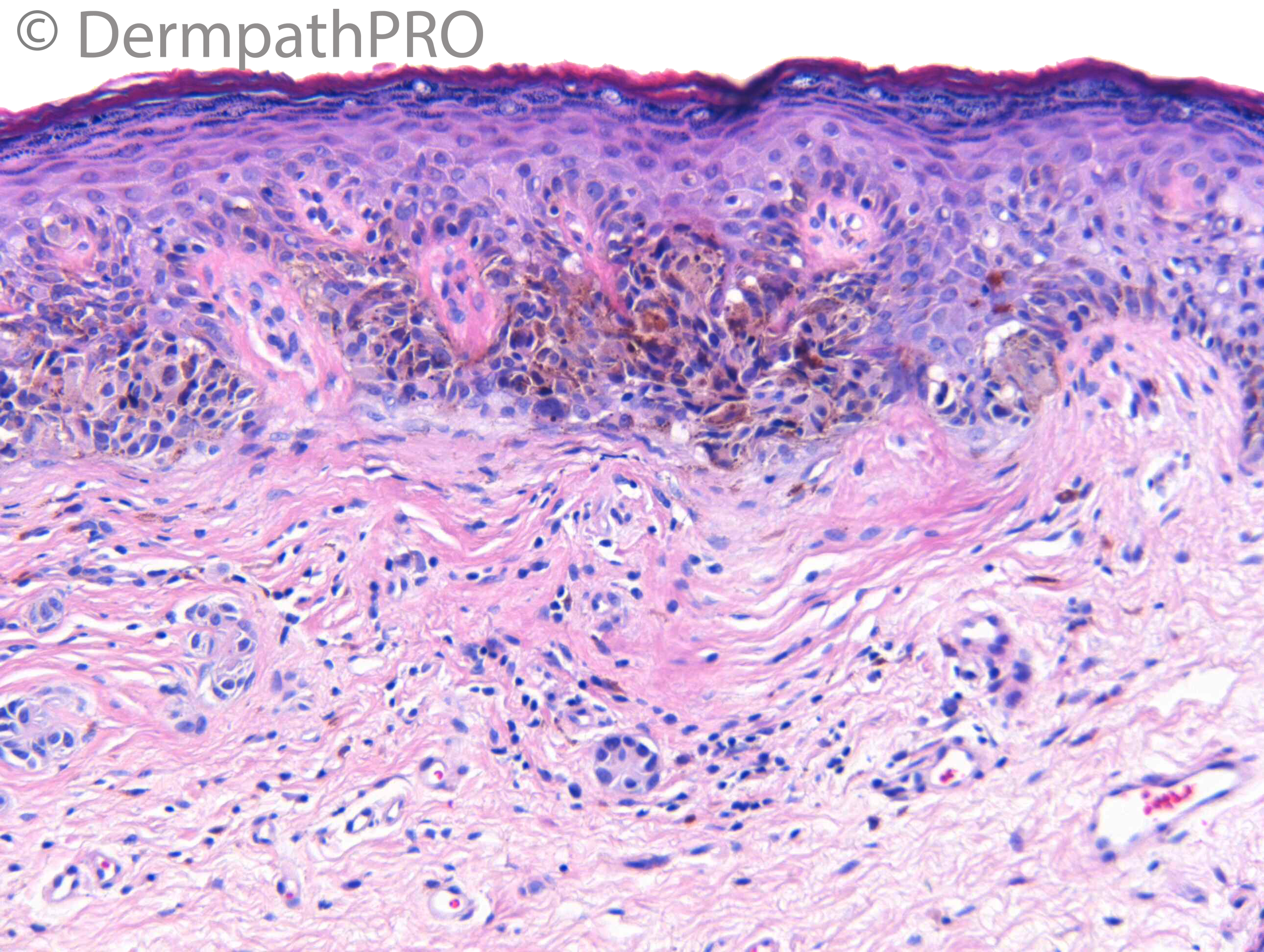
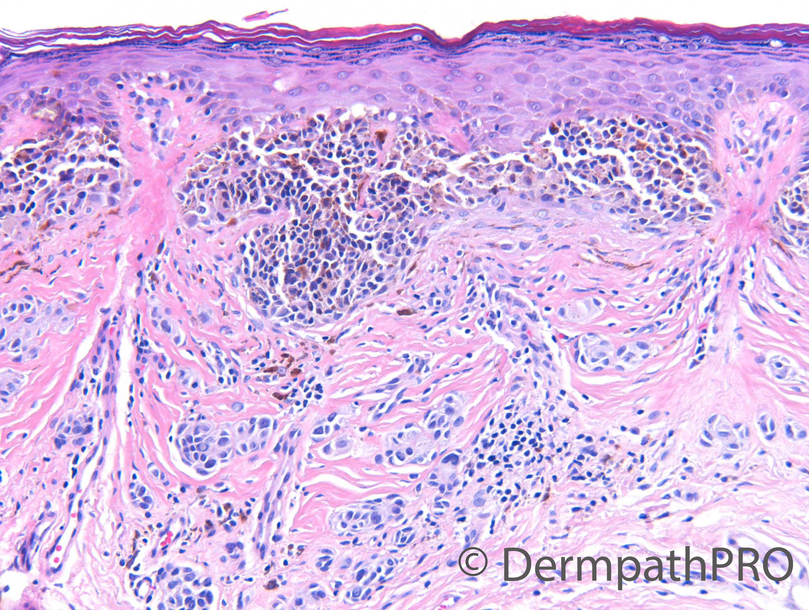
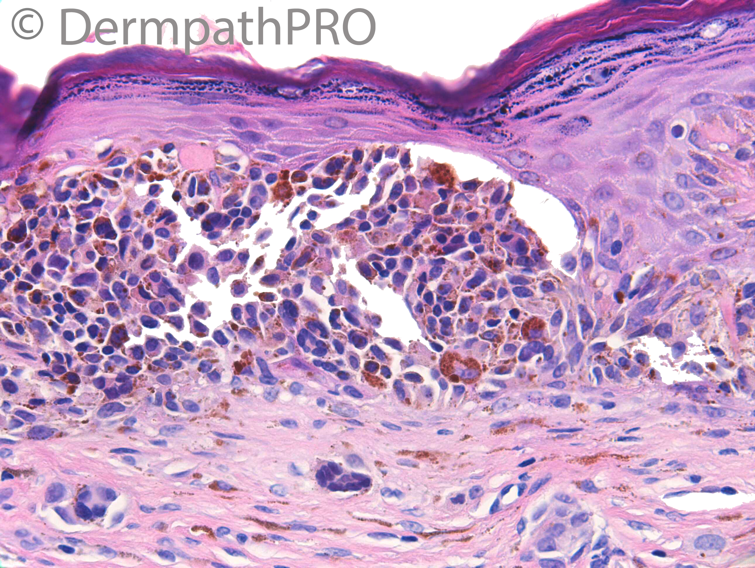
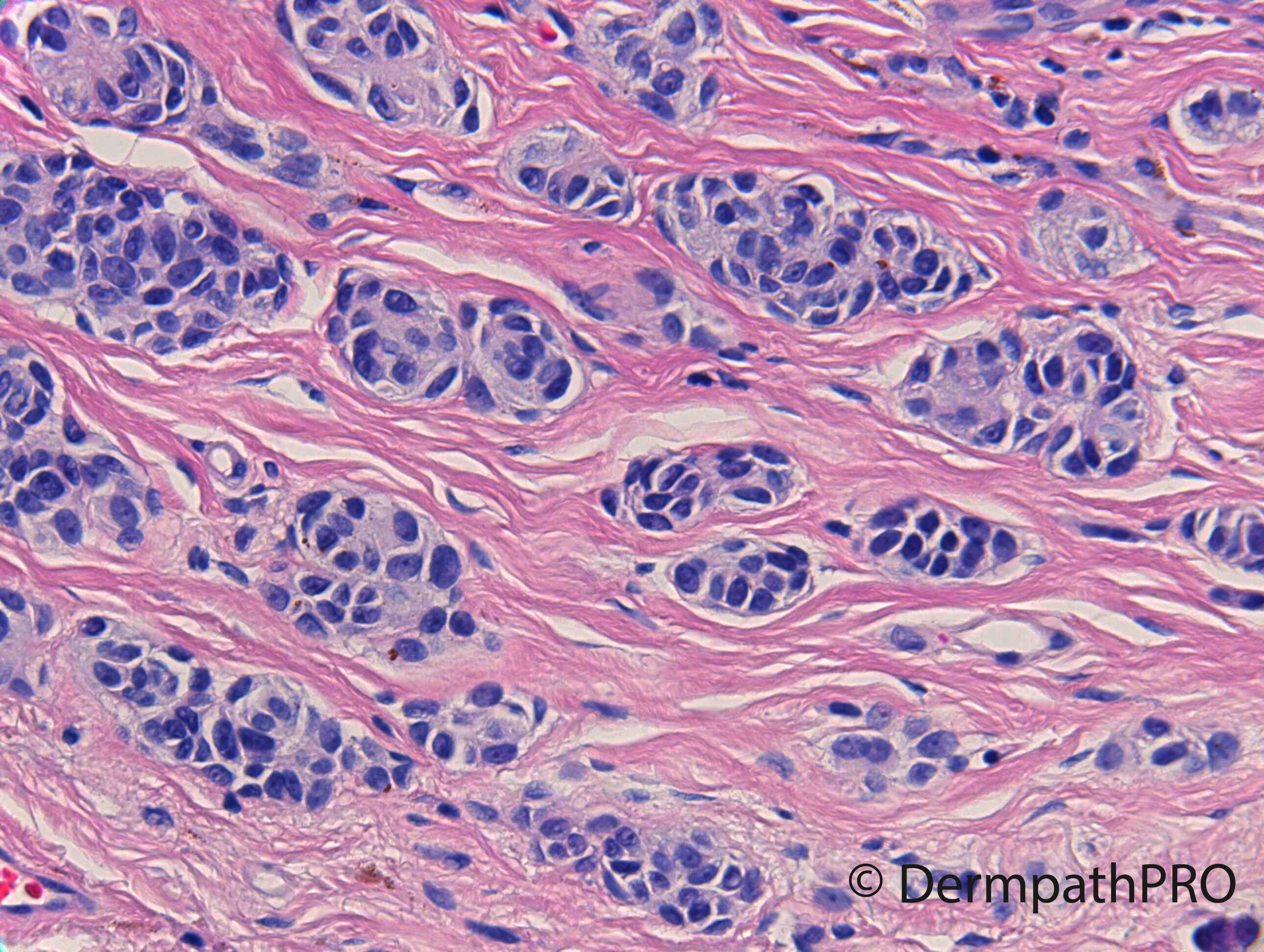
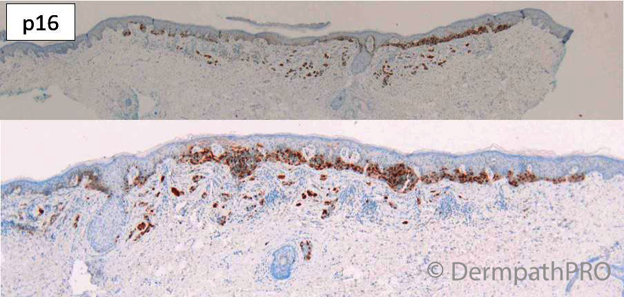
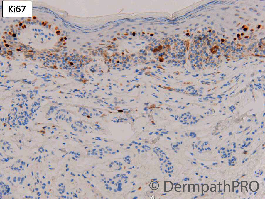
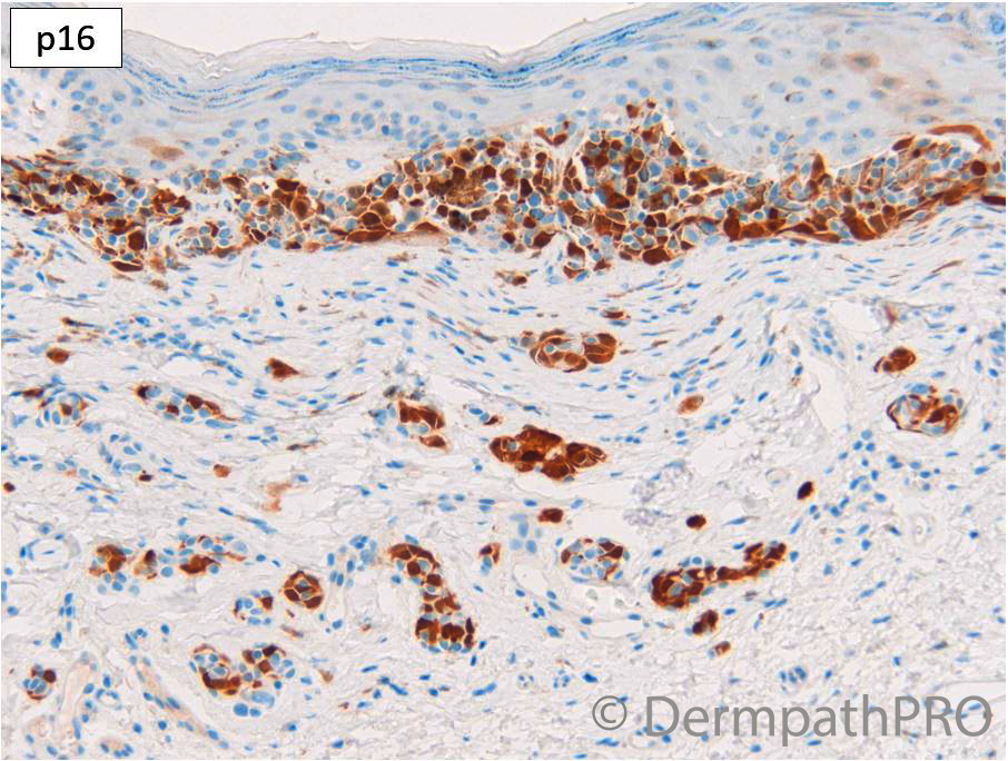
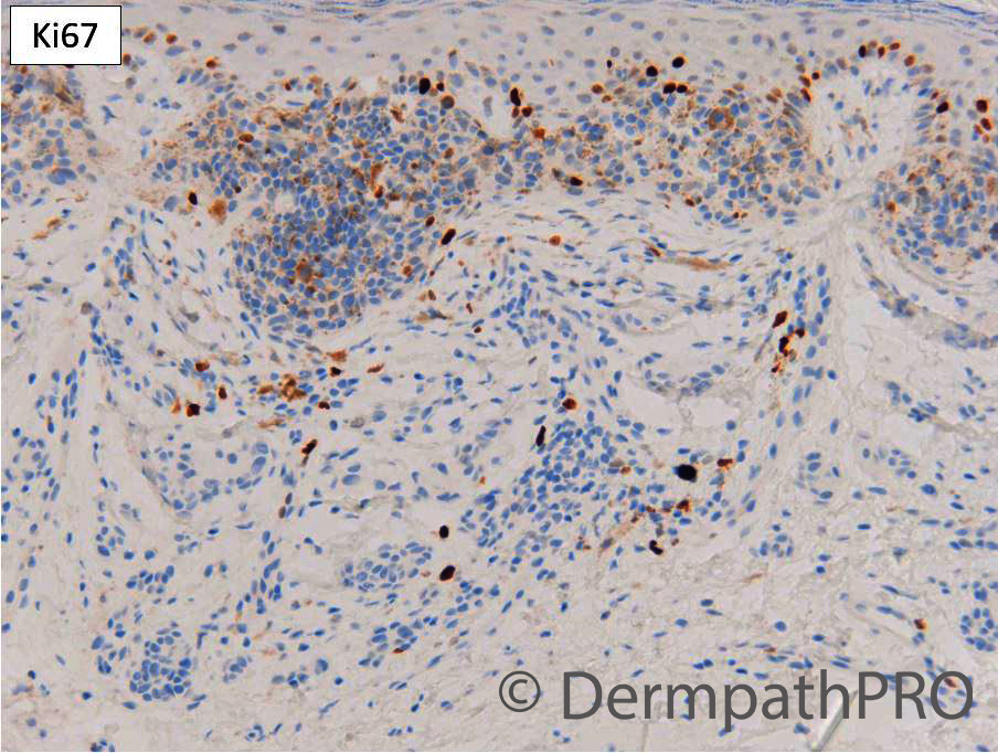
F70 Vulval skin. Pigmented lesion. Previous history of melanoma.


  Report Record

User Feedback


Eman El-Nabarawy

Posted

I'm inclined to think this is a recurrent/traumatised/sclerosed naevus (of special site). 

Share this comment


Link to comment
share_externally

Special site Nevus, with some traumatic effect. 

Share this comment


Link to comment
share_externally

Dr. Richard Carr

Posted

This case was challenging but I favoured a special site naevus and advised watchful waiting. Nice symmetry, lack of pagetoid spread, good lateral demarcation, no significantly worrying features in the dermal component. p16 preserved mosaic and low Ki67 proliferation index. A highly respected UK colleague had favoured a melanoma in situ for the impressive junctional proliferation and I was sent the case for a further opinion. It's normal to get a range of different opinions on such cases but on balance I favoured it to be all benign / atypical special site naevus.

Share this comment


Link to comment
share_externally

Krishnakumar subramanian

Posted

Dear sir,

 Atypical genital nevi are rare melanocytic lesions that most commonly arise on the vulva of young women. they behave in a benign manner. Here the lady is aged 70 years and we do not have information how long the lady had the nevus. so clinically melanoma is high on mind. with IHC we can conclude this. But this your years of wisdom. Now is it mandatory any atypical nevus or melanoma in doubt we have to do these markers, what is the role of PRAME here. Also, is there any age limit to consider nevus at special sites

please clarify Sir


Share this comment


Link to comment
share_externally

18 hours ago, Krishnakumar subramanian said:

Dear sir,

 Atypical genital nevi are rare melanocytic lesions that most commonly arise on the vulva of young women. they behave in a benign manner. Here the lady is aged 70 years and we do not have information how long the lady had the nevus. so clinically melanoma is high on mind. with IHC we can conclude this. But this your years of wisdom. Now is it mandatory any atypical nevus or melanoma in doubt we have to do these markers, what is the role of PRAME here. Also, is there any age limit to consider nevus at special sites

please clarify Sir


I also have similar concerns as you for this case. at least it is dysplastic nevus if the junctional not reaching to the degree of melanoma in situ. the dyhesive nature of those melanocytes maybe duet to special site. but the relative florid growth is worrisome, I would do PRAME in this case, P16 only meaningful to me if there is complete loss in the melanocytes. melanoma can have intact preserved P16. 

Share this comment


Link to comment
share_externally

Dr. Richard Carr

Posted

Thank you for the additional discussions. PRAME could be helpful. I don't have it available locally but could have requested it to be sent away and waited for it. When assessing an IHC one has to have a feel for the prior probability and interpret the result in the light of this. My prior probability is relatively low but a positive PRAME could move the goal posts more into the uncertain end i.e. cannot rule out early  melanoma. The discussion then becomes what margins are acceptable for thin somewhat challenging uncertain lesions and I think we already have that problem. Watchful waiting (and I mean careful follow-up given the location with informed consent to the pros and cons) should remain a safe and viable option if the patient prefers (the location and lack of history cause problems for guiding management. I think we have to remember that harm can come from over-diagnosis and over-treatment and from under-diagnosis and under-treatment and that for thin and inherently subjective lesions there is no "zero harm" option.

Share this comment


Link to comment
share_externally

msofopoulos

Posted (edited)

Very interesting case, I thought  it was in situ SSM due to "Contiguous proliferation of uniformly (e.g. > 50%) moderately to severely atypical melanocytes
in an area ≥ 0.5 mm2" as WHO indicates, however atypia is somehow subjective. I am glad I don't have to sign this one.

Regarding PRAME stain I must say that we haven't had great results,so far, in our lab, however we are using alkaline phosphatase (Red) instead DAB.

Again thanks

Edited by msofopoulos
typos

Share this comment


Link to comment
share_externally


Join the conversation

You can post now and register later. If you have an account, sign in now to post with your account.

Guest
Add a comment...

×   Pasted as rich text.   Paste as plain text instead

  Only 75 emoji are allowed.

×   Your link has been automatically embedded.   Display as a link instead

×   Your previous content has been restored.   Clear editor

×   You cannot paste images directly. Upload or insert images from URL.

Loading...

×
×
  • Create New...