Jump to content
In this section we have spot diagnoses posted on a daily basis since June 2010, now over 4000! You can review the archived cases and read the suggested diagnoses by users and the final comment by the contributors.
Case are uploaded each week day by 10 am UK time with the correct diagnosis will generally be posted at 8 pm UK time. Why not view the most recent spot diagnosis and proffer a diagnosis?

Case Number : Case 2882 - 23 July 2021 Posted By: Dr. Richard Carr

Please read the clinical history and view the images by clicking on them before you proffer your diagnosis.
Submitted Date :
   (0 reviews)

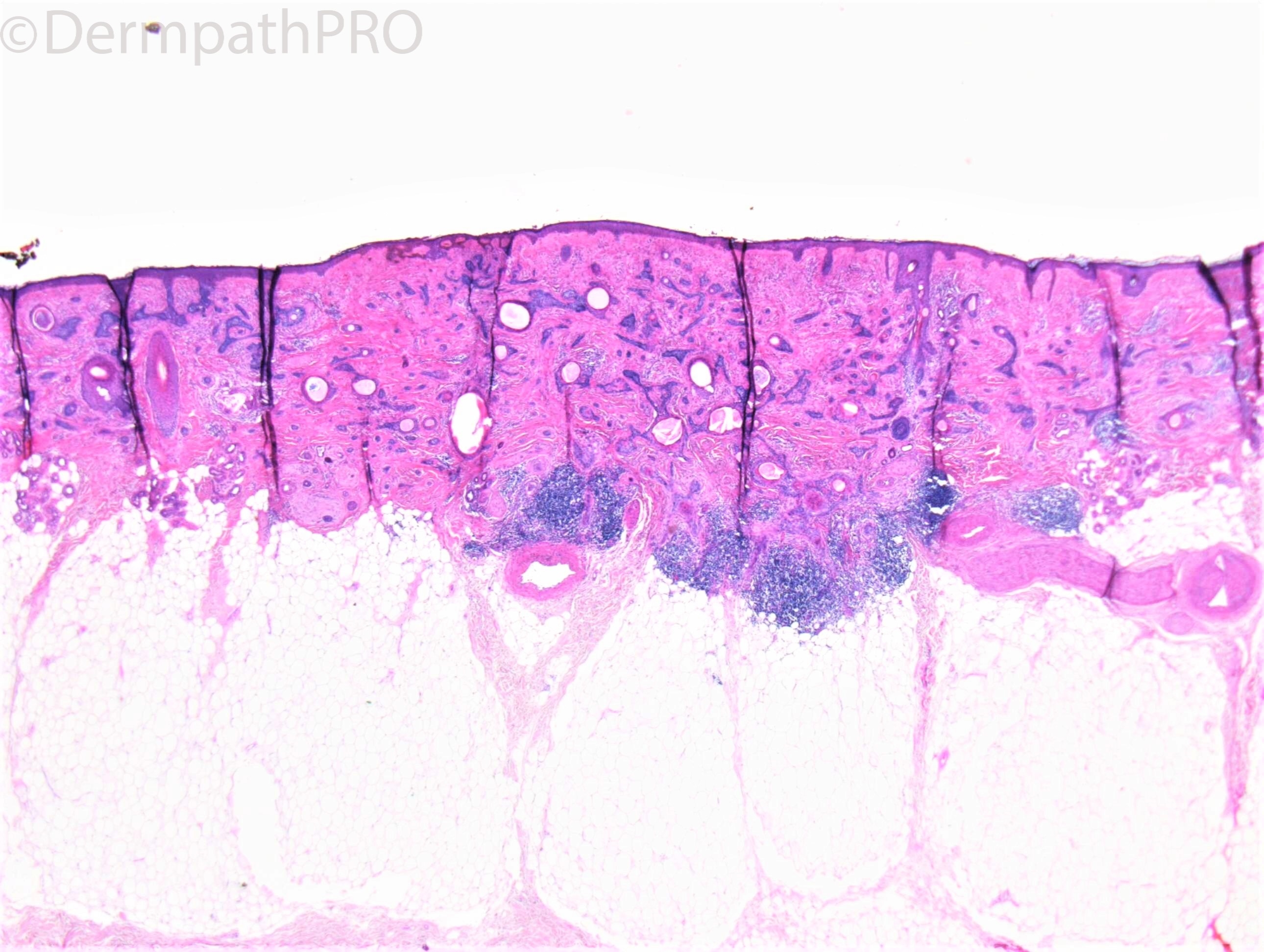
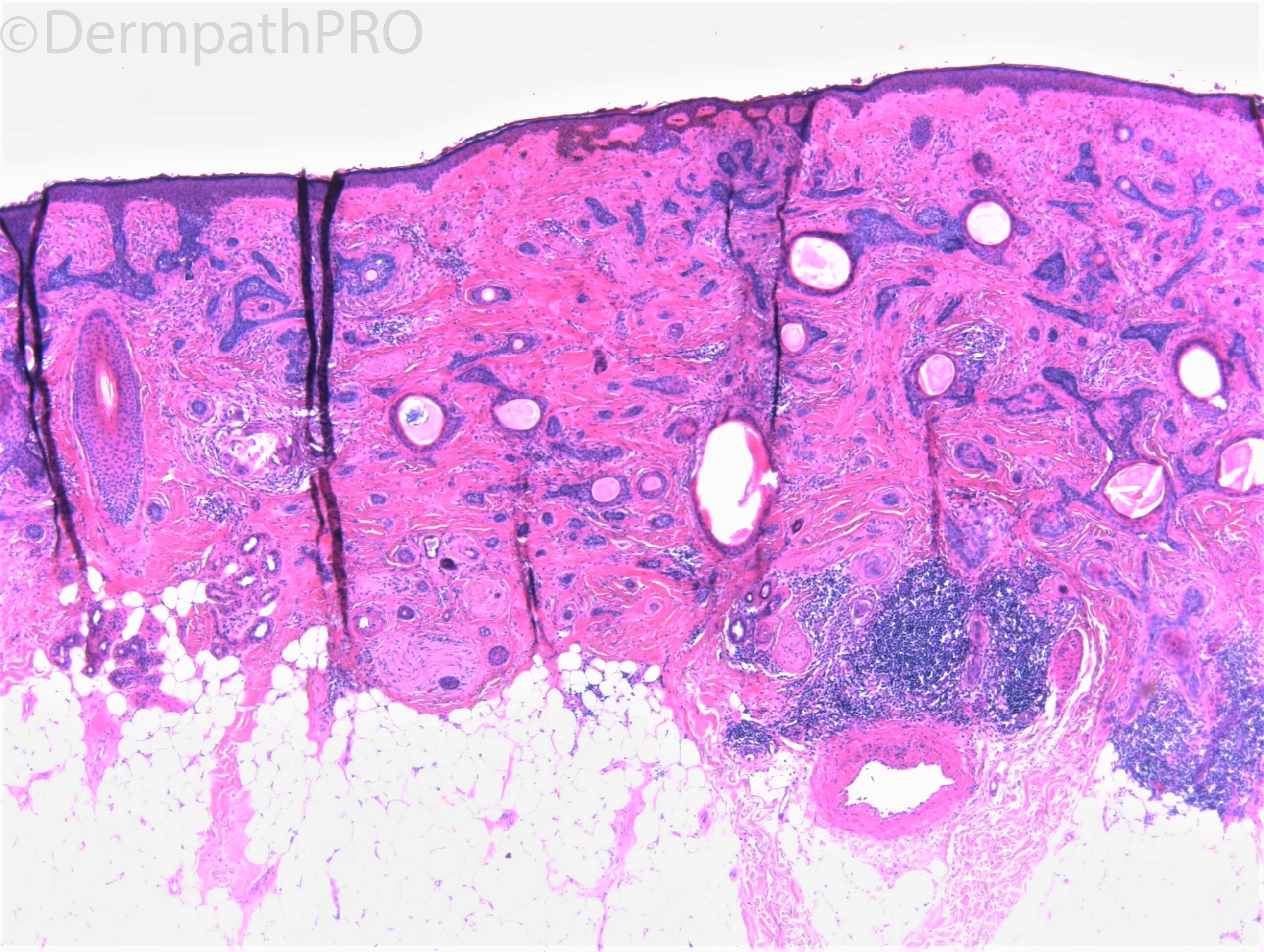
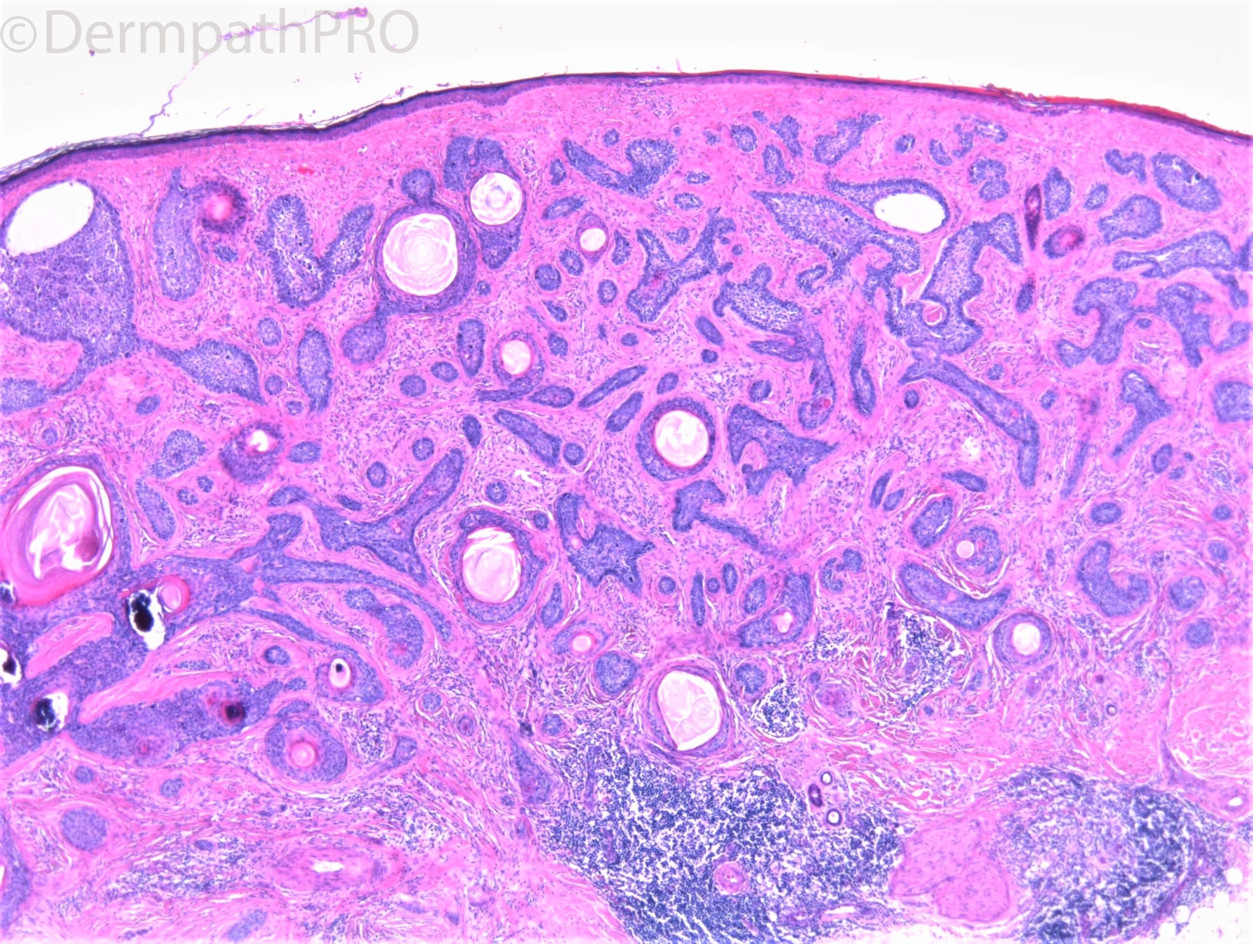
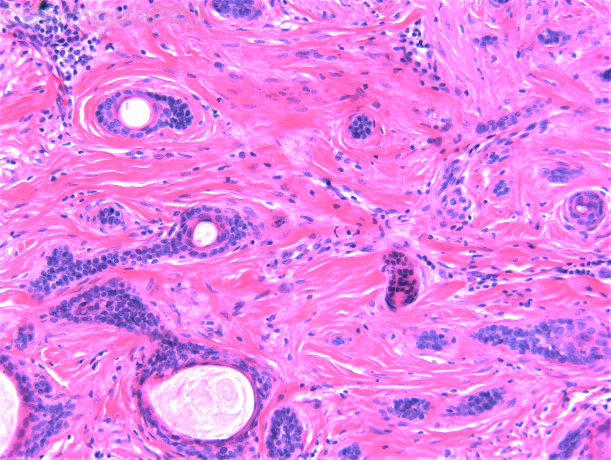
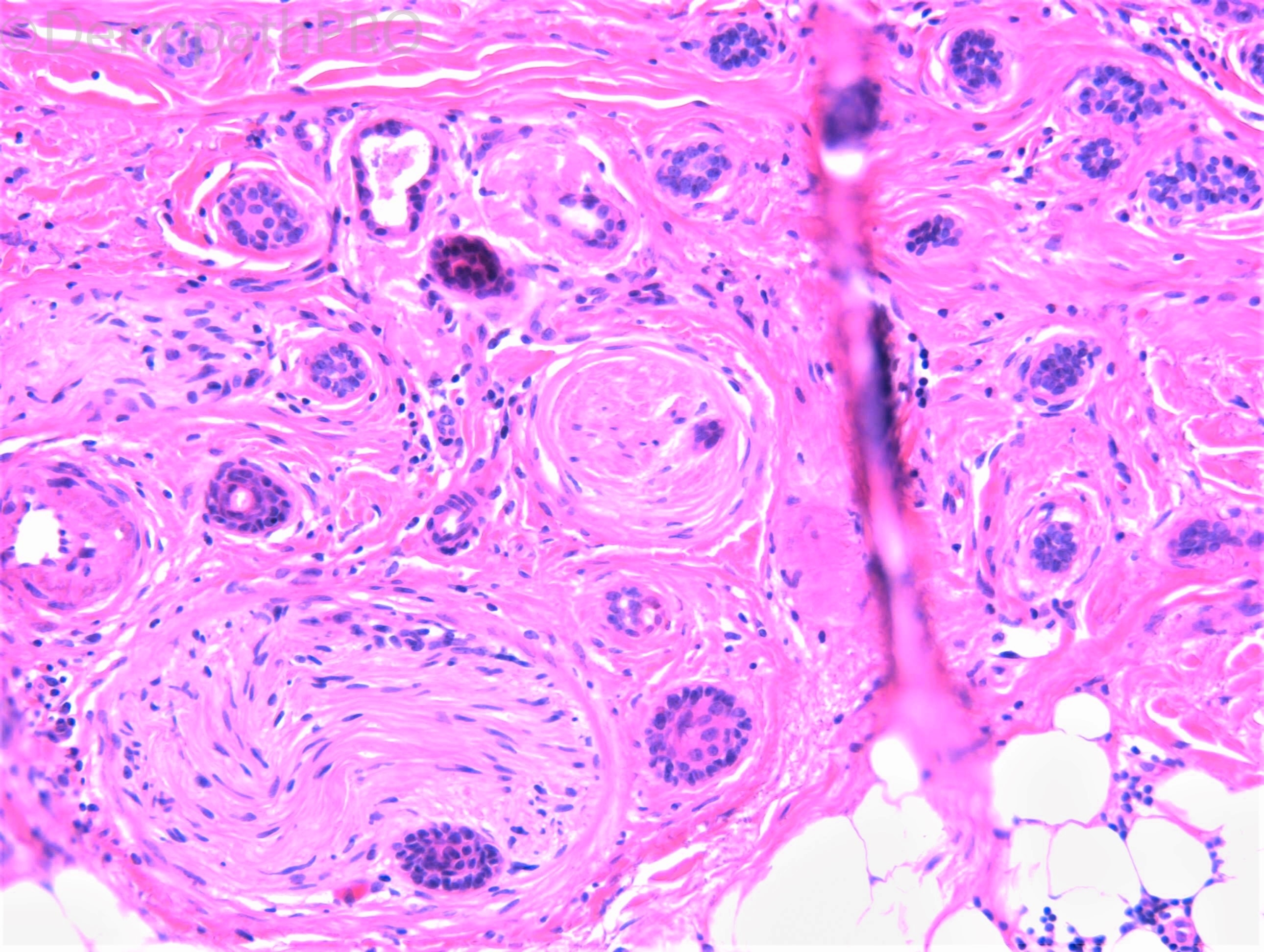
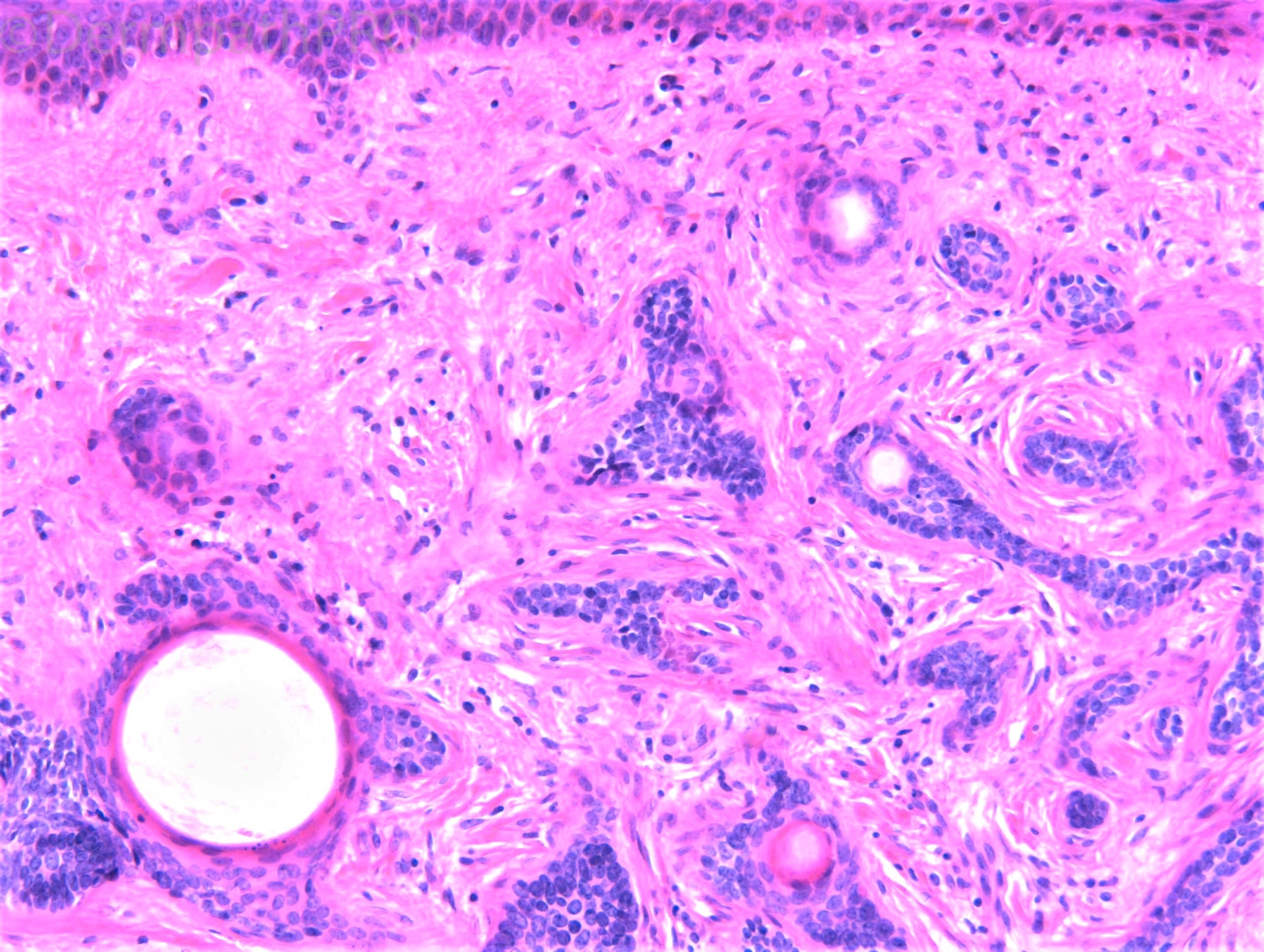
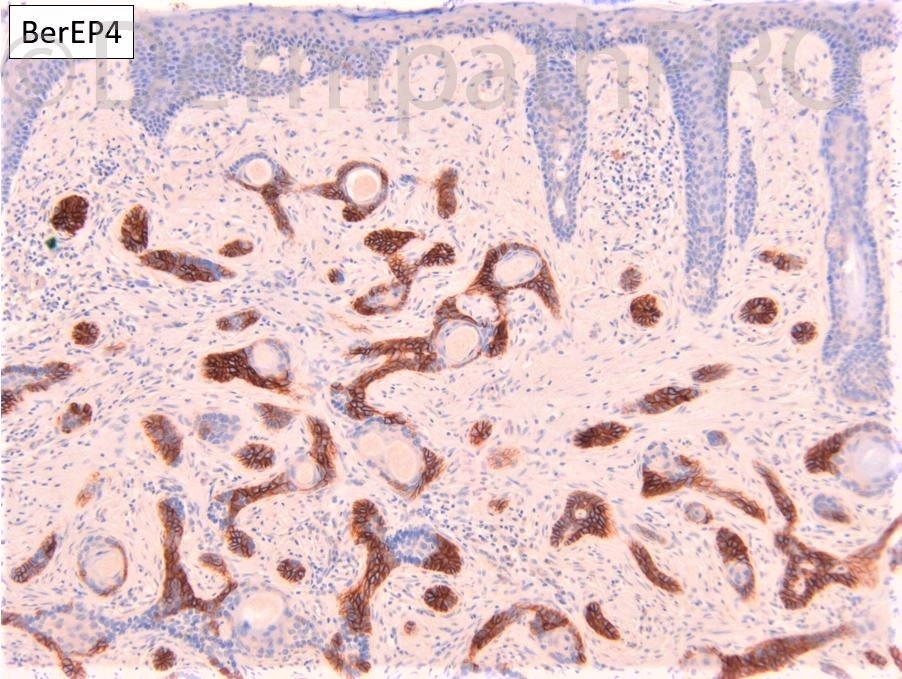
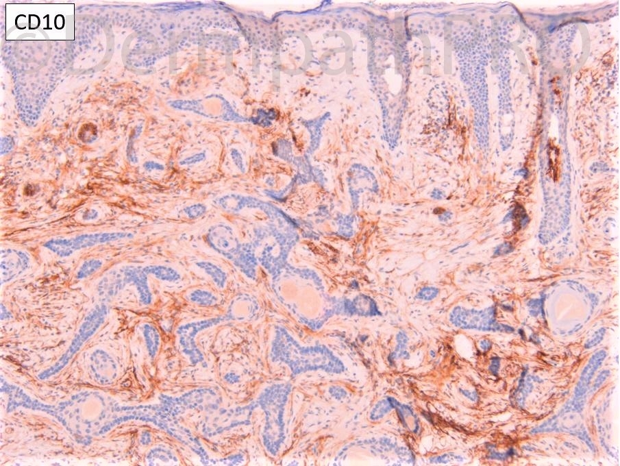
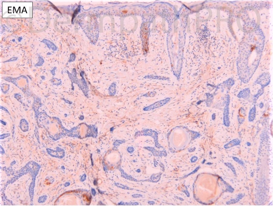
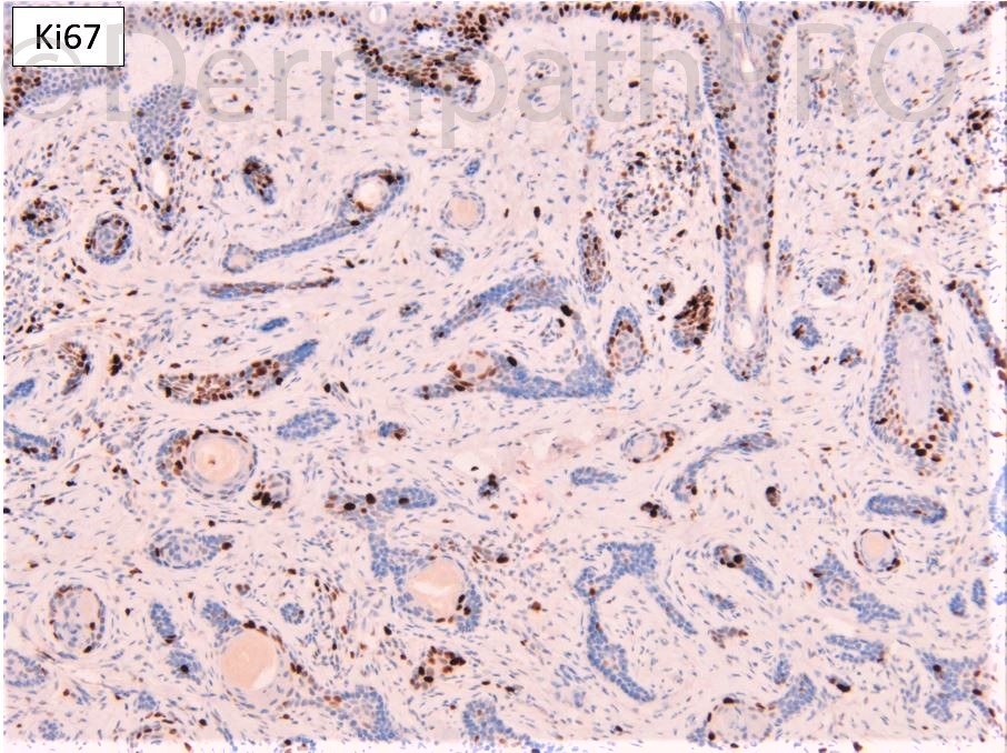
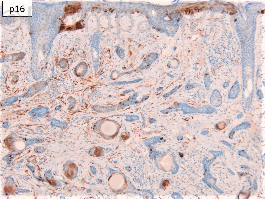
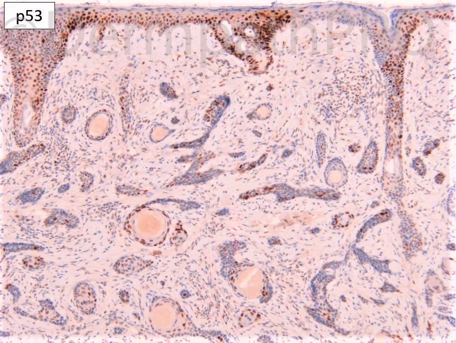
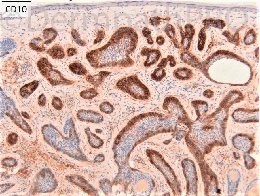
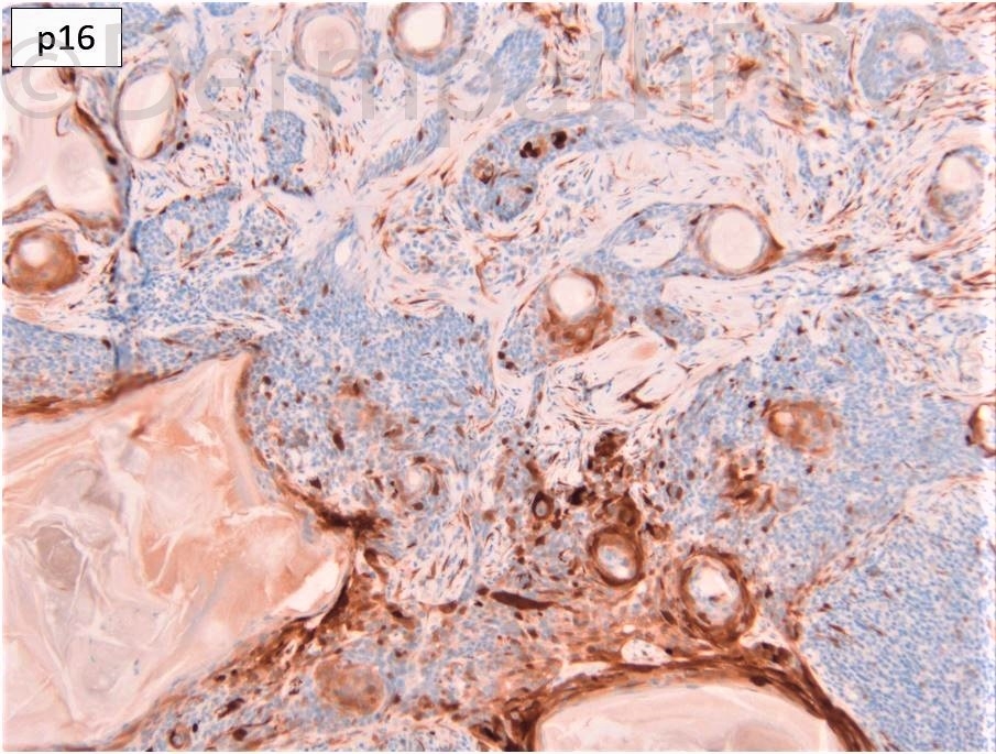
F85. Forehead. Nodular BCC?


  • Like 1
  Report Record

User Feedback


Richard Logan

Posted

At first I thought this might be cutaneous lymphadenoma, especially from the appearance in image 3.  However, on balance I agree with Cem Leblebici that this is more likely microcystic adnexal carcinoma.

Share this comment


Link to comment
share_externally

Eman El-Nabarawy

Posted

Well. Positive BerEp4 excludes MAC..CD10 is positive in part stromal and epithelial in another part.. When I saw the case for the first time I was really struggling between BCC and TE! Is this a papillary mesenchymal body in fig 6? Now I am thinking why not it's BCC arising in TE? 

Nice case report!

https://www.ncbi.nlm.nih.gov/pmc/articles/PMC6793645/#!po=16.6667

Share this comment


Link to comment
share_externally

Cem Leblebici

Posted

After evaluating immunhistochemistry, I considered basal cell carcinoma with adnexal differentiation. Maybe, it can arise in TE, as Eman El-Nabarawy wrote. 

Share this comment


Link to comment
share_externally

This neoplasia is enough infiltrative, but radially rather than vertically, as expected in MAC. It’s difficult because there aren’t any epithelio-stromal m. ucin material or clear spaces. At a glance it seemed a lymphoadenoma/TE. However the malignant growth pattern makes me thinking of BCC. 

Share this comment


Link to comment
share_externally

Dr. Richard Carr

Posted

This was a BCC with some DTE-like areas. Interesting that I did not consider MAC because typical areas of BCC were present (note the nice palisading in 2nd image down on left. It's unusual to see an infiltrative BCC so closely mimic DTE with nice keratocysts and relatively evenly cellular stroma. I suppose we can add also add a nice mimic MAC in view of the responses. BerEP4 was a little variable in the DTE-like areas but more strong and diffuse in the more typical BCC areas (not highlighting lumina only as can be seen in MAC). CD10 shows areas with minimal epithelial staining and other areas with epithelial predominant pattern typical for BCC. No convincing reactive dendritic Merkel cells (which are typically common in DTE). I did not see extra-tumoural perineural invasion in this lesion (just cords sat next to nerves) and it's well to remember DTE may also occassionaly show apparent intra-tumoural perineural invasion (but not extra-tumoural in my experience). For me p16 and p53 were not "aberrant", seems BCC are a little variable in the patterns but do show some nulls for p16 and p53 and some aberrant patterns but also some with wild type patterns. Usually the latter are not required for diagnosis as EMA and BerEP4 sorts out >95% of cases.

Share this comment


Link to comment
share_externally

Eman El-Nabarawy

Posted

59 minutes ago, Dr. Richard Carr said:

This was a BCC with some DTE-like areas. Interesting that I did not consider MAC because typical areas of BCC were present (not the nice palisading in 2nd image down on left. It's unusual to see an infiltrative BCC so closely mimic DTE with nice keratocysts and relatively evenly cellular stroma. I suppose we can add also add a nice mimic MAC in view of the responses. BerEP4 was a little variable in the DTE-like areas but more strong and diffuse in the more typical BCC areas (not highlighting lumina only as can be seen in MAC). CD10 shows areas with minimal epithelial staining and other areas with epithelial predominant pattern typical for BCC. No convincing reactive dendritic Merkel cells (which are typically common in DTE). I did not see extra-tumoural perineural invasion in this lesion (just cords sat next to nerves) and it's well to remember DTE may also occassionally show apparent intra-tumoural perineural invasion (but not extra-tumoural in my experience). For me p16 and p53 were not "aberrant", seems BCC are a little variable in the patterns but do show some nulls for p16 and p53 and some aberrant patterns but also some with wild type patterns. Usually the latter are not required for diagnosis as EMA and BerEP4 sorts out >95% of cases.

Thank u for the interesting case Sir.  I wonder about Bcl2 staining pattern?!

Share this comment


Link to comment
share_externally

Dr. Richard Carr

Posted

I stopped using Bcl2 around 10+ ago now, CK20 is the most helpful marker for me and careful attention to the 26 criteria of Ackerman. See discussion for case 813 (8 years ago!). In my spreadsheet which I still maintain for interest this case scored -2 (i.e. slight balance of features for DTE) but typical DTE score around -15 on average (all at least -10). DTE-like BCC's score between -2 and +8 (latter a collision of BCC and DTE) i.e. different ball-park. In rare cases when there is genuine significant doubt, recommend complete excision which may require margin control.


Share this comment


Link to comment
share_externally


Join the conversation

You can post now and register later. If you have an account, sign in now to post with your account.

Guest
Add a comment...

×   Pasted as rich text.   Paste as plain text instead

  Only 75 emoji are allowed.

×   Your link has been automatically embedded.   Display as a link instead

×   Your previous content has been restored.   Clear editor

×   You cannot paste images directly. Upload or insert images from URL.

Loading...

×
×
  • Create New...