Jump to content
In this section we have spot diagnoses posted on a daily basis since June 2010, now over 4000! You can review the archived cases and read the suggested diagnoses by users and the final comment by the contributors.
Case are uploaded each week day by 10 am UK time with the correct diagnosis will generally be posted at 8 pm UK time. Why not view the most recent spot diagnosis and proffer a diagnosis?

Case Number : Case 2886 - 29 July 2021 Posted By: Saleem Taibjee

Please read the clinical history and view the images by clicking on them before you proffer your diagnosis.
Submitted Date :
   (0 reviews)

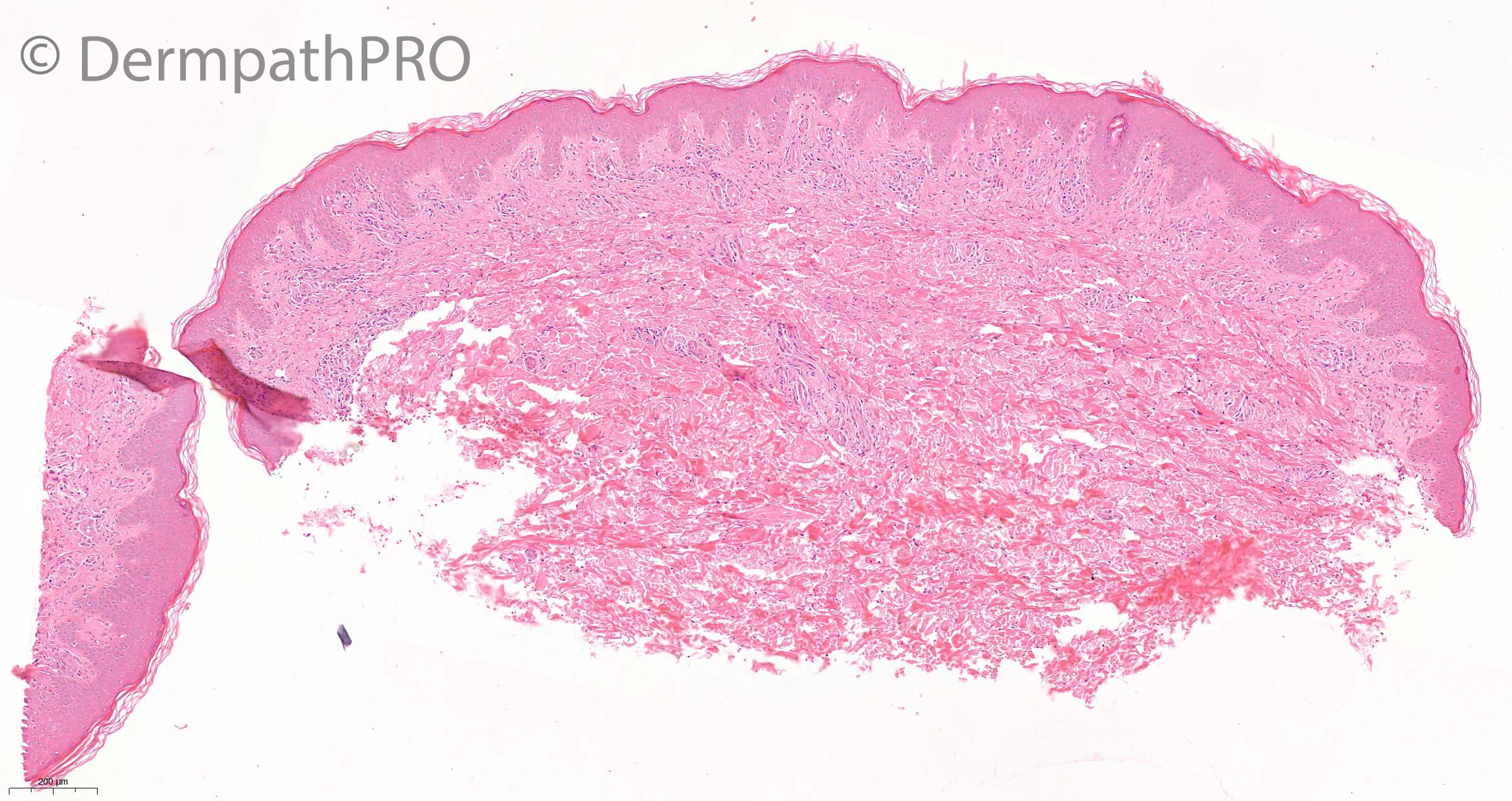
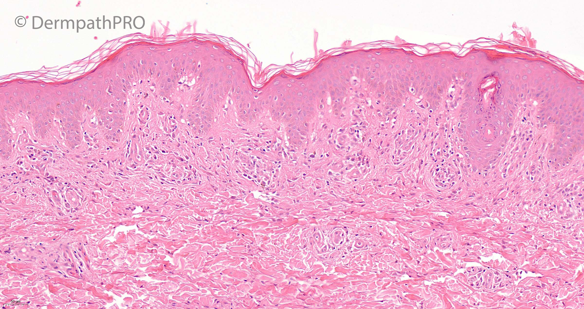
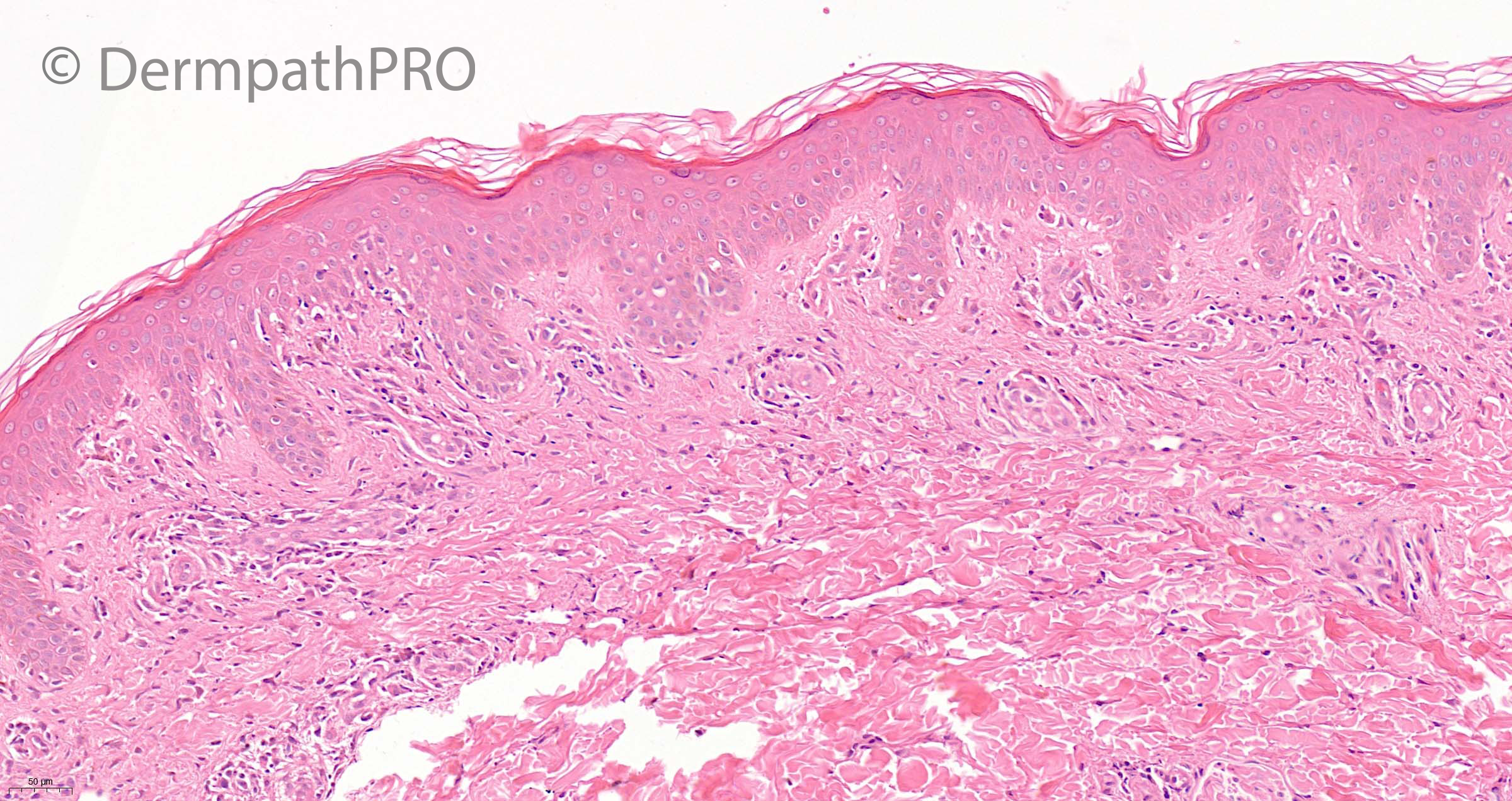
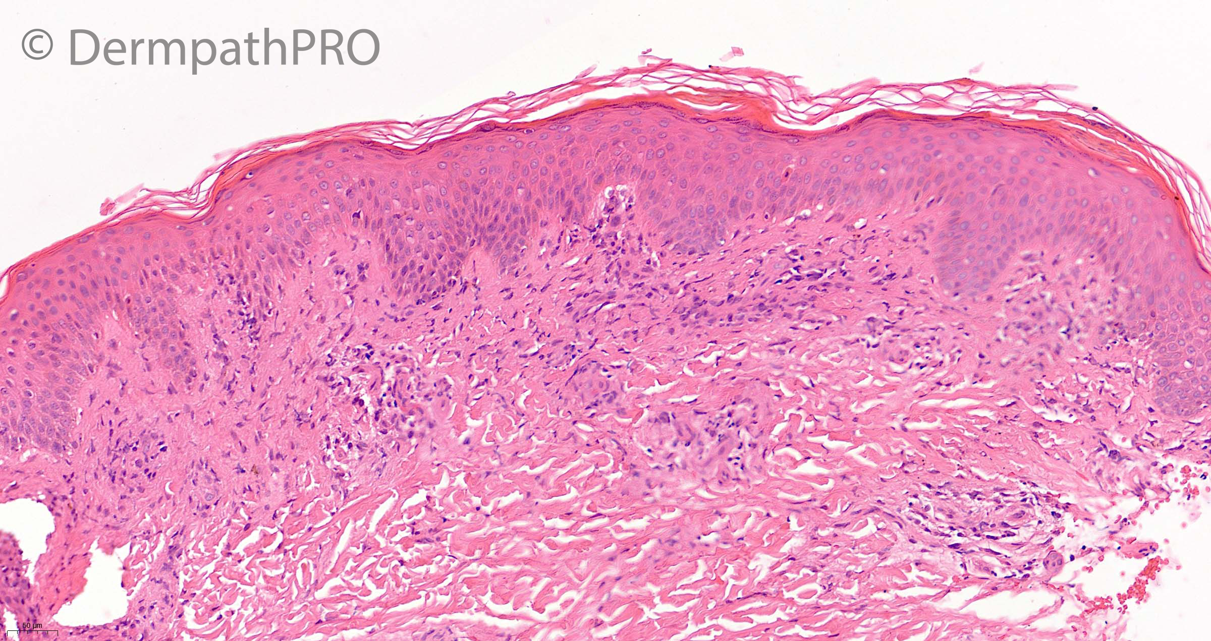
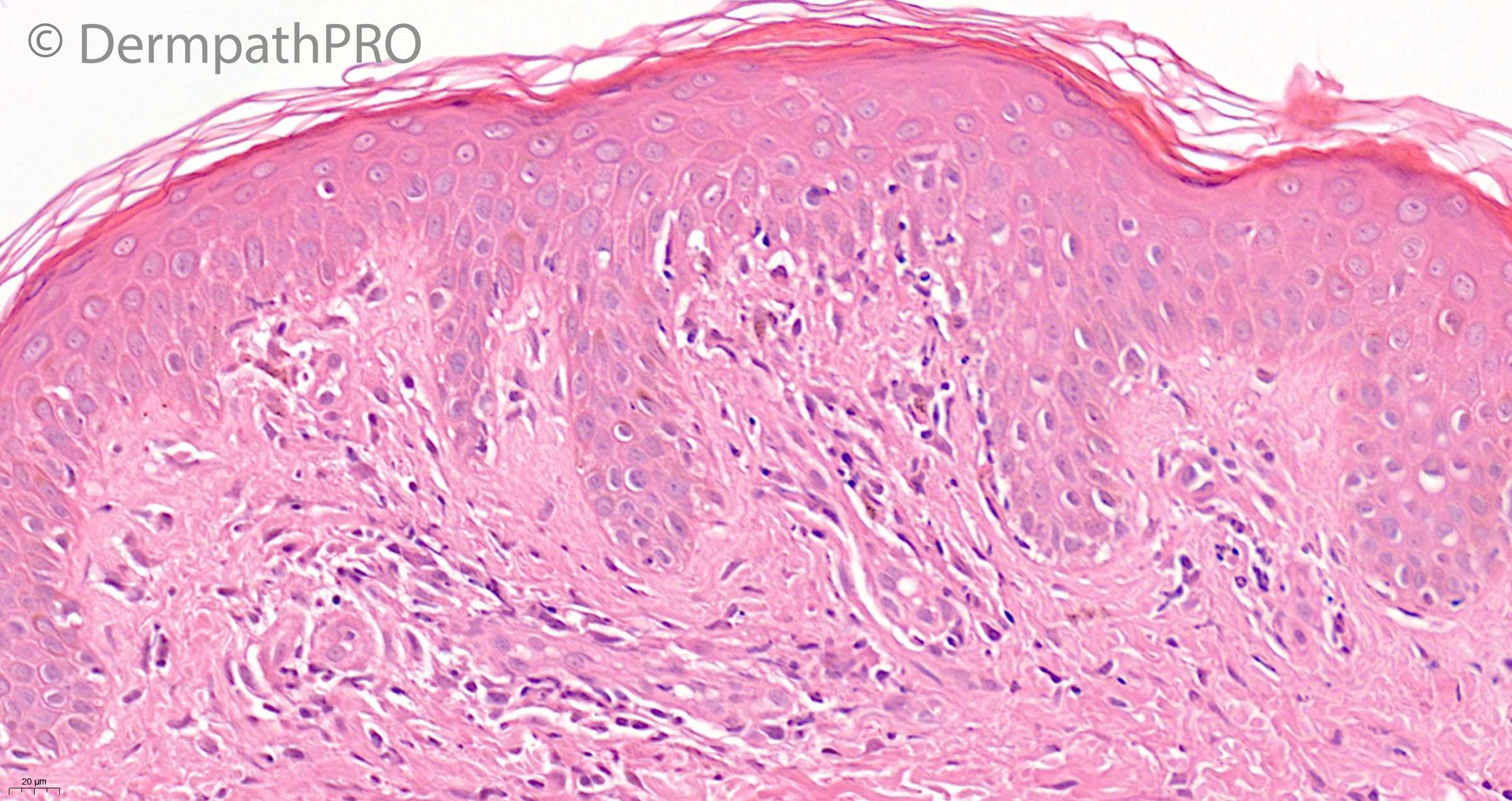
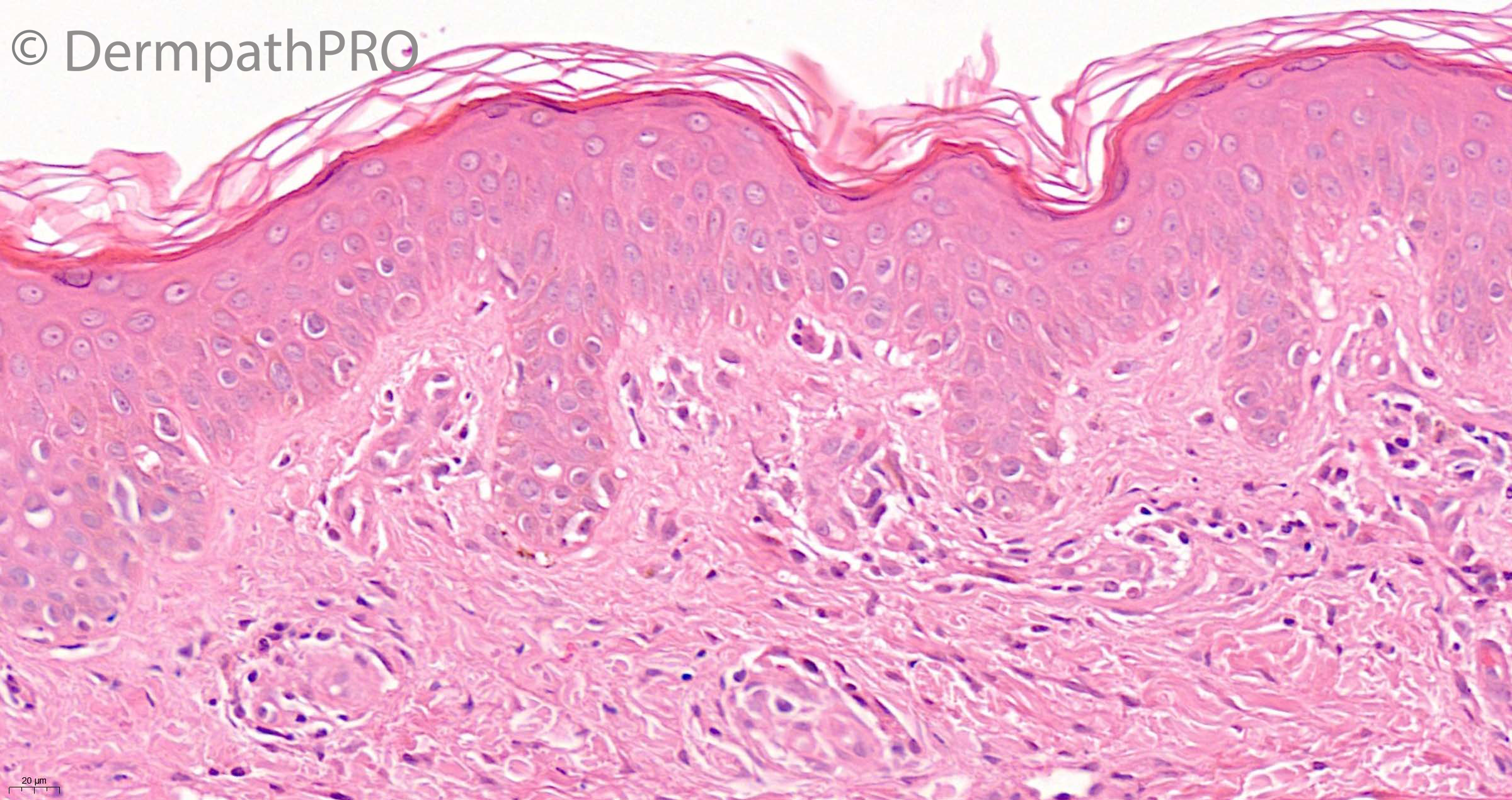
7 year old girl, punch biopsy back. Acute rash head, neck & trunk


  Report Record

User Feedback


Eman El-Nabarawy

Posted

This needs clinical correlation..Focal interface changes with high up dyskeratotic kcs and hint of neutrophilic dermal infiltrate in a child.. To start the discussion what about Still's disease?

Share this comment


Link to comment
share_externally

A parakeratotic thin layer fits with an acute but basically parakeratotic disorder.  There is an interface dyskeratotic but also vacuolar lesion pattern. So it’s a Mucha Haberman for me. 

Share this comment


Link to comment
share_externally

Beautiful case. Saw those vacuolar interface changes with keratinocytes dyskeratosis and focal "lymphocytes satellitosis". dermal pigment incontinence is noted. Clearly need clinical history, but I guess this kid may have had stem cell transplant and now have acute GVHD. waiting for your answer, Saleem.  Always enjoy your cases. 

Share this comment


Link to comment
share_externally

Saleem Taibjee

Posted

Just a a few participant responses on this case, but nonetheless very interesting to see these responses which illustrate the wide histological differential diagnosis and challenging nature of such cases, and need for clinicopathological correlation ....

Well done, Henry. Yes this was indeed a recent case of GVHD which developed approximately 3 weeks after stem cell transplantation for leukaemia. Of course, the difficulty in such cases is that the clinical differential diagnosis often lies between GVHD and drug reaction, and the histology often cannot allow for confident distinction, although we might look for eosinophils, etc. But I put this case in because I thought it was indeed a nice example of satellite cell necrosis (as Henry also spotted, illustrated in the final 2 images above) which as we know can be a feature/clue to GVHD.

BW

Saleem

Share this comment


Link to comment
share_externally


Join the conversation

You can post now and register later. If you have an account, sign in now to post with your account.

Guest
Add a comment...

×   Pasted as rich text.   Paste as plain text instead

  Only 75 emoji are allowed.

×   Your link has been automatically embedded.   Display as a link instead

×   Your previous content has been restored.   Clear editor

×   You cannot paste images directly. Upload or insert images from URL.

Loading...

×
×
  • Create New...