Jump to content
In this section we have spot diagnoses posted on a daily basis since June 2010, now over 4000! You can review the archived cases and read the suggested diagnoses by users and the final comment by the contributors.
Case are uploaded each week day by 10 am UK time with the correct diagnosis will generally be posted at 8 pm UK time. Why not view the most recent spot diagnosis and proffer a diagnosis?

Case Number : Case 2846- 03 June 2021 Posted By: Saleem Taibjee

Please read the clinical history and view the images by clicking on them before you proffer your diagnosis.
Submitted Date :
   (0 reviews)

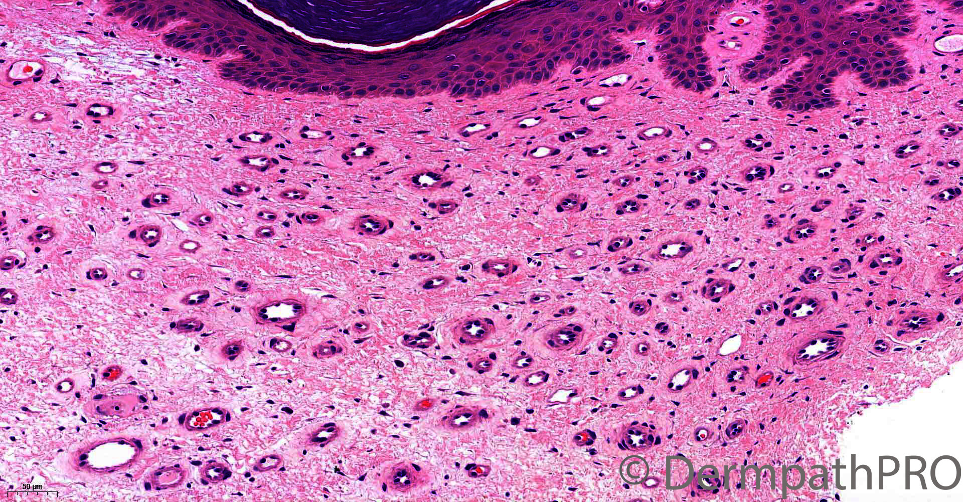
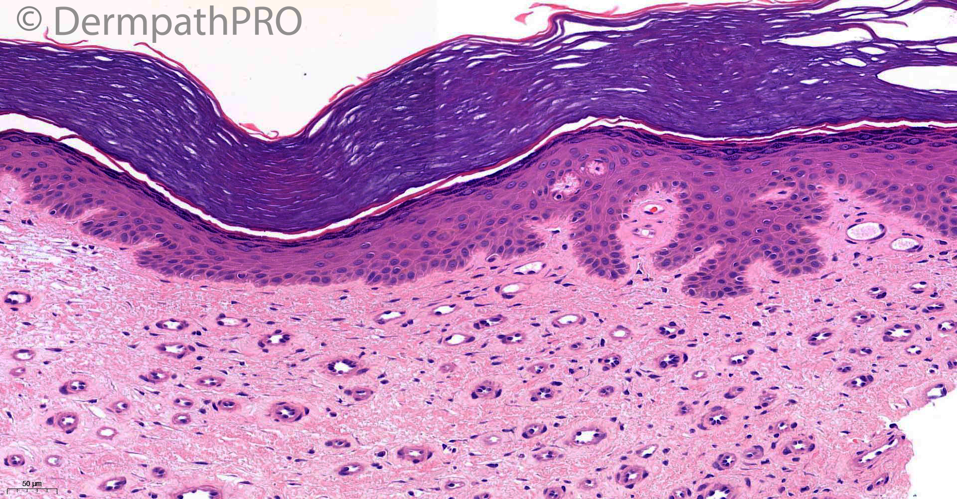
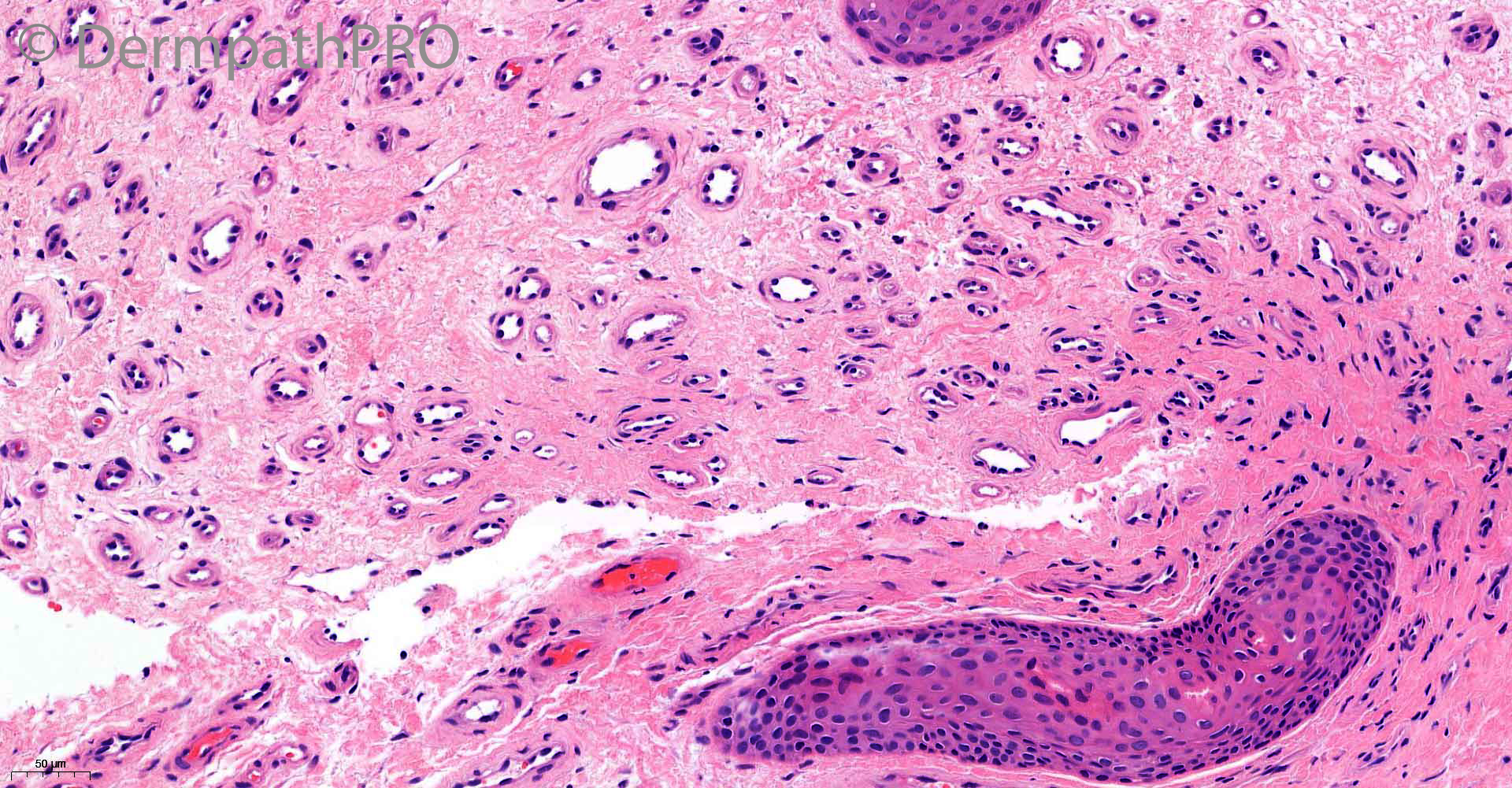
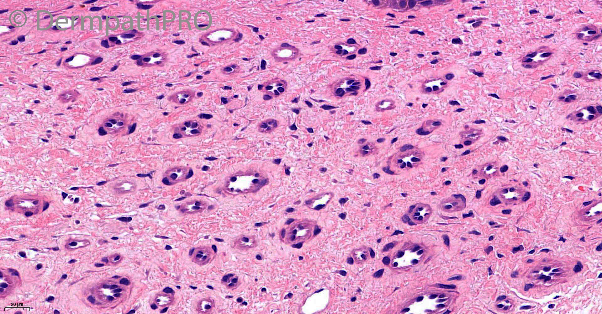
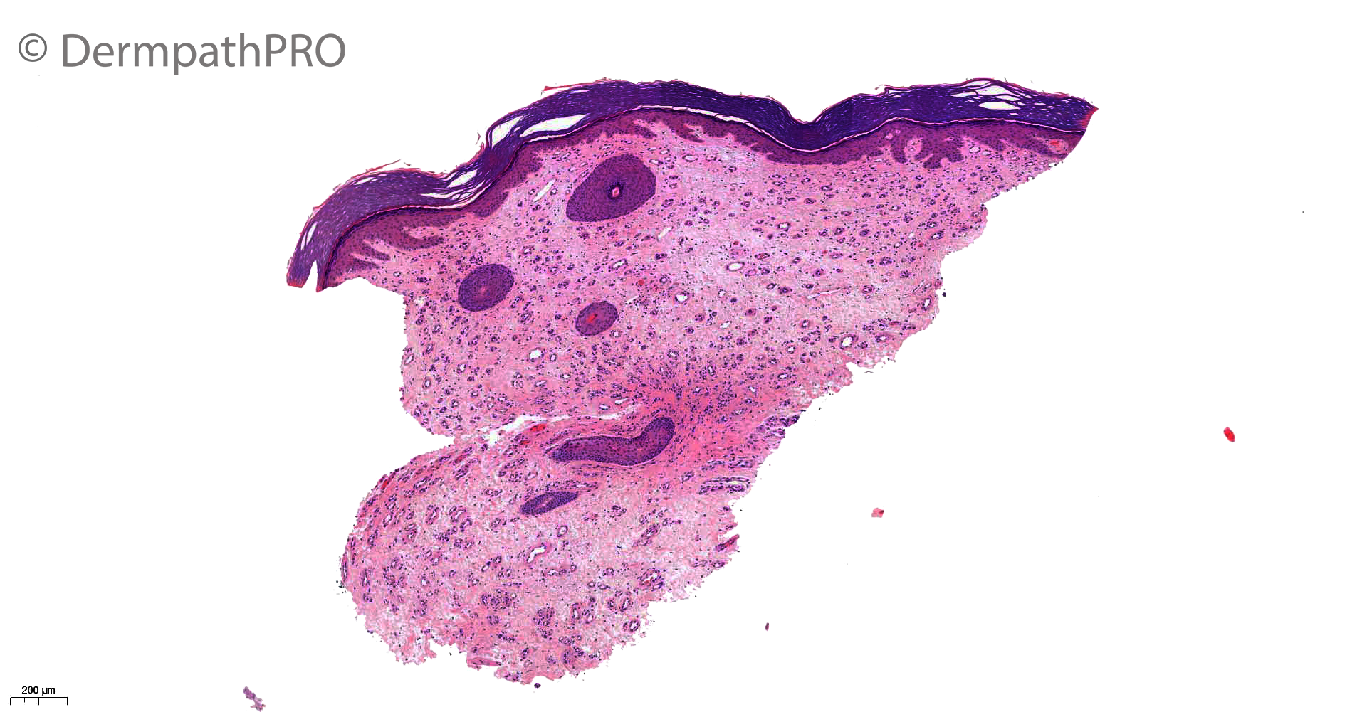
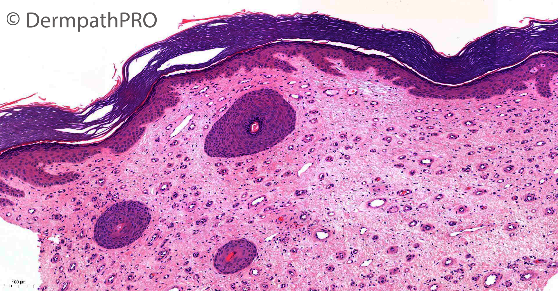
93M, biopsy right lower leg. 4x3cm blue-purple plaque ?acroangiodermatitis pseudo-Kaposi? Kaposi? angiosarcoma? Lymphoma

  Report Record

User Feedback


Richard Logan

Posted

I think this is a very nice example of the effects of chronic venous hypertension, with capillary loss, dermal fibrosis, and reduplication and tortuosity of the remaining capillaries giving the impression of increased vascularity.  There is also overlying hyperkeratosis.

I guess acroangiodermatitis is one of several names applied to it.

Share this comment


Link to comment
share_externally

Saleem Taibjee

Posted

Just to let you know that I have now posted a few immunos on last Thursday's case 27 May.

Share this comment


Link to comment
share_externally

Krishnakumar subramanian

Posted

There is arrangement of blood vessels in a band like pattern, can we call it Poikilodermatous plaque-like hemangioma

Share this comment


Link to comment
share_externally

Saleem Taibjee

Posted

Just to let you know that I have now posted the 'answer' for last Thursday's case 27 May, case number 2841.


Share this comment


Link to comment
share_externally

vincenzo

Posted

On 03/06/2021 at 20:13, Richard Logan said:

I think this is a very nice example of the effects of chronic venous hypertension, with capillary loss, dermal fibrosis, and reduplication and tortuosity of the remaining capillaries giving the impression of increased vascularity.  There is also overlying hyperkeratosis.

I guess acroangiodermatitis is one of several names applied to it.

Agree. 

Share this comment


Link to comment
share_externally

Saleem Taibjee

Posted

Well done, Krishnakumar. This recent case is a good example of the entity which we published, referred to as polkilodermatous plaque-like hemangioma. See case series:https://www.jaad.org/article/S0190-9622(19)30513-4/pdf

Although the publication refers to hemangioma, and implies a neoplasm, some of us think this is likely to be more of a reactive (but distinct) vascular proliferation. Many of the cases are on the legs, especially close to the knees, and we speculated this might be a response to trauma, particularly since histologically there is often a suggestion of surface lichenification as in this example. Curiously, this contrasts with the clinical appearance which tends to look more atrophic (and poikilodermic, hence the name), and in some cases mycosis fungoides is suggested clinically. Of course, venous hypertension may also be relevant in the aetiology. But I don't think this is the same as acroangiodermatitis, in which the capillary proliferation looks quite different and tends to be more lobular.

BW

Saleem

Share this comment


Link to comment
share_externally

John Zhang

Posted

On 09/06/2021 at 02:49, Saleem Taibjee said:

Well done, Krishnakumar. This recent case is a good example of the entity which we published, referred to as polkilodermatous plaque-like hemangioma. See case series:https://www.jaad.org/article/S0190-9622(19)30513-4/pdf

Although the publication refers to hemangioma, and implies a neoplasm, some of us think this is likely to be more of a reactive (but distinct) vascular proliferation. Many of the cases are on the legs, especially close to the knees, and we speculated this might be a response to trauma, particularly since histologically there is often a suggestion of surface lichenification as in this example. Curiously, this contrasts with the clinical appearance which tends to look more atrophic (and poikilodermic, hence the name), and in some cases mycosis fungoides is suggested clinically. Of course, venous hypertension may also be relevant in the aetiology. But I don't think this is the same as acroangiodermatitis, in which the capillary proliferation looks quite different and tends to be more lobular.

BW

Saleem

Fascinating. Thanks Saleem. How does this entity differ from acquired elastotic hemangioma?

Share this comment


Link to comment
share_externally

Saleem Taibjee

Posted

acquired elastotic hemangioma occurs in the context of chronic sun damage. The vascular proliferation tends to be more dissecting.

Share this comment


Link to comment
share_externally

Richard Logan

Posted

On 09/06/2021 at 10:49, Saleem Taibjee said:

Well done, Krishnakumar. This recent case is a good example of the entity which we published, referred to as polkilodermatous plaque-like hemangioma. See case series:https://www.jaad.org/article/S0190-9622(19)30513-4/pdf

Although the publication refers to hemangioma, and implies a neoplasm, some of us think this is likely to be more of a reactive (but distinct) vascular proliferation. Many of the cases are on the legs, especially close to the knees, and we speculated this might be a response to trauma, particularly since histologically there is often a suggestion of surface lichenification as in this example. Curiously, this contrasts with the clinical appearance which tends to look more atrophic (and poikilodermic, hence the name), and in some cases mycosis fungoides is suggested clinically. Of course, venous hypertension may also be relevant in the aetiology. But I don't think this is the same as acroangiodermatitis, in which the capillary proliferation looks quite different and tends to be more lobular.

BW

Saleem

Thank you Saleem.  I was unaware of your publication and I stand corrected.

I take your point about the more even distribution of the capillary proliferation and the lack of a lobular pattern that would be expected in venous hypertension.  Also, in your case the endothelial cells are rather more prominent, almost hobnail in appearance.

Share this comment


Link to comment
share_externally

I learned another pathological chapter , today. Thanks, Saleem. 

Share this comment


Link to comment
share_externally


Join the conversation

You can post now and register later. If you have an account, sign in now to post with your account.

Guest
Add a comment...

×   Pasted as rich text.   Paste as plain text instead

  Only 75 emoji are allowed.

×   Your link has been automatically embedded.   Display as a link instead

×   Your previous content has been restored.   Clear editor

×   You cannot paste images directly. Upload or insert images from URL.

Loading...

×
×
  • Create New...