Jump to content
In this section we have spot diagnoses posted on a daily basis since June 2010, now over 4000! You can review the archived cases and read the suggested diagnoses by users and the final comment by the contributors.
Case are uploaded each week day by 10 am UK time with the correct diagnosis will generally be posted at 8 pm UK time. Why not view the most recent spot diagnosis and proffer a diagnosis?

Case Number : Case 2852- 11 June 2021 Posted By: Dr. Richard Carr

Please read the clinical history and view the images by clicking on them before you proffer your diagnosis.
Submitted Date :
   (0 reviews)

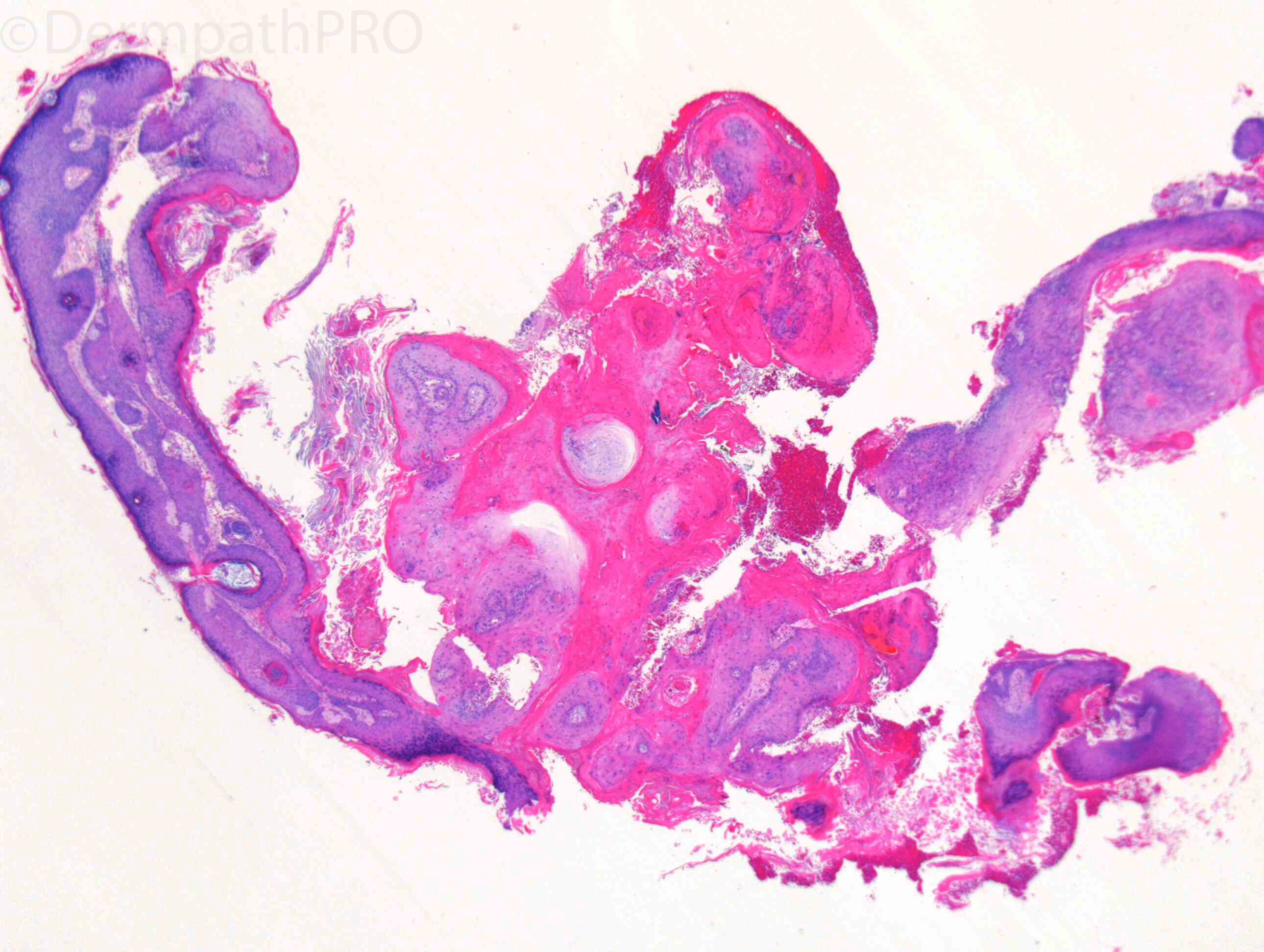
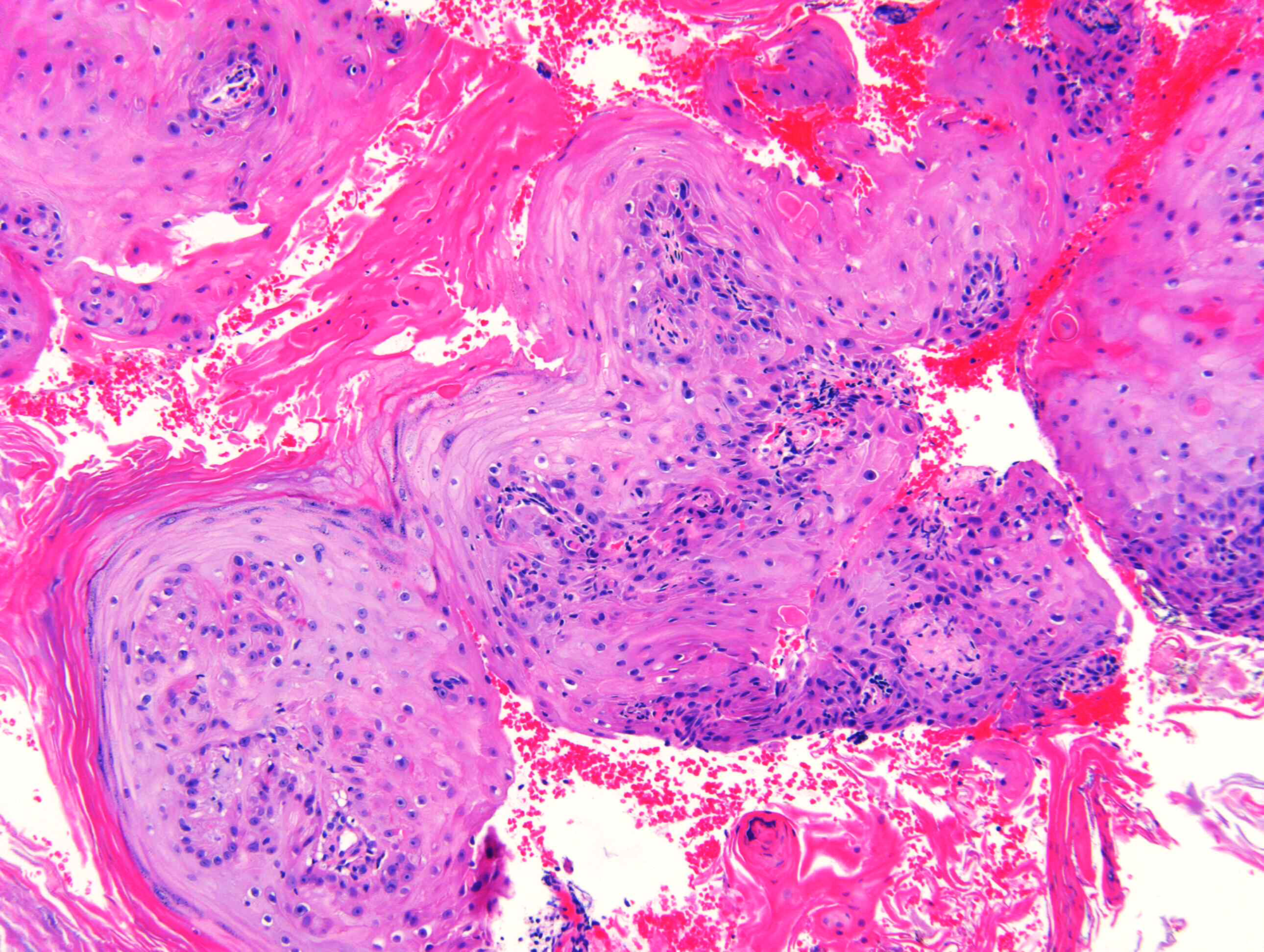
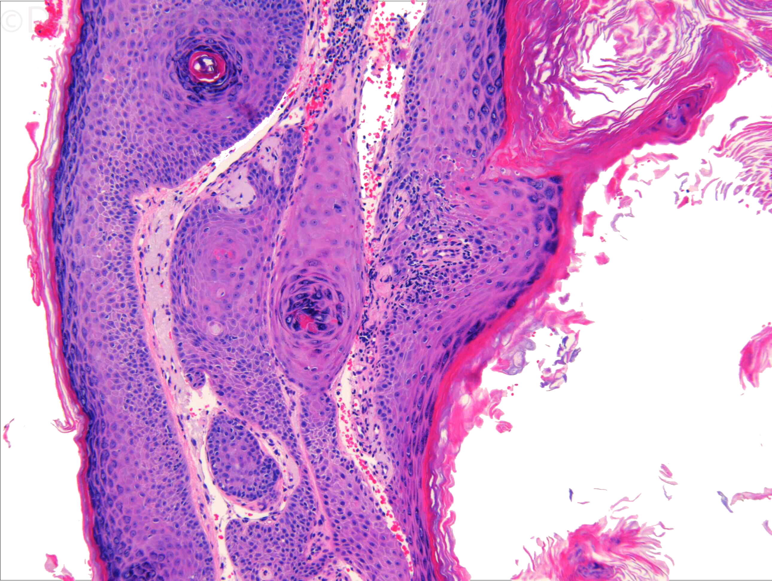
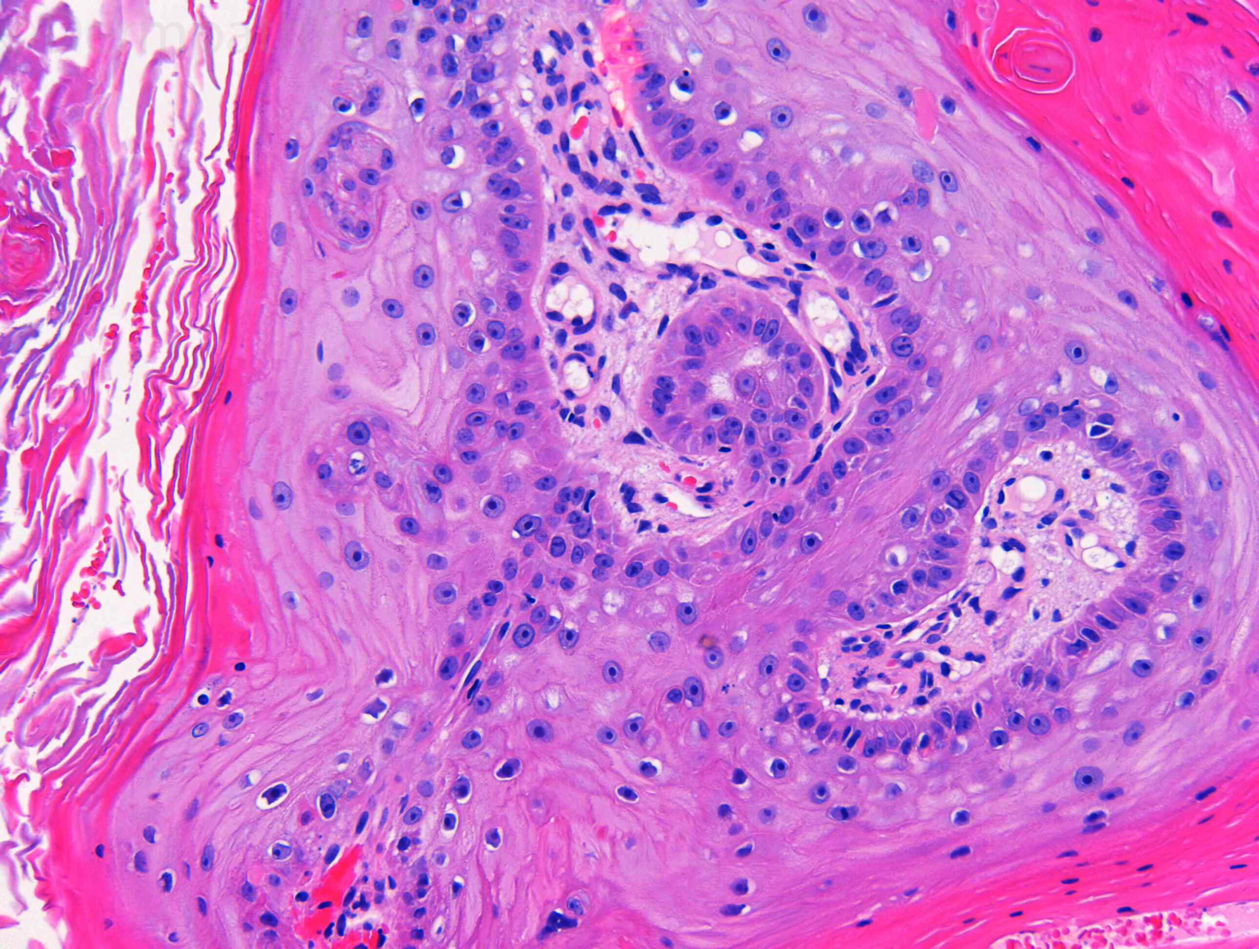
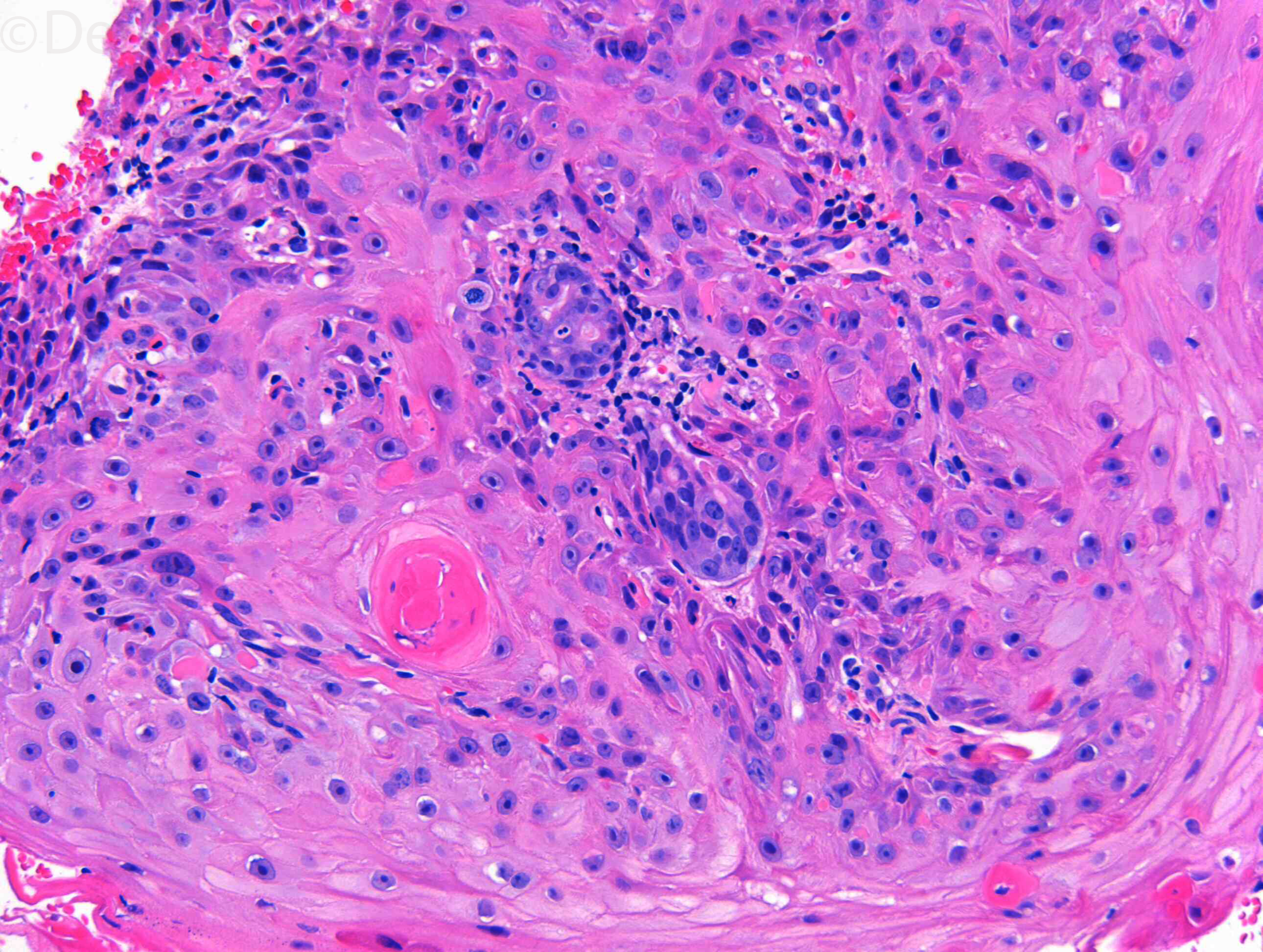
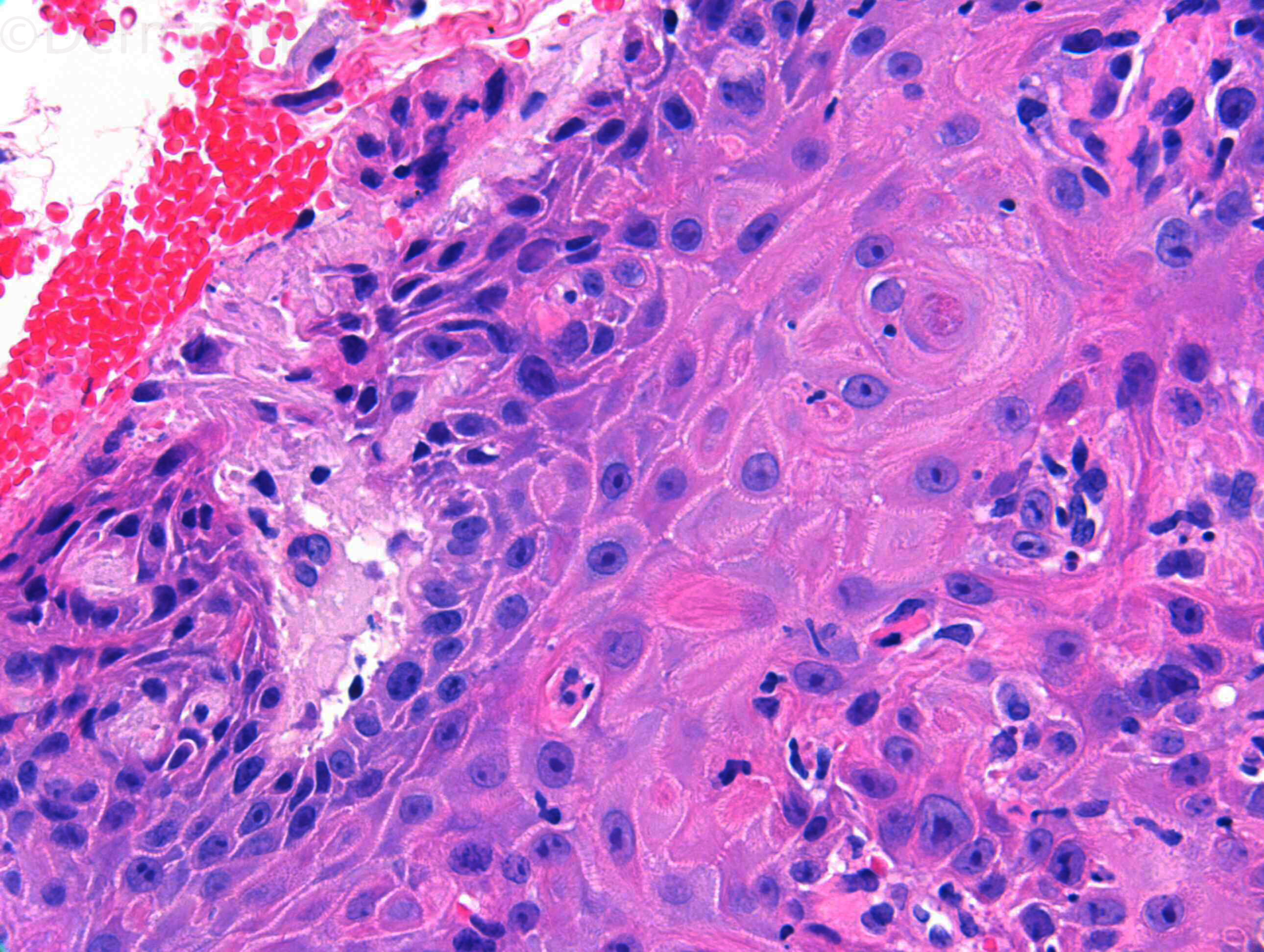
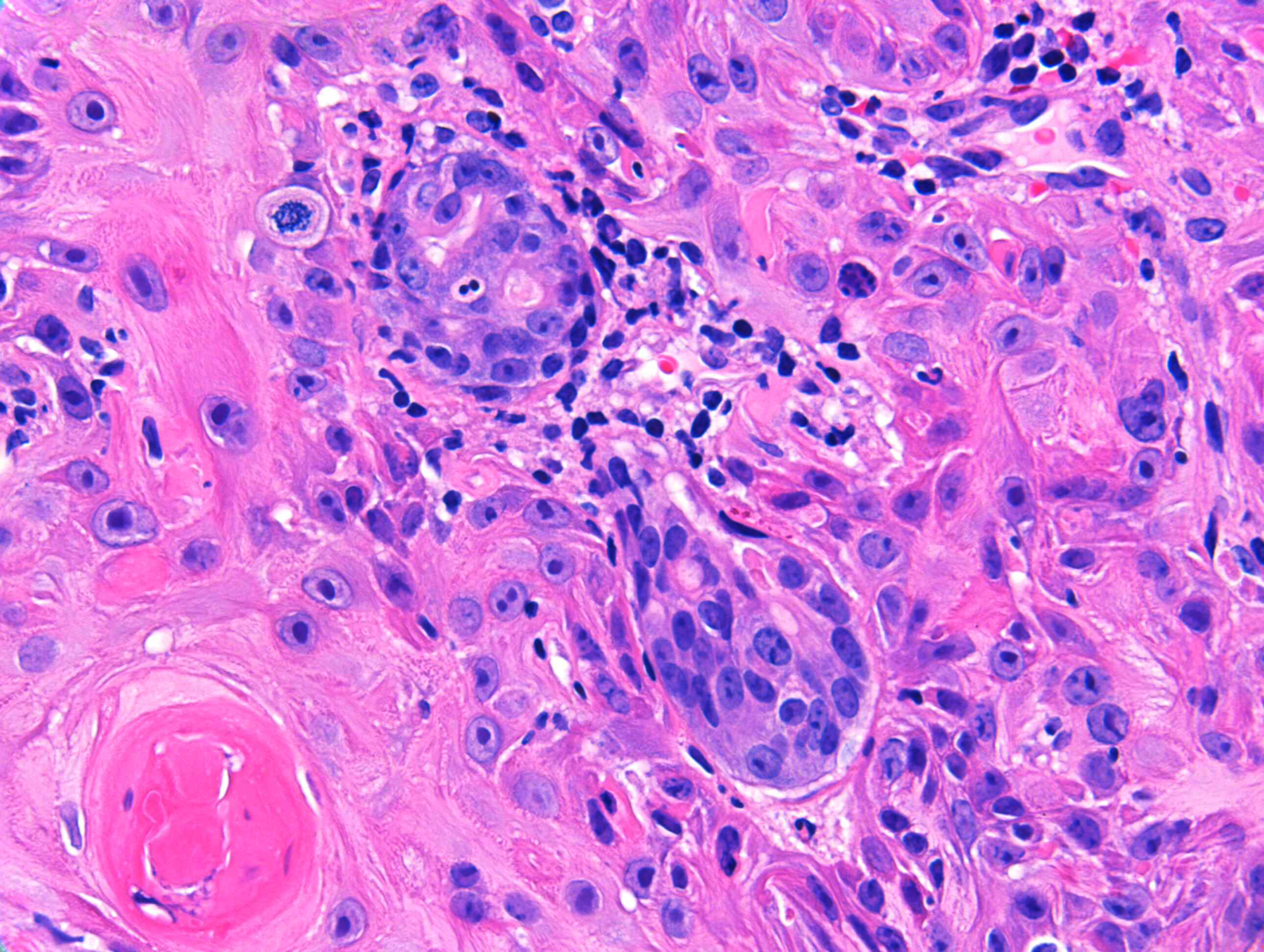
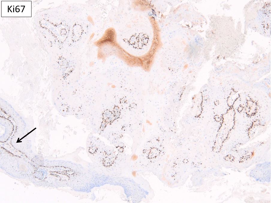
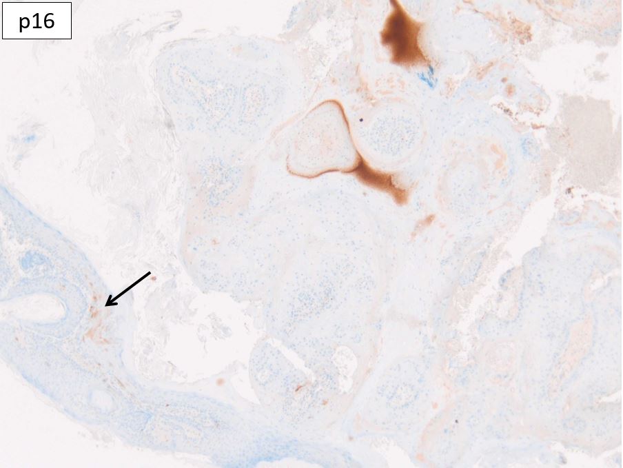
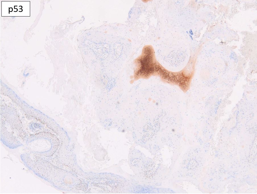
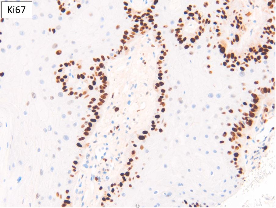
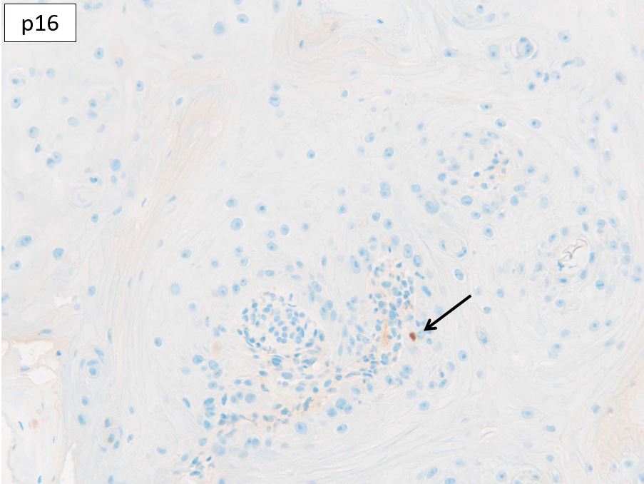
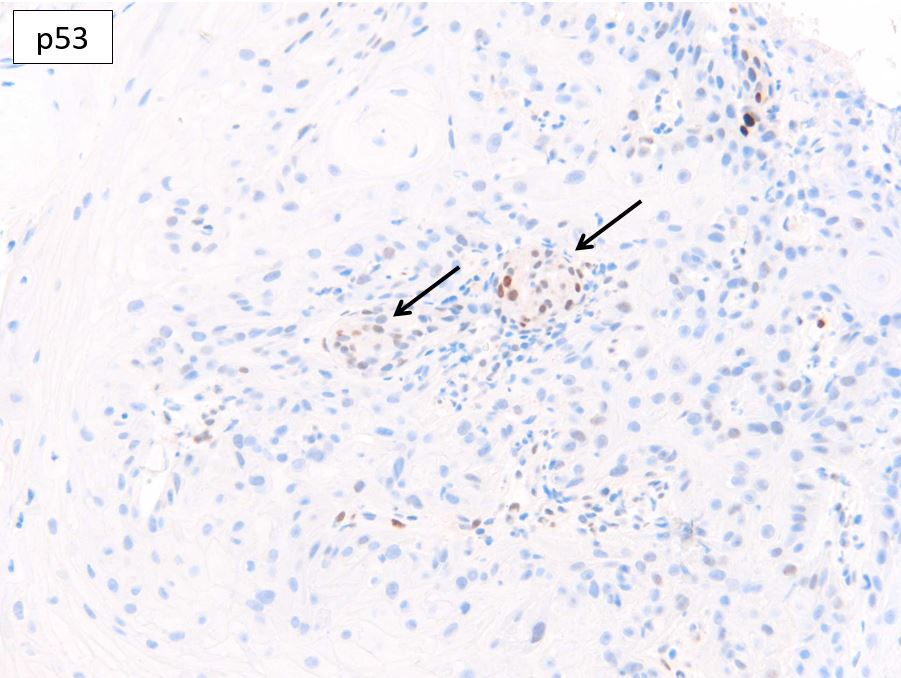
F79. Nose. 3/52 keratotic lesion. Biopsied by a general practitioner.


  Report Record

User Feedback


Irritated SK with actinic atypical changes. 

Share this comment


Link to comment
share_externally

Eman El-Nabarawy

Posted

Difficult..My ideas: Regressing KA (but too atypical) Follicular AK or fSCC..

Share this comment


Link to comment
share_externally

Cem Leblebici

Posted

At least AK. Need excision to rule out SCC.

Share this comment


Link to comment
share_externally

Dr. Richard Carr

Posted

Okay, some sensible suggestions. What about the IHC?

Share this comment


Link to comment
share_externally

Krishnakumar subramanian

Posted

p16 few staining in basal cells. no full p16 staining seen in epidermis. this rules out carcinoma. Ki 67 seen in basal cells. Not much p53 staining. So shall go for actinic keratosis

Share this comment


Link to comment
share_externally

P53 wild type. Ki67 increased in basal layer. p16..difficult to understand for me. So: why not a SK with actinic changes?

Share this comment


Link to comment
share_externally

Dr. Richard Carr

Posted

Okay thanks. This is a null p16 (mosaic in the adjacent epithelial lip) and an inflammatory cell in the epithelium. You will practically never see a benign lesion (on sun-exposed skin) with the extent of null staining for p16 as this lesion. p53 is also null, staining is limited to the sweat duct and inflammatory cells, note the reactive pattern in the epithelial lip on the low power. So we have a proliferative lesion with an adjacent reactive epithelial lip with a differential on H&E of KA and proliferating follicular actinic keratosis or SCC (probably the latter as the architecture is somewhat complex - EVG can be helpful to show elastic and collagen entrapment of a frankly invasive lesion). The double highly aberrant p16/p53 at a sun-exposed site (superficial lesion) is incompatible with a benign lesion. I therefore concluded a follicular SCC and advised referral for MDM discussion. If the lesion was fully removed and healing well watchful waiting is an option. The Ki67 showing fairly continuous peripheral pattern and could be compatible with KA or a well differentiated SCC. These are everyday problems in sign out and the immunostains can be very useful but need some experience to interpret and of course hopefully I'll be providing the publication on their use in due course. Warm regards to all.

Share this comment


Link to comment
share_externally

Krishnakumar subramanian

Posted

Thanks sir, but  i was wondering p16 would be more in carcinomas  in sun exposed areas. Please correct me sir

Eur J Dermatol

. Sep-Oct 2006;16(5):518-22.

P16 is overexpressed in cutaneous carcinomas located on sun-exposed areas

Isabelle Conscience 1, Nicolas Jovenin, Christelle Coissard, Marianne Lorenzato, Anne Durlach, Florent Grange, Philippe Birembaut, Christine Clavel, Philippe Bernard

Share this comment


Link to comment
share_externally

Dr. Richard Carr

Posted

I don't know why they found this. I do have this paper and will read it again. They had only 30 SCC. They found p16 over-expression in 60% of SCC. If they had a high rate of bowenoid SCC (the sort that occur in non-sun exposed skin or patients with immunosuppression that could explain a high rate of p16 over-expression. Also it's common to see reactive follicular pseudoepithehlious hyperplasia in FSCC and that could be mis-construed as over-expression. Finally if KA were included as SCC then they do show prominent p16 expression. Taking a quick look at my database 83% of follicular SCC have a null p16. Only 3% had a wild type mosaic pattern. Of 20 AK and BAK 85.7% had null p16 and zero wild type mosaic pattern.

Share this comment


Link to comment
share_externally


Join the conversation

You can post now and register later. If you have an account, sign in now to post with your account.

Guest
Add a comment...

×   Pasted as rich text.   Paste as plain text instead

  Only 75 emoji are allowed.

×   Your link has been automatically embedded.   Display as a link instead

×   Your previous content has been restored.   Clear editor

×   You cannot paste images directly. Upload or insert images from URL.

Loading...

×
×
  • Create New...