Jump to content
In this section we have spot diagnoses posted on a daily basis since June 2010, now over 4000! You can review the archived cases and read the suggested diagnoses by users and the final comment by the contributors.
Case are uploaded each week day by 10 am UK time with the correct diagnosis will generally be posted at 8 pm UK time. Why not view the most recent spot diagnosis and proffer a diagnosis?

Case Number : Case 2857- 18 June 2021 Posted By: Dr. Richard Carr

Please read the clinical history and view the images by clicking on them before you proffer your diagnosis.
Submitted Date :
   (0 reviews)

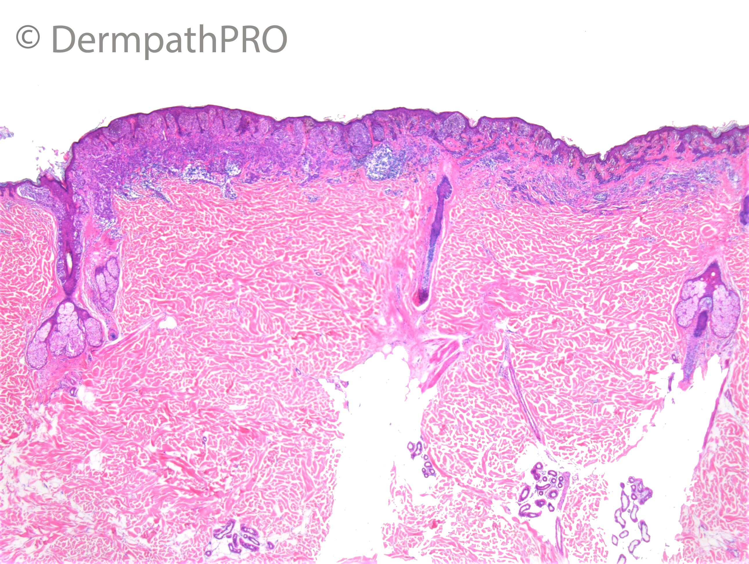
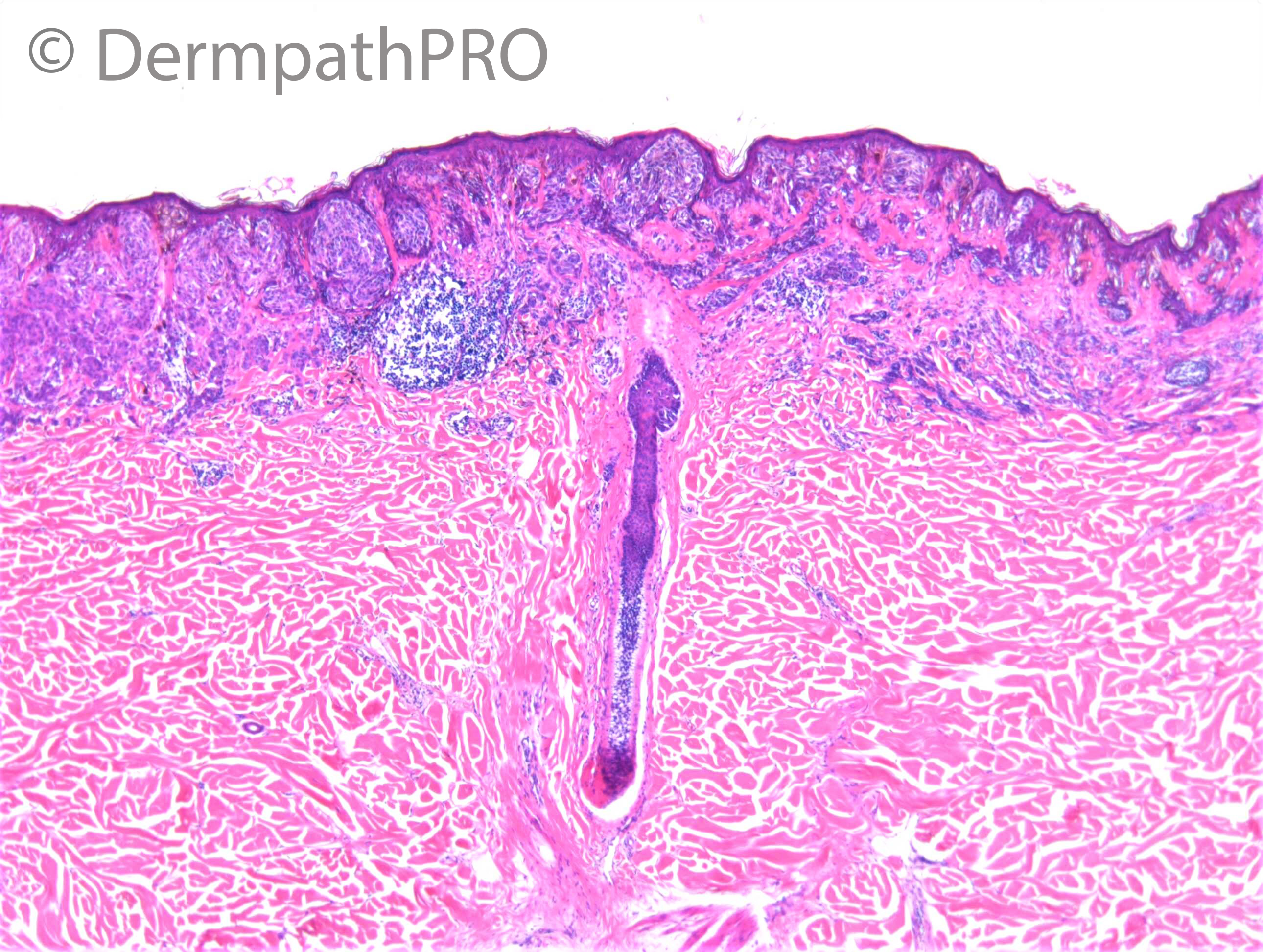
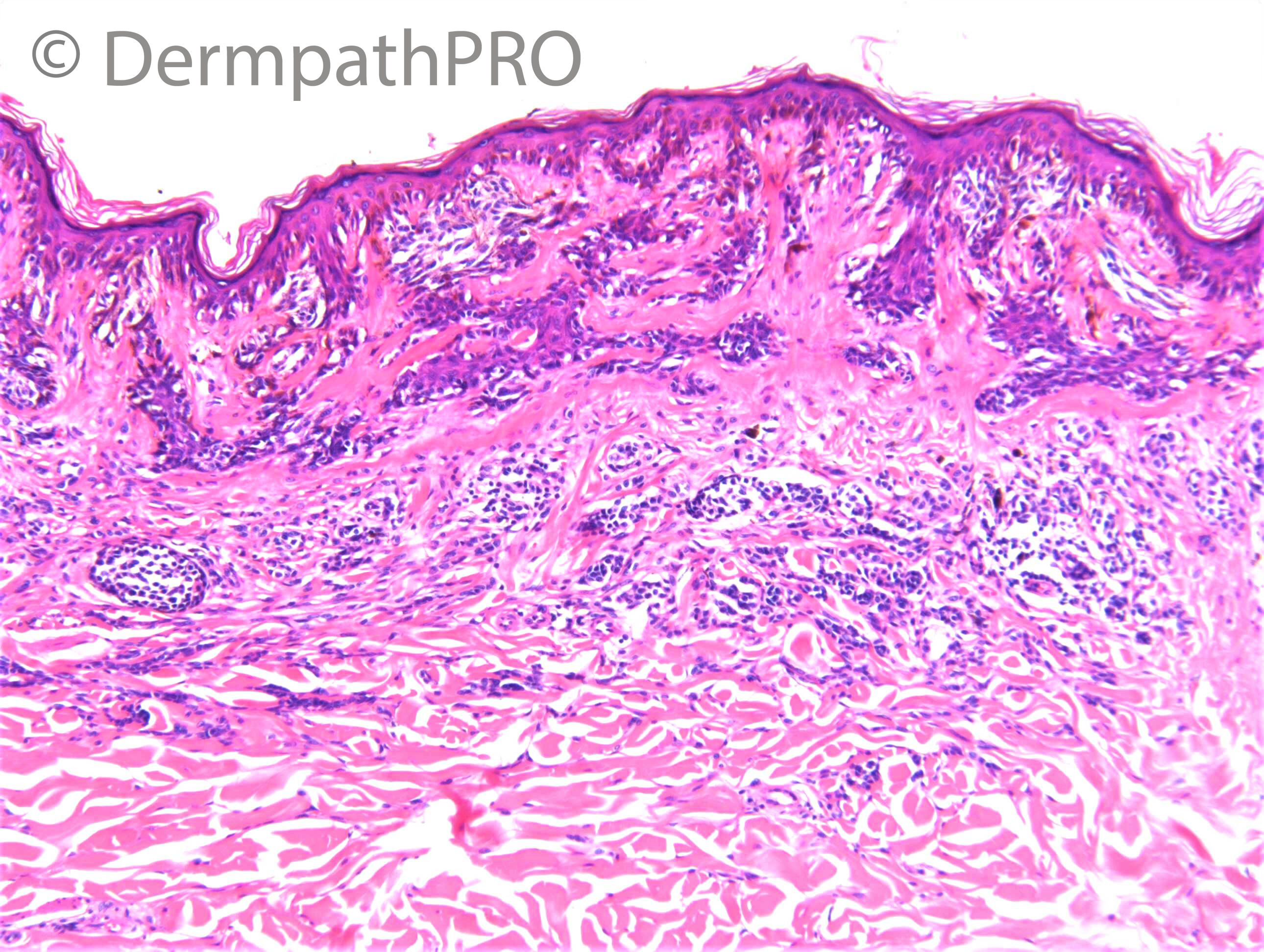
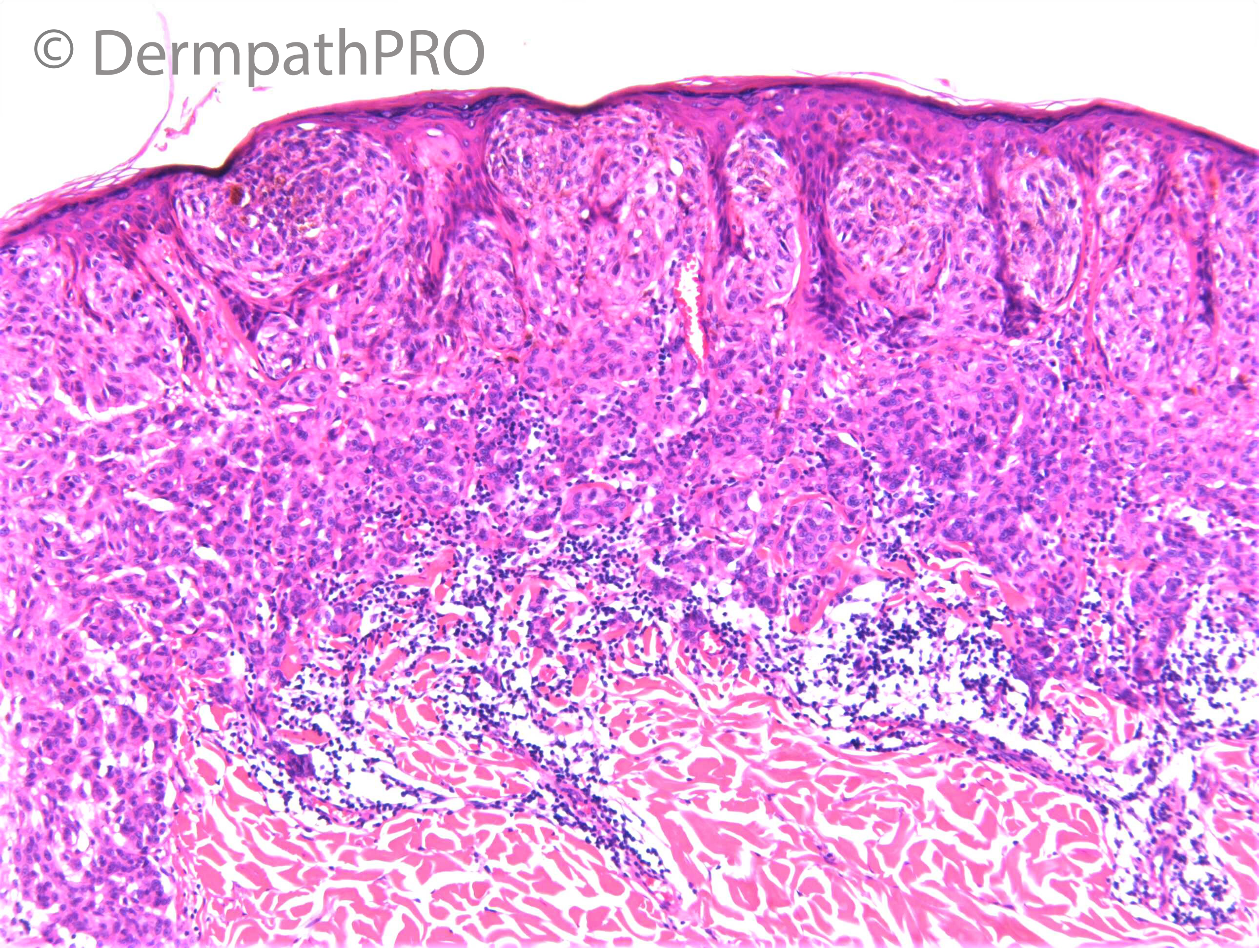
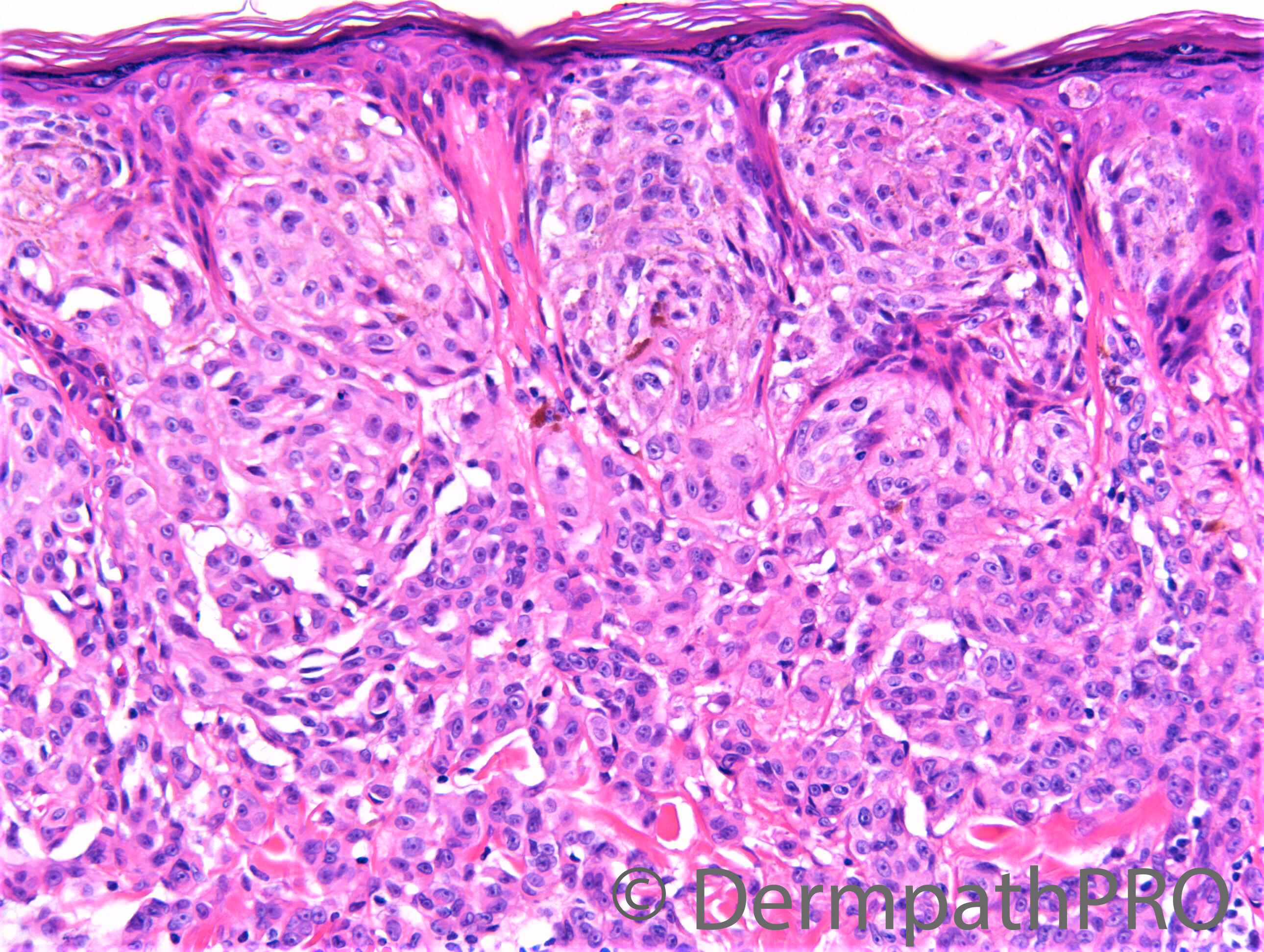
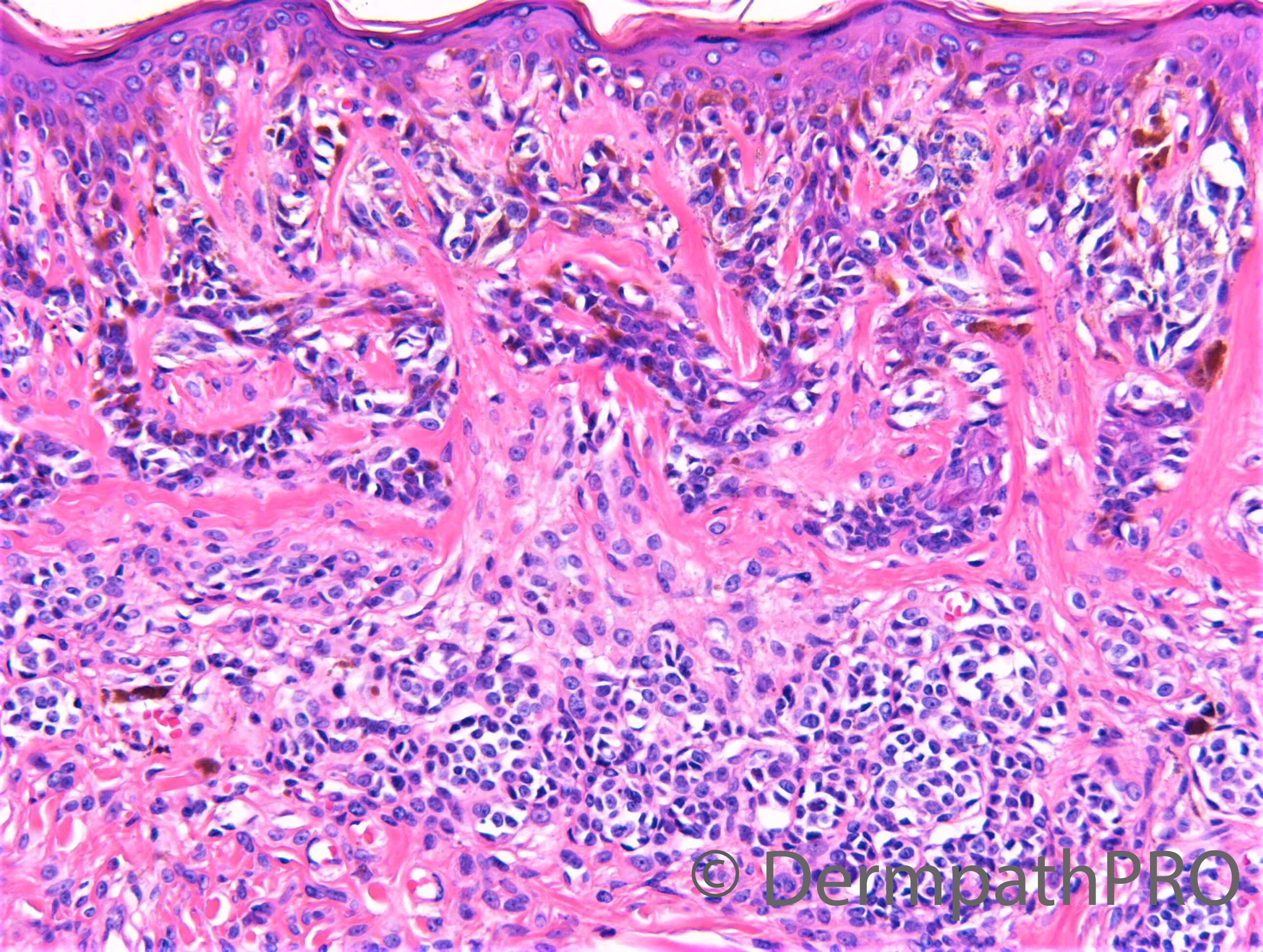
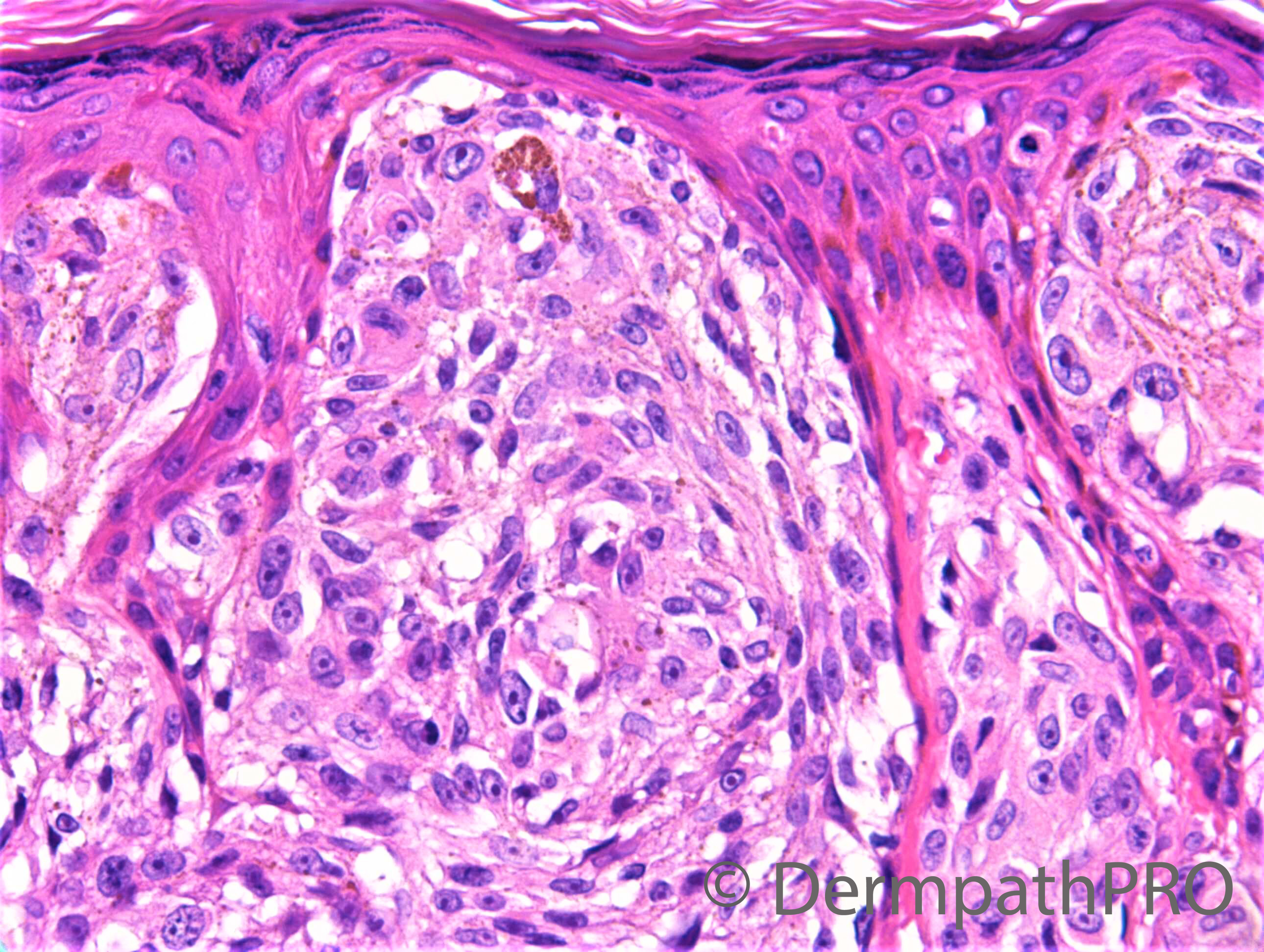
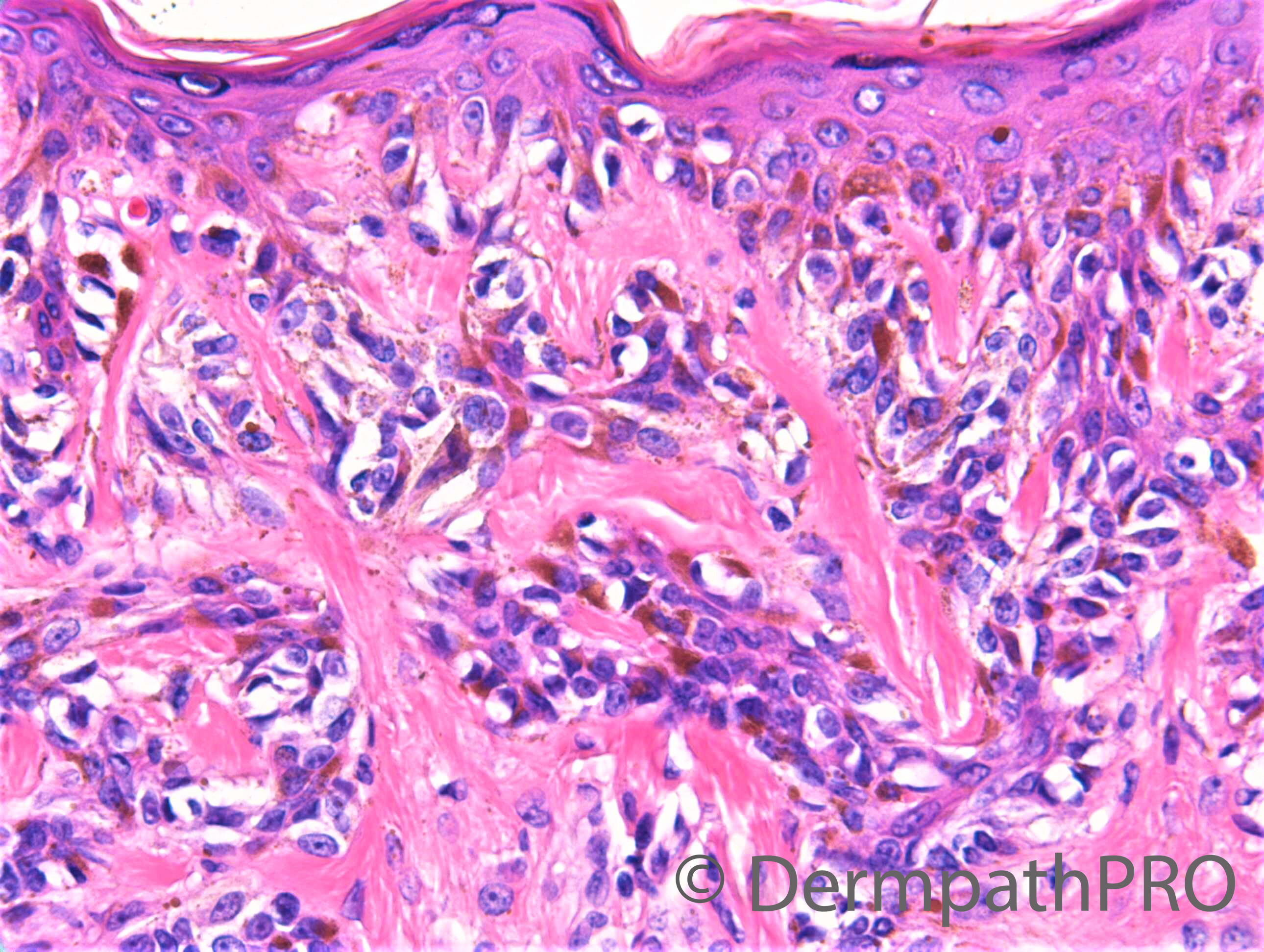
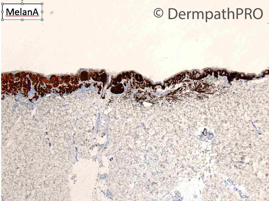
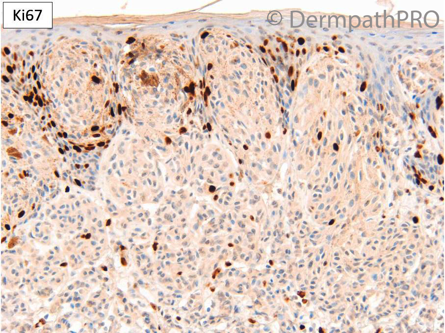
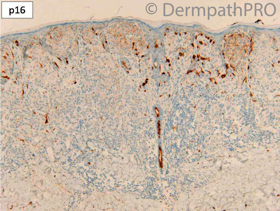
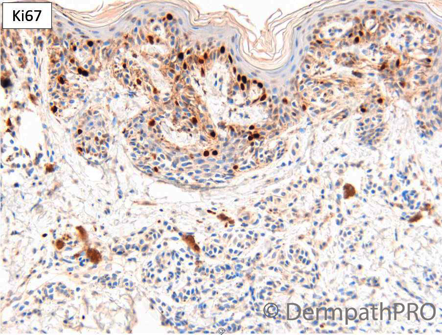
F50. Back, upper central, 9 x 6 mm, dark brown dermoscopically chaotic mole – longstanding but has become darker - ? atypical, ?MM


  • Like 1
  Report Record

User Feedback


Krishnakumar subramanian

Posted

spindle cell nevus of Reed

Share this comment


Link to comment
share_externally

Meenakshi Batrani

Posted

Seems like melanoma arising in a dysplastic nevus. One part is very epithelioid, while the other part shows small nevic cells.  

Share this comment


Link to comment
share_externally

8 hours ago, Meenakshi Batrani said:

Seems like melanoma arising in a dysplastic nevus. One part is very epithelioid, while the other part shows small nevic cells.  

Yes. The left part should be a melanoma, because of a nodular-expansive intradermal growth. The spindle/epithelioid cells are very atypical. And p16 loss is in keeping with melanoma. Useful a Prame stain.

Share this comment


Link to comment
share_externally

msofopoulos

Posted

I think it is a (spitzoid?) melanoma. It is asymmetric with atypical cells and (localized) loss of p16. Can we attribute the fibrosis in pic 3 and 5 on dysplastic nevus, regression or location (back)?

Share this comment


Link to comment
share_externally

Dr. Richard Carr

Posted

I agree with a "Spitzoid" or Spitz-like melanoma arising in a dysplastic naevus. The clue in image 5 is the expansile nests that squeeze the elongated rete (umbrella sign = narrow compressed rete with thinned curving epidermis on each side) or also referred to as the puffy shirt sign (rounded expansile nests that bulge the epidermis). The null p16 is supportive of melanoma arising in a naevus and I believe is seen in around 70% of naevoid melanomas. 

Share this comment


Link to comment
share_externally


Join the conversation

You can post now and register later. If you have an account, sign in now to post with your account.

Guest
Add a comment...

×   Pasted as rich text.   Paste as plain text instead

  Only 75 emoji are allowed.

×   Your link has been automatically embedded.   Display as a link instead

×   Your previous content has been restored.   Clear editor

×   You cannot paste images directly. Upload or insert images from URL.

Loading...

×
×
  • Create New...