Jump to content
In this section we have spot diagnoses posted on a daily basis since June 2010, now over 4000! You can review the archived cases and read the suggested diagnoses by users and the final comment by the contributors.
Case are uploaded each week day by 10 am UK time with the correct diagnosis will generally be posted at 8 pm UK time. Why not view the most recent spot diagnosis and proffer a diagnosis?

Case Number : Case 2858 - 21 June 2021 Posted By: Dr. Mona Abdel-Halim

Please read the clinical history and view the images by clicking on them before you proffer your diagnosis.
Submitted Date :
   (0 reviews)

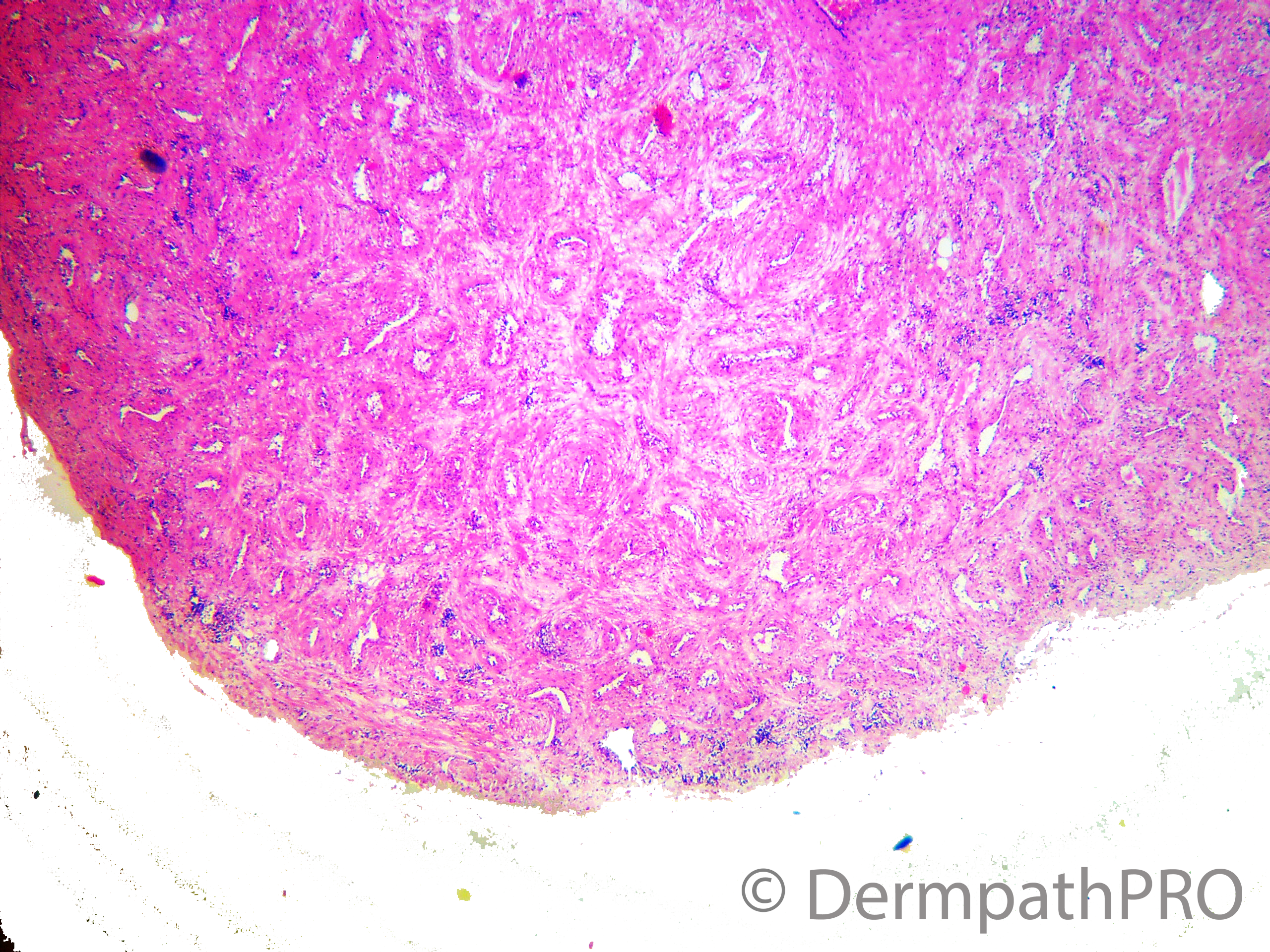
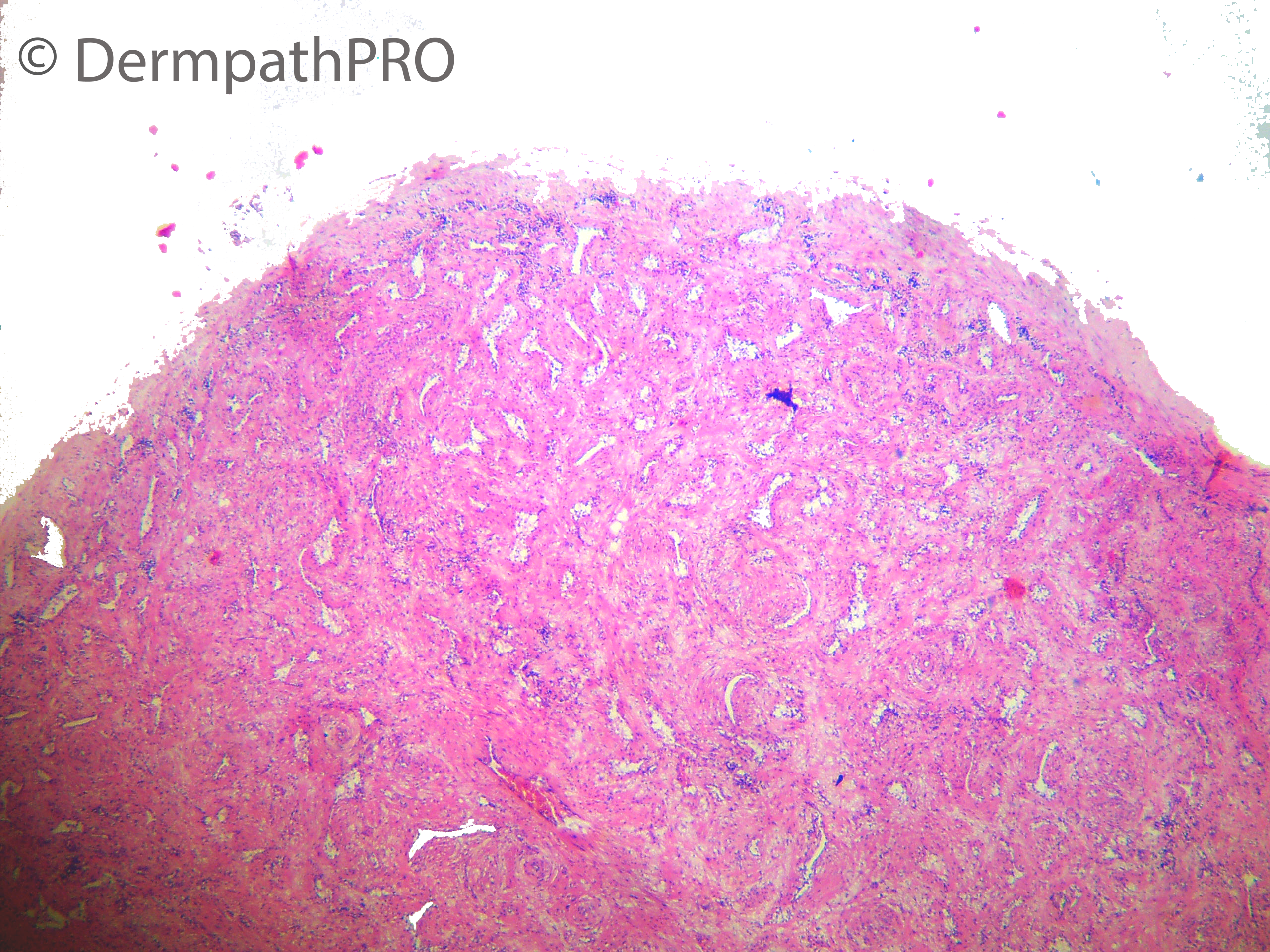
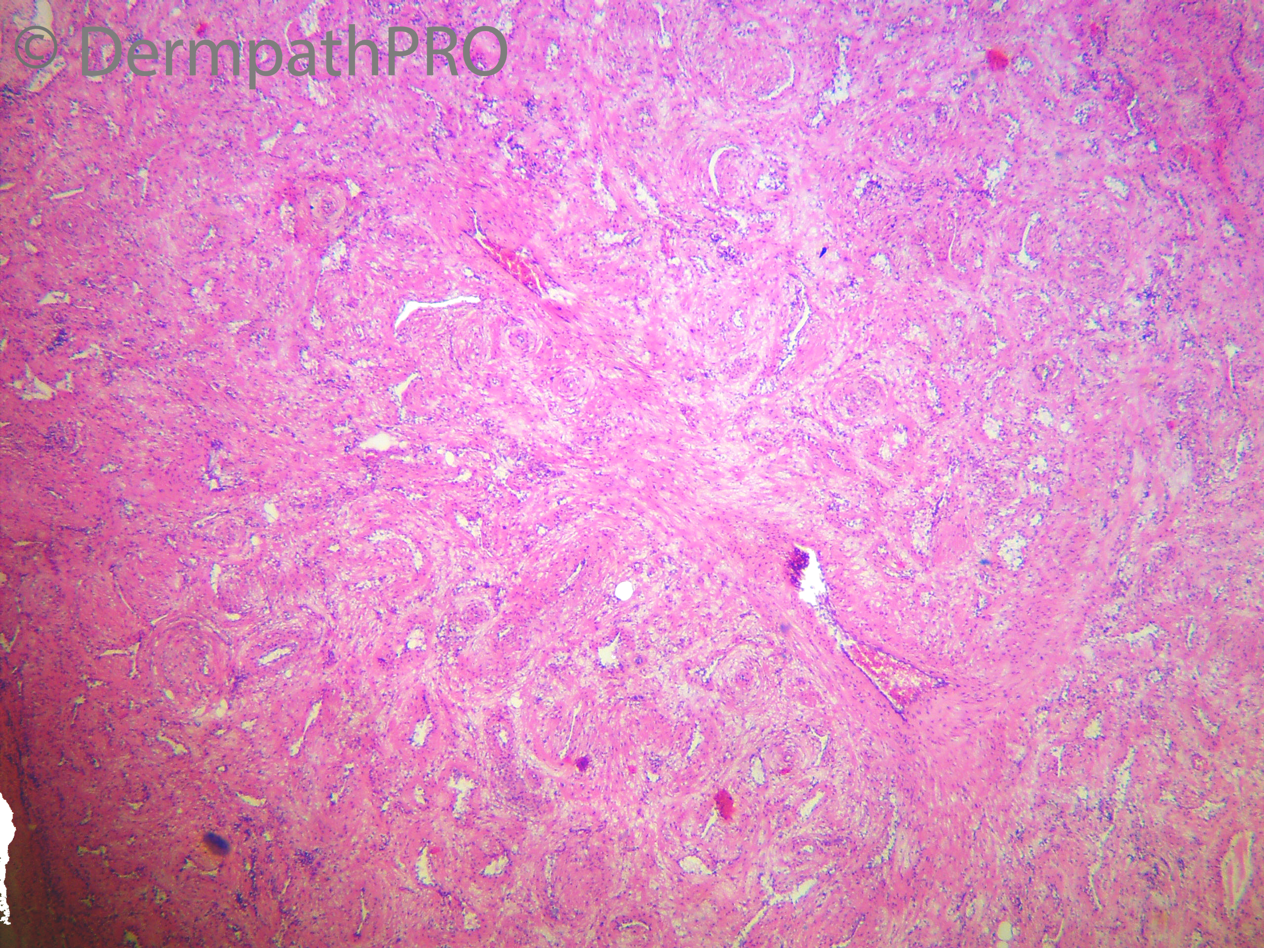
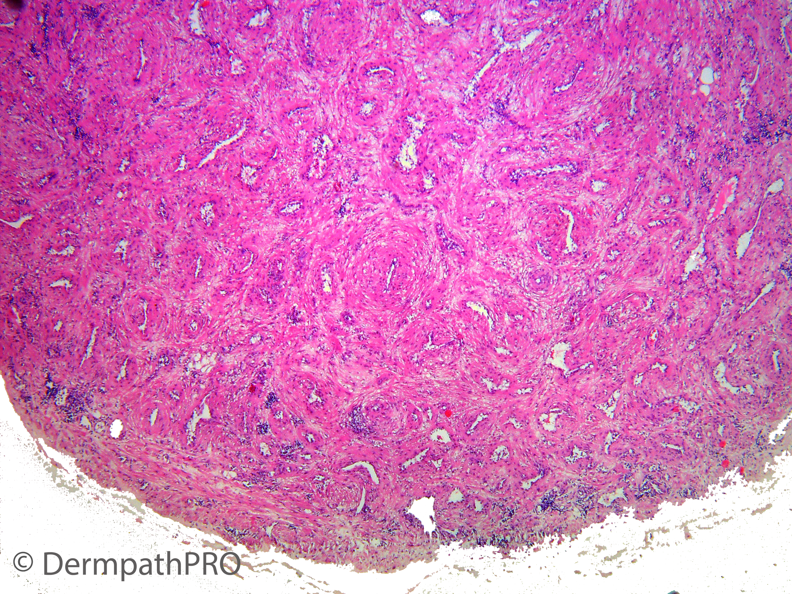
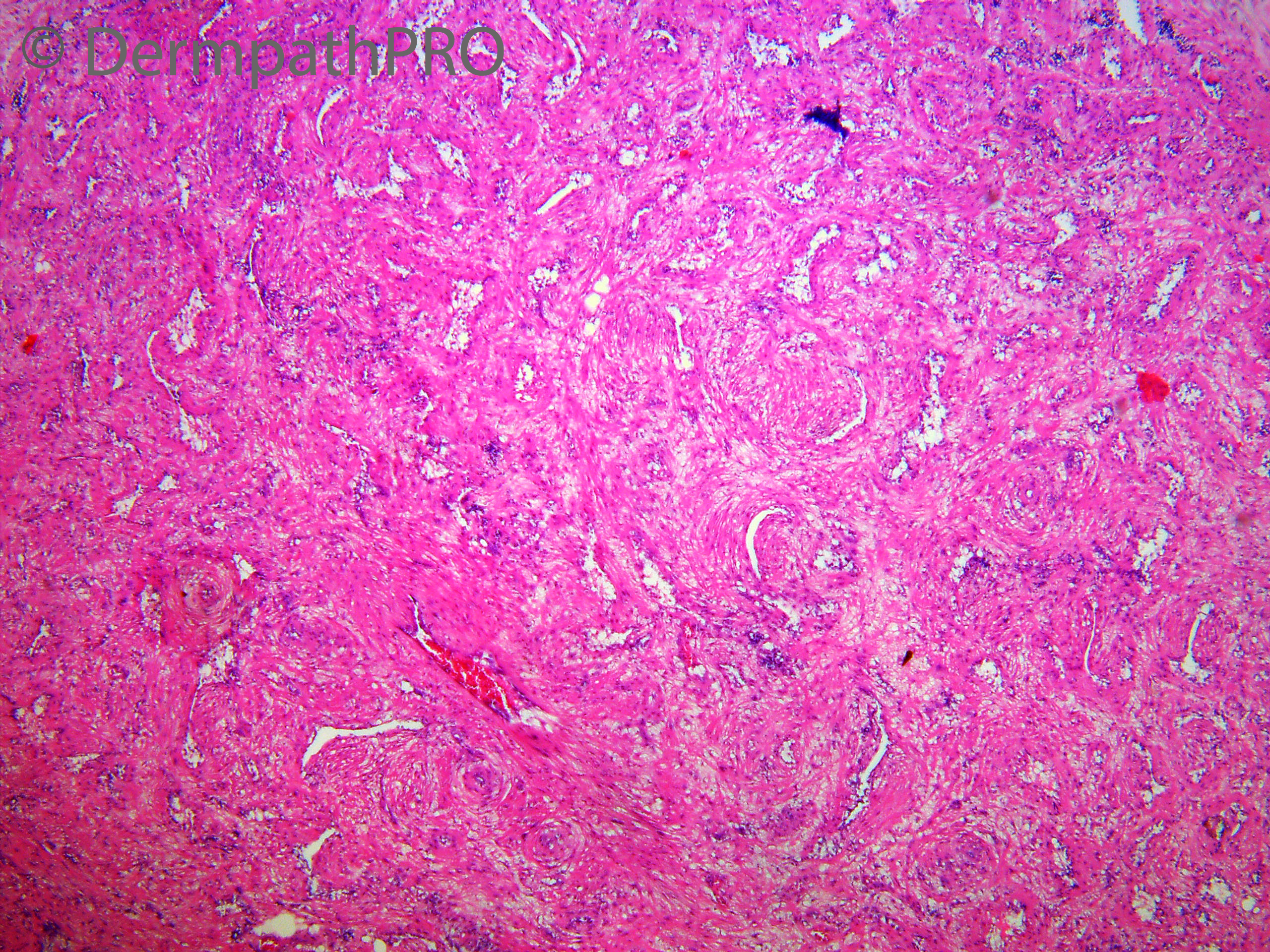
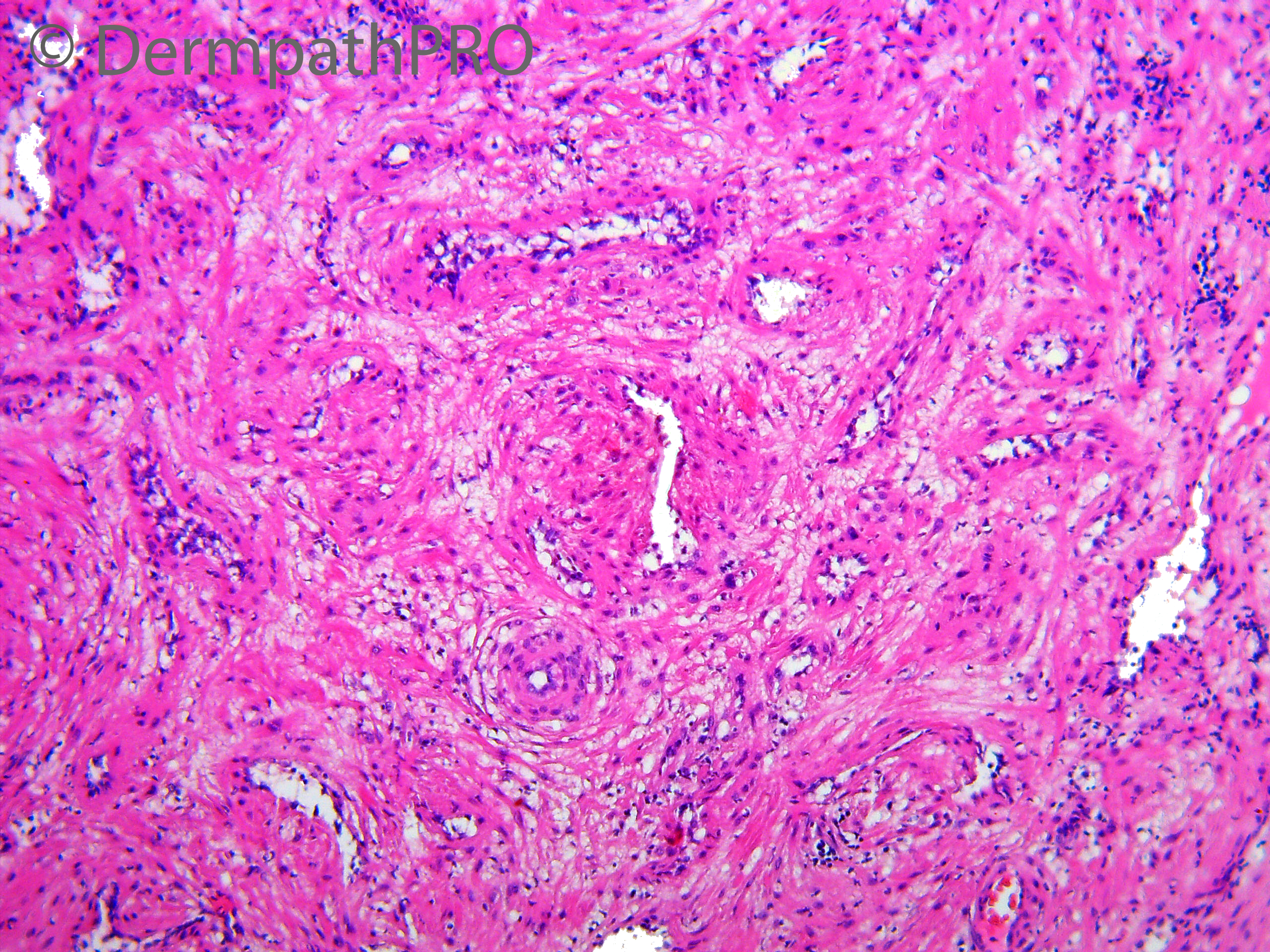
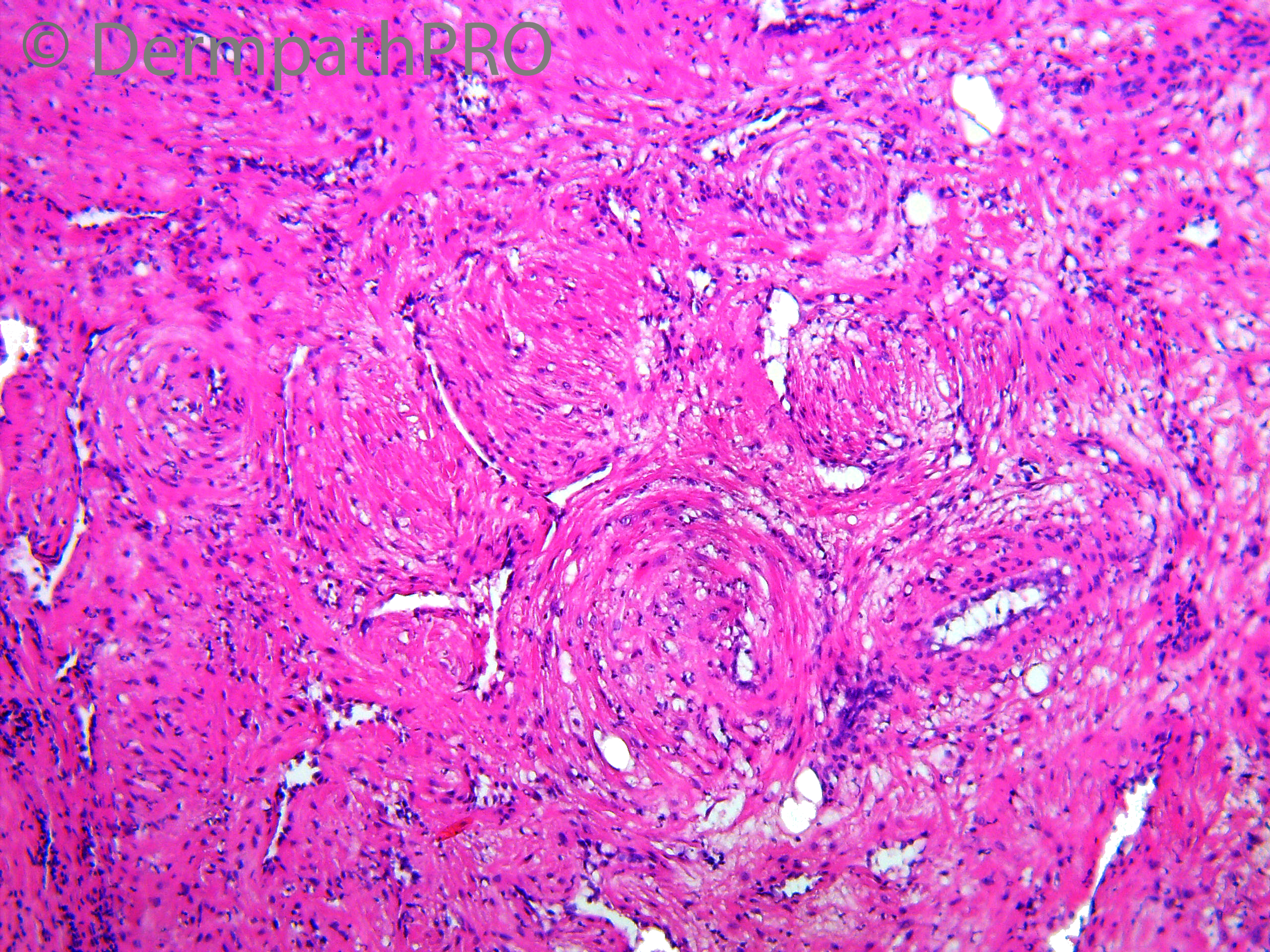
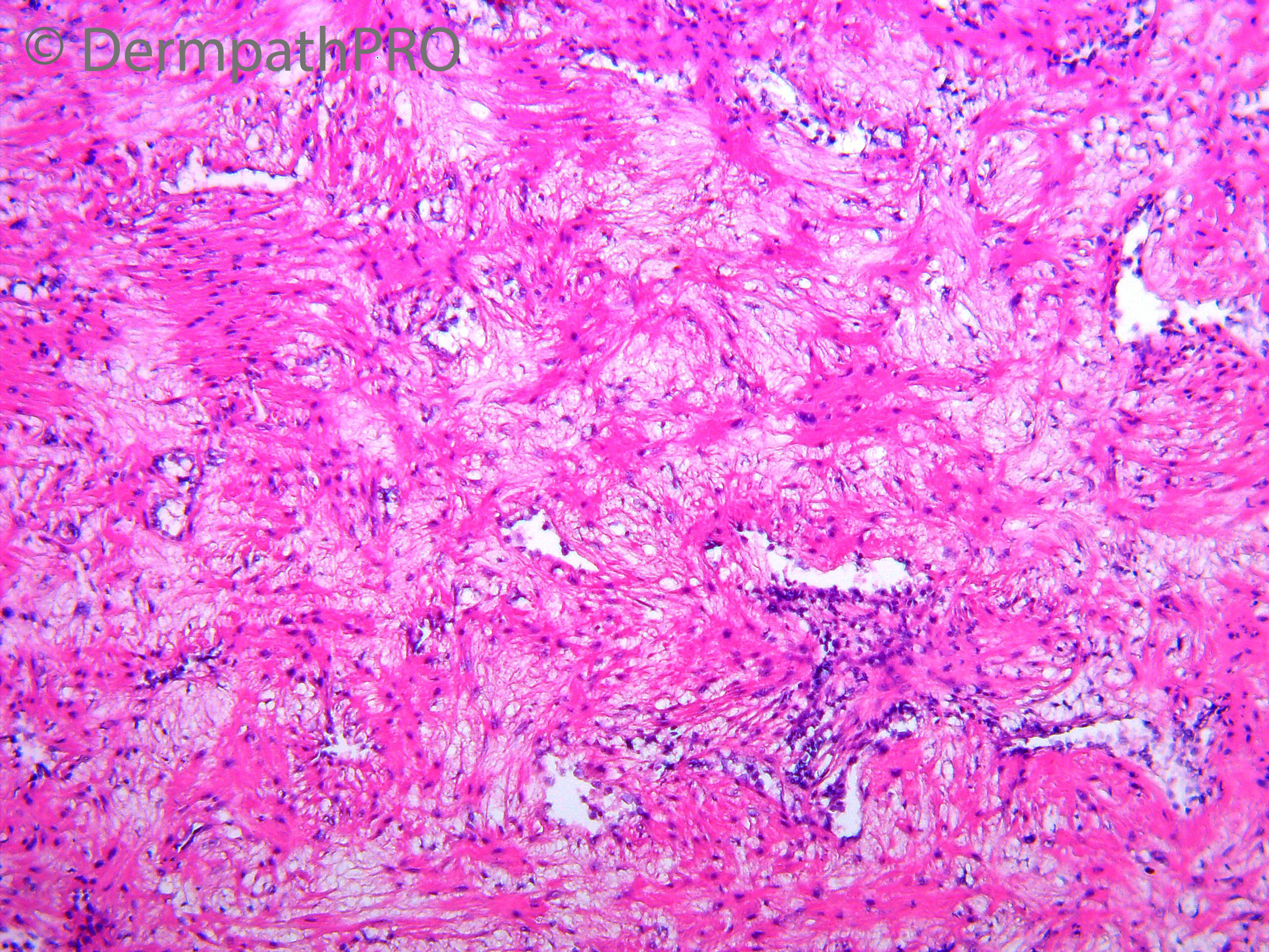
M70, Nodule on the auricle of the left ear. 3 years duration.
Stationary course - removed for cosmetic purposes.


  Report Record

User Feedback


Krishnakumar subramanian

Posted

agree withy myopericytoma

Share this comment


Link to comment
share_externally

Meenakshi Batrani

Posted

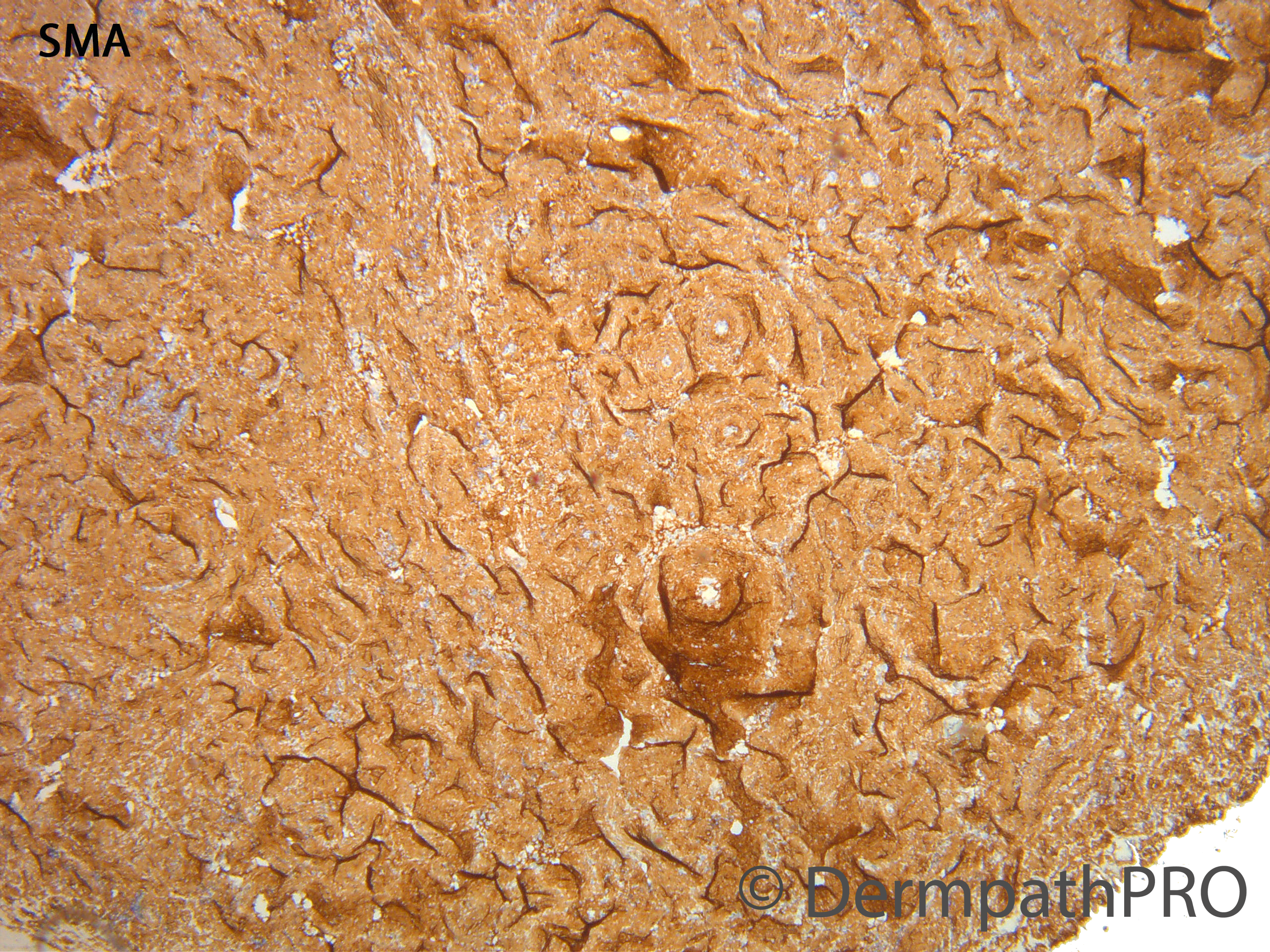
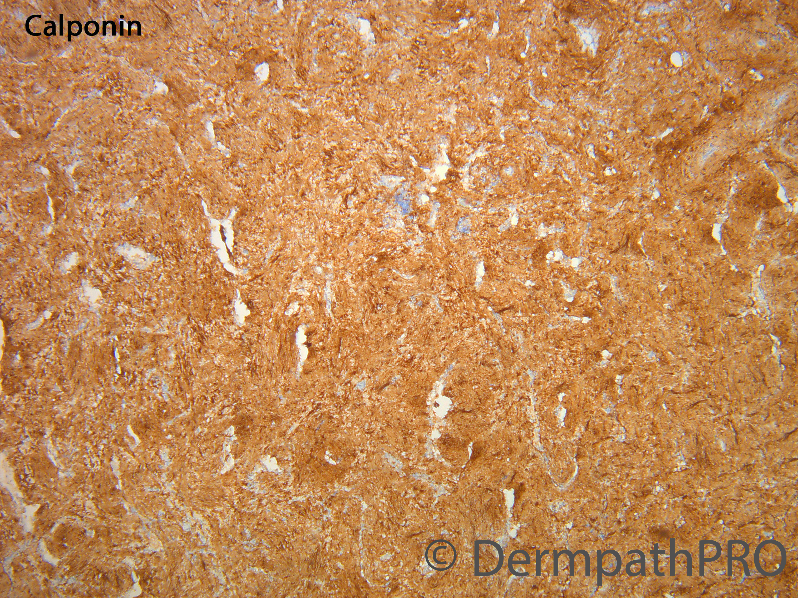
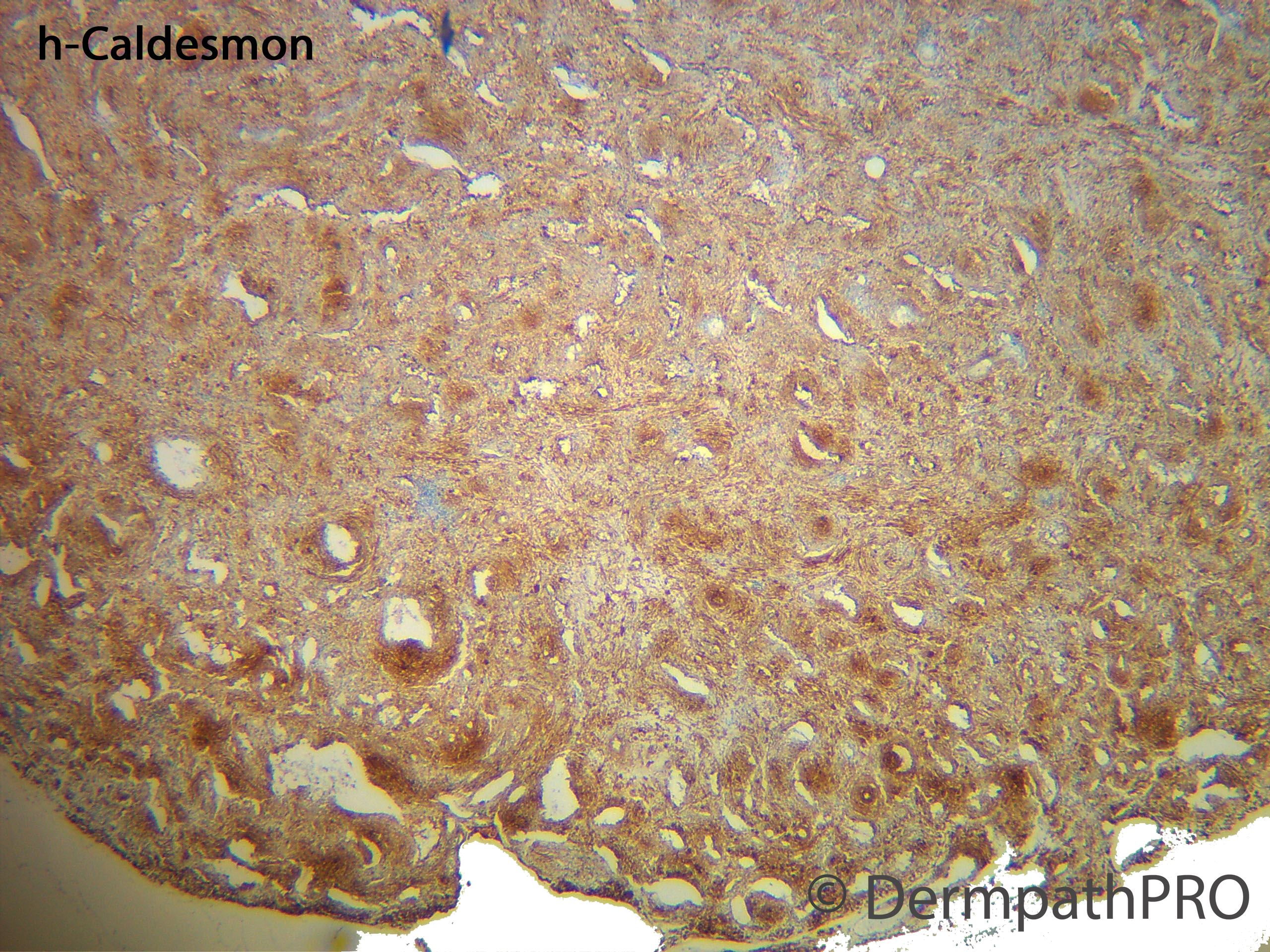
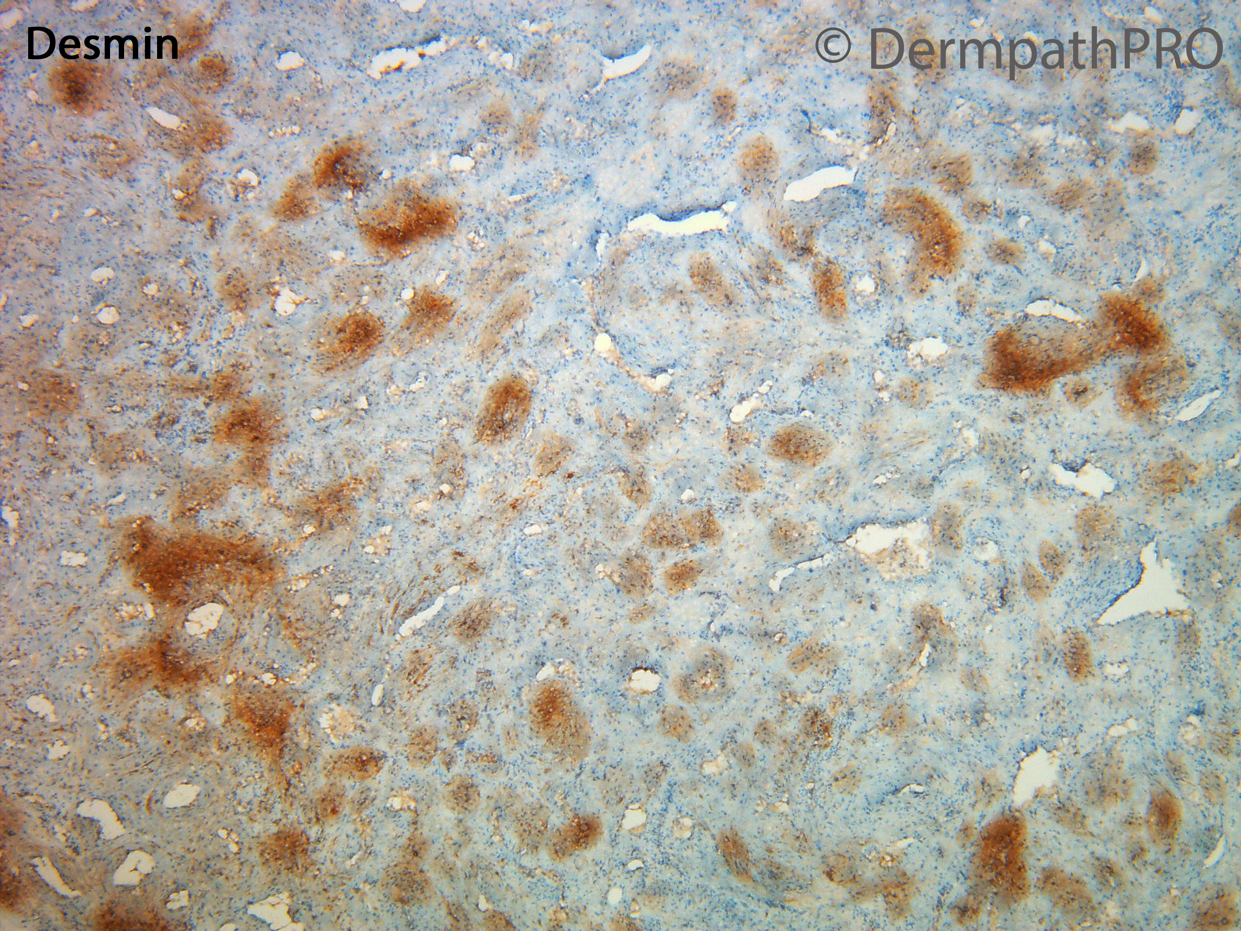
There is lot of overlap between angioleiomyoma and myopericytoma which are considered a part of spectrum. In this case some smooth muscle bundles in between the vessels, and patchy Desmin positivity, may be favors angioleiomyoma over myopericytoma. 

Share this comment


Link to comment
share_externally

2 hours ago, Meenakshi Batrani said:

There is lot of overlap between angioleiomyoma and myopericytoma which are considered a part of spectrum. In this case some smooth muscle bundles in between the vessels, and patchy Desmin positivity, may be favors angioleiomyoma over myopericytoma. 

Agree

Share this comment


Link to comment
share_externally

Dr. Mona Abdel-Halim

Posted

Thank you all!

I found the prominent concentric arrangement of the short spindled ovoid cells around the blood vessels (onion peel pattern) together with the focal (rather than diffuse desmin staining pattern) more in keeping with the classic description of myopericytoma. A classic angioleiomyoma will have a less prominent concentric arrangement of the tumor cells around the blo0d vessels and a diffuse desmin positivity. But as Meenakshi said, in recent years the term myopericytoma is used to describe a spectrum of tumors that also includes angioleiomyoma and myofibroma. Whatever you prefer to call it, it is benign and no harm is expected to the patient! 

Share this comment


Link to comment
share_externally

Meenakshi Batrani

Posted

16 hours ago, Dr. Mona Abdel-Halim said:

Thank you all!

I found the prominent concentric arrangement of the short spindled ovoid cells around the blood vessels (onion peel pattern) together with the focal (rather than diffuse desmin staining pattern) more in keeping with the classic description of myopericytoma. A classic angioleiomyoma will have a less prominent concentric arrangement of the tumor cells around the blo0d vessels and a diffuse desmin positivity. But as Meenakshi said, in recent years the term myopericytoma is used to describe a spectrum of tumors that also includes angioleiomyoma and myofibroma. Whatever you prefer to call it, it is benign and no harm is expected to the patient! 

Agree. 

Share this comment


Link to comment
share_externally


Join the conversation

You can post now and register later. If you have an account, sign in now to post with your account.

Guest
Add a comment...

×   Pasted as rich text.   Paste as plain text instead

  Only 75 emoji are allowed.

×   Your link has been automatically embedded.   Display as a link instead

×   Your previous content has been restored.   Clear editor

×   You cannot paste images directly. Upload or insert images from URL.

Loading...

×
×
  • Create New...