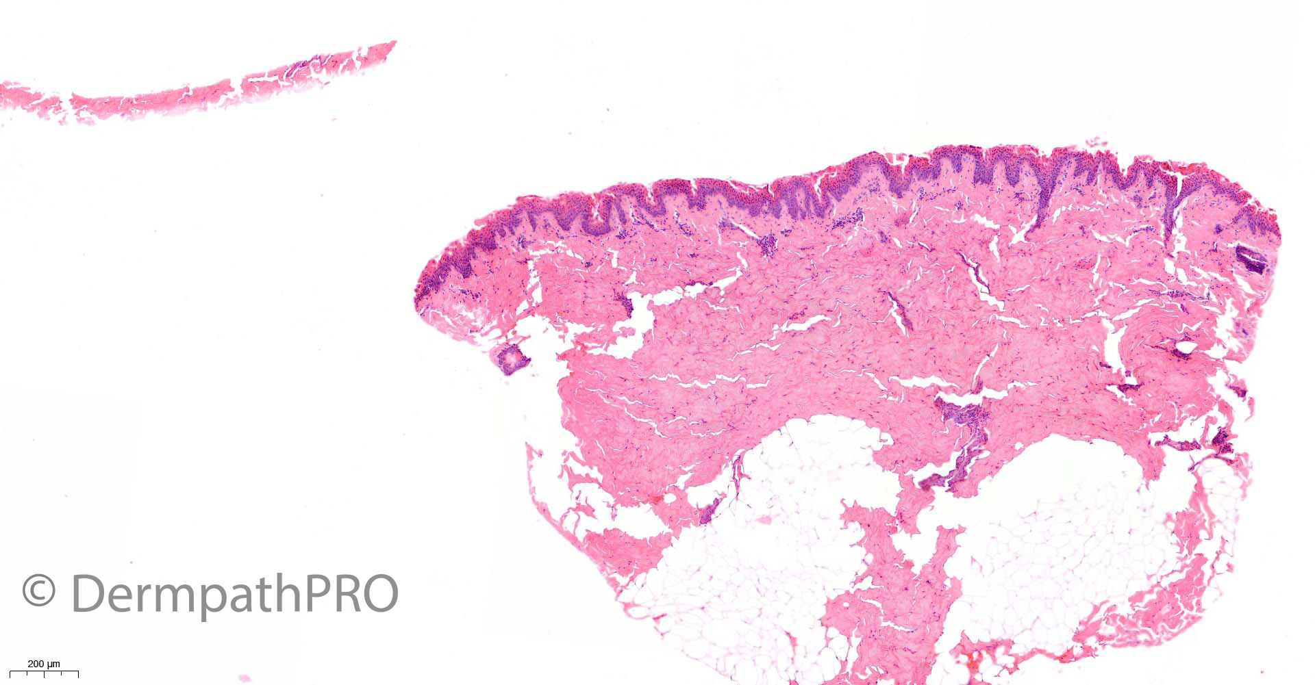
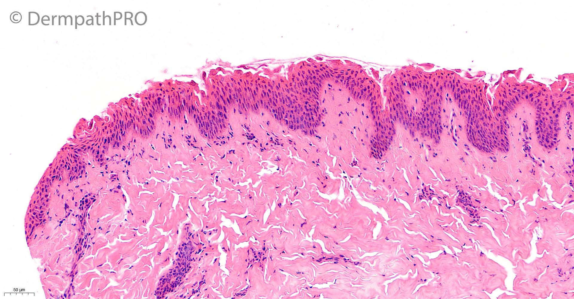
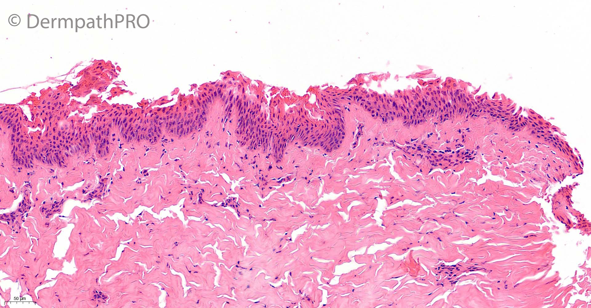
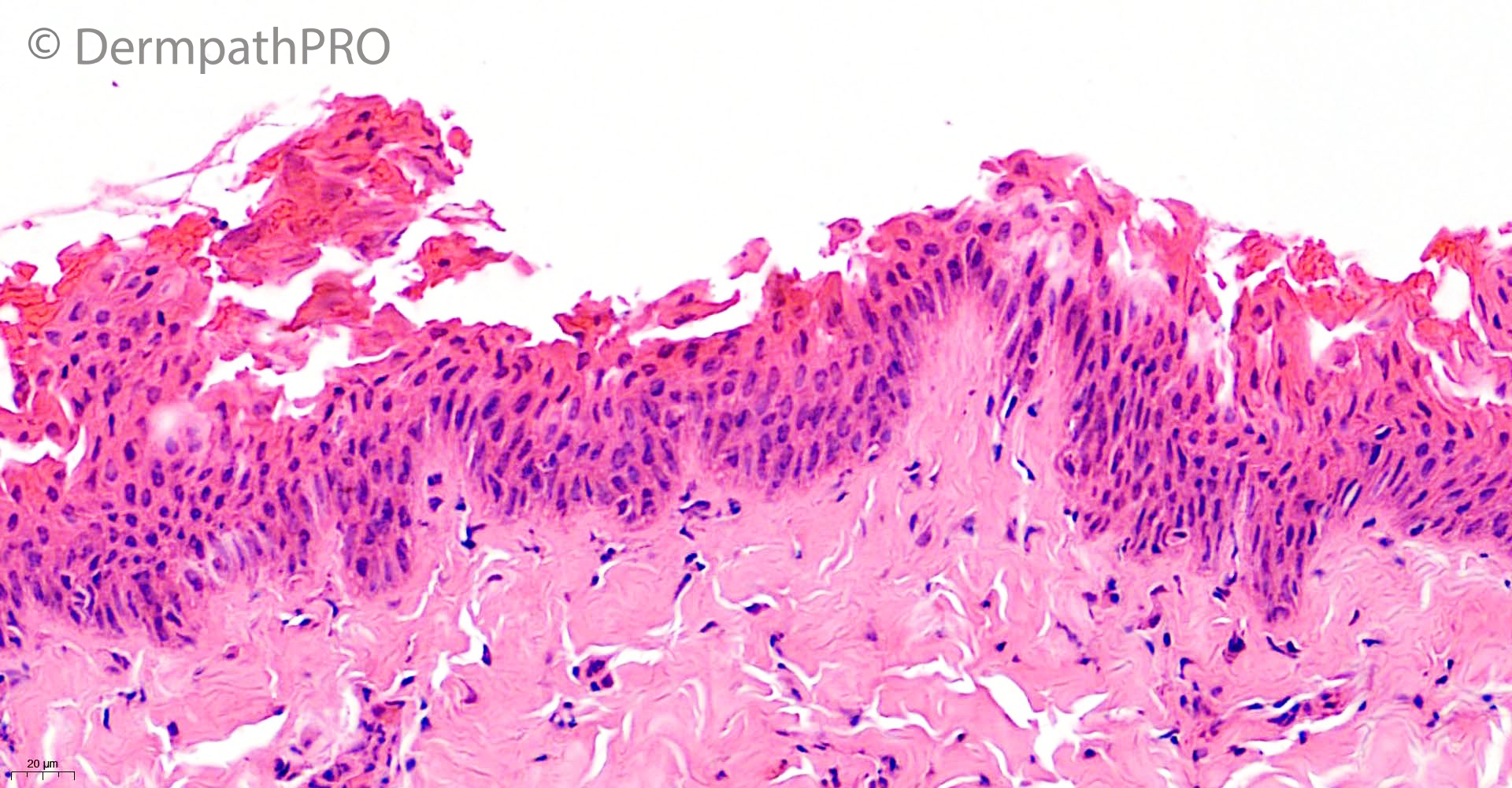
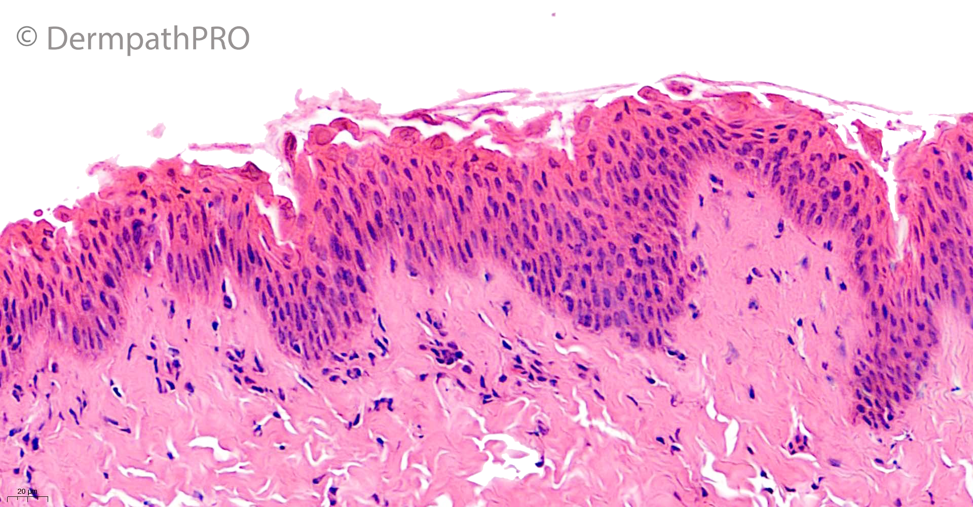
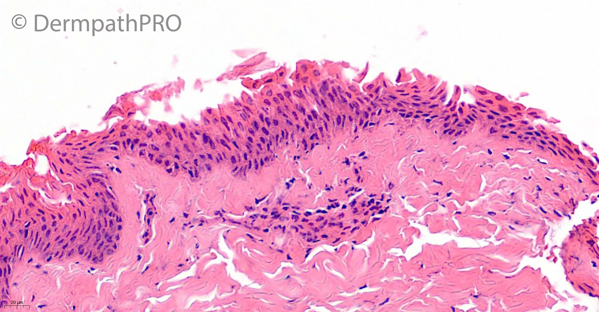
Jump to content
In this section we have spot diagnoses posted on a daily basis since June 2010, now over 4000! You can review the archived cases and read the suggested diagnoses by users and the final comment by the contributors.
Case are uploaded each week day by 10 am UK time with the correct diagnosis will generally be posted at 8 pm UK time. Why not view the most recent spot diagnosis and proffer a diagnosis?

Case Number : Case 2861 - 24 June 2021 Posted By: Saleem Taibjee

Please read the clinical history and view the images by clicking on them before you proffer your diagnosis.
Submitted Date :
   (0 reviews)

5 year old boy with widespread painful blistering rash


  • Like 1
  Report Record

User Feedback


daniellindsay

Posted

Appears to be intraepidermal separation at level of the granular layer. Would consider a staphylococcal scalded skin syndrome. 

Share this comment


Link to comment
share_externally

Subcorneal separation of cells, coagulative necrosis in the superior epidermis with dyskeratosis resembling pemphigus foliaceus, epidermal nuclear elongation and streaming, pyknotic tightly packed epidermal nuclei, dark staining of epidermal nuclei, homogenization of dermal collagen..thinking of some external traumatic agent, like burns...

Share this comment


Link to comment
share_externally

Krishnakumar subramanian

Posted

superficial epidermal split. No inflammation. No epidermal necrosis and no dermmal inflamation

-staphylococcal scalded skin syndrome. 

Share this comment


Link to comment
share_externally

Richard Logan

Posted

2 hours ago, Victor Delgado said:

Pemphigus Foliaceus

Histologically yes, but pemphigus is virtually unheard of in this age group.  I agree with SSSS.

Share this comment


Link to comment
share_externally

Saleem Taibjee

Posted

Yes, well done. This was a recent case in which the suggested clinical diagnosis was Staphylococcal scalded skin syndrome. I haven't seen many examples of this histologically because it tends to be a clinical diagnosis and only rarely biopsied, and of course more common in children. In adults it is sometimes seen in the context of patients who are immunosuppressed or have renal impairment (which may impair clearance of the circulating toxin).

I have submitted this case to illustrate it as a nice example of the Floating (or absent) stratum corneum sign (see below). This is an important histological clue to a superficial (acantholytic) blistering process, and may be encountered in SSSS, bullous impetigo as well as pemphigus foliaceous.

BW

Saleem

floating stratum corneum sign.jpg


Share this comment


Link to comment
share_externally

SSSS, subcorneal separation and no significant eosinophilic or neutrophilic infiltrate, together with the history favor it 

Share this comment


Link to comment
share_externally


Join the conversation

You can post now and register later. If you have an account, sign in now to post with your account.

Guest
Add a comment...

×   Pasted as rich text.   Paste as plain text instead

  Only 75 emoji are allowed.

×   Your link has been automatically embedded.   Display as a link instead

×   Your previous content has been restored.   Clear editor

×   You cannot paste images directly. Upload or insert images from URL.

Loading...

×
×
  • Create New...