Jump to content
In this section we have spot diagnoses posted on a daily basis since June 2010, now over 4000! You can review the archived cases and read the suggested diagnoses by users and the final comment by the contributors.
Case are uploaded each week day by 10 am UK time with the correct diagnosis will generally be posted at 8 pm UK time. Why not view the most recent spot diagnosis and proffer a diagnosis?

Case Number : Case 2862 - 25 June 2021 Posted By: Dr. Richard Carr

Please read the clinical history and view the images by clicking on them before you proffer your diagnosis.
Submitted Date :
   (0 reviews)

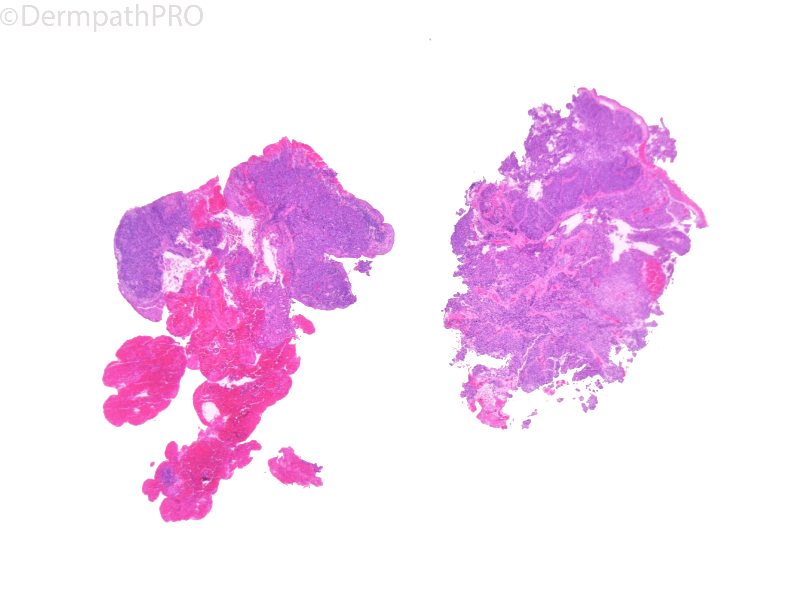
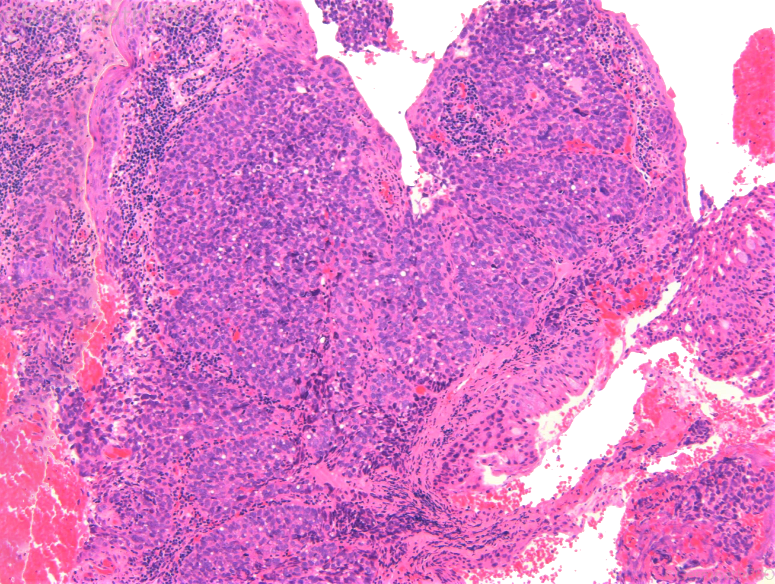
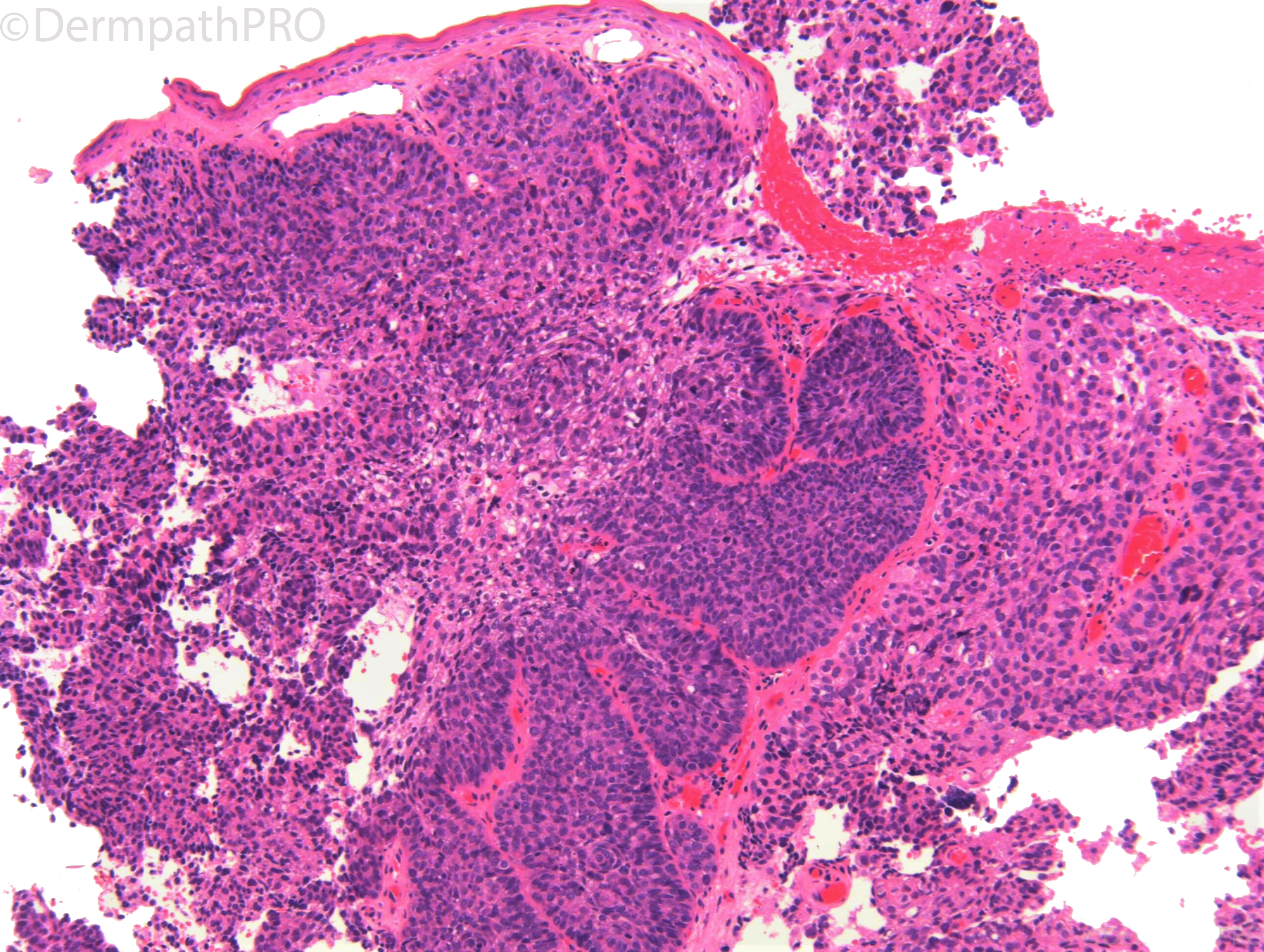
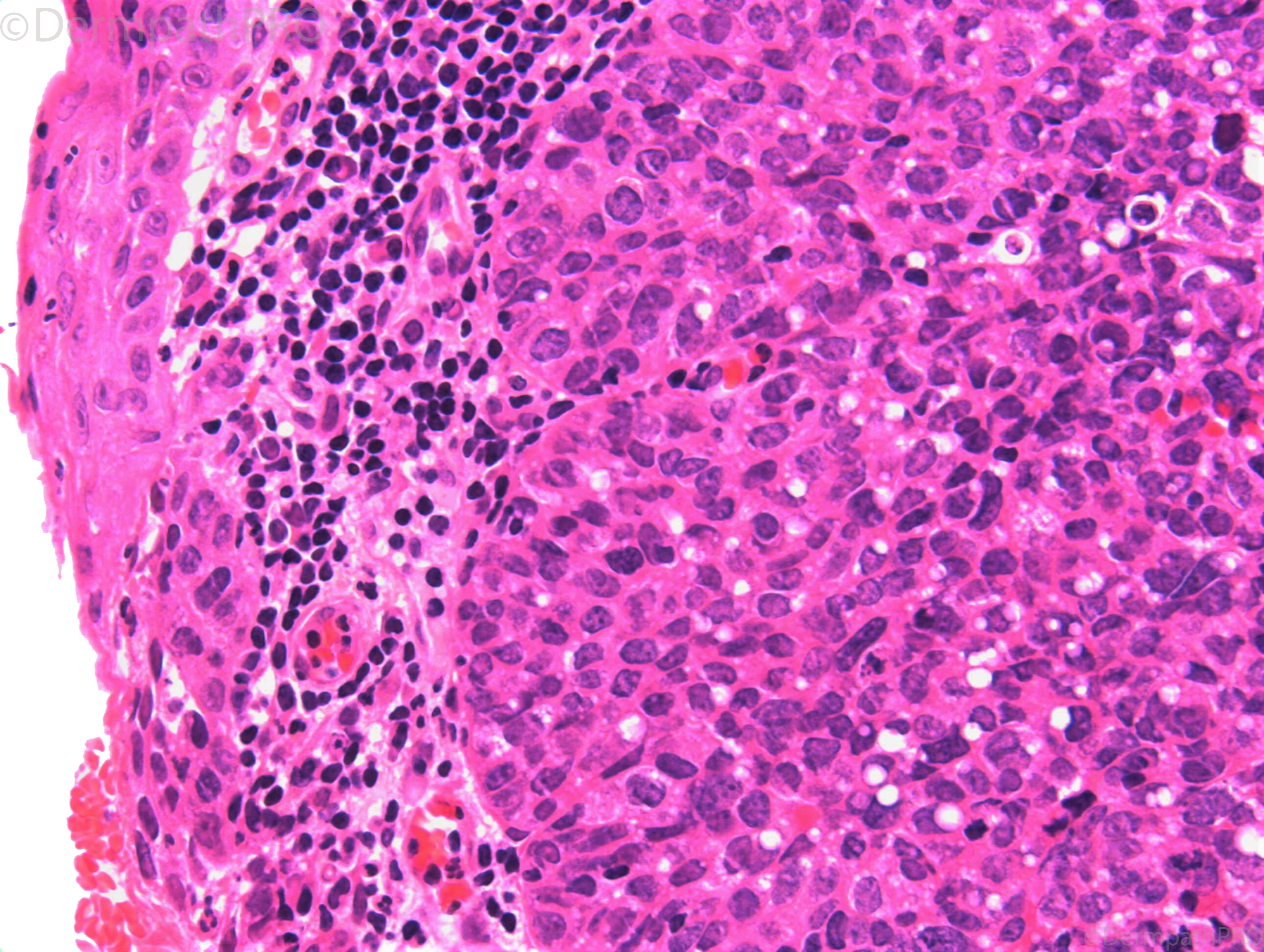
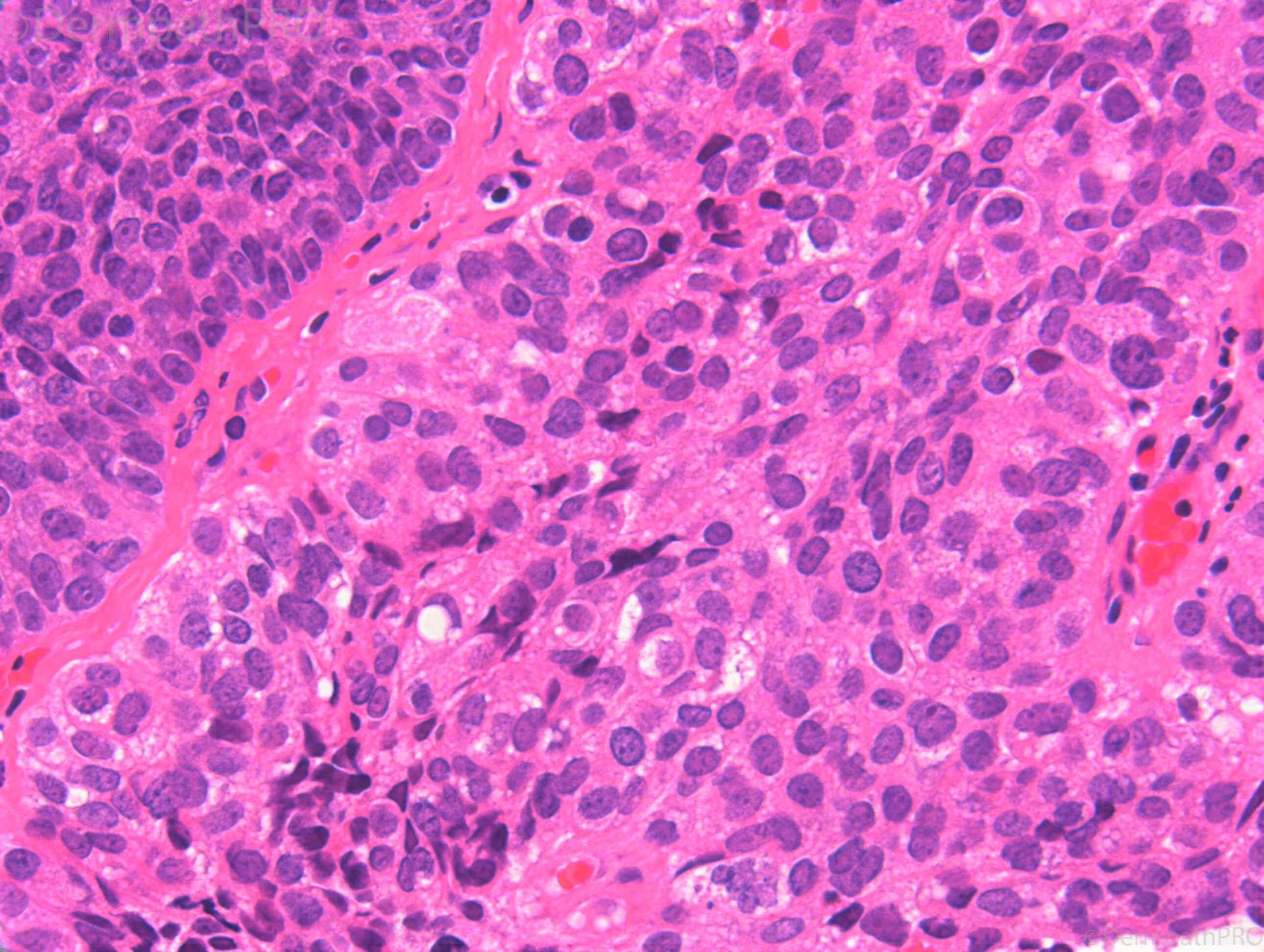
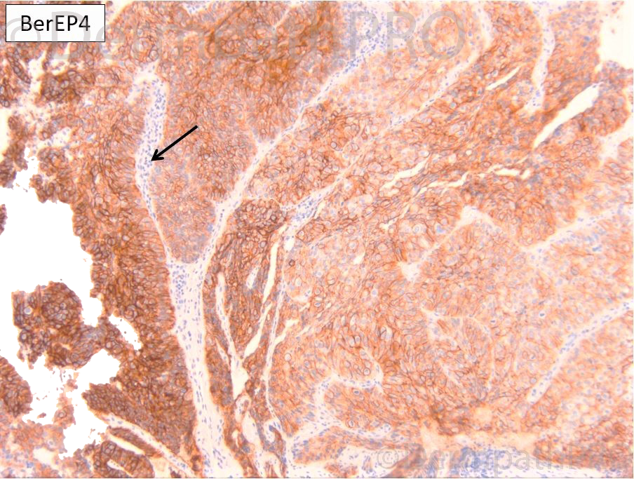
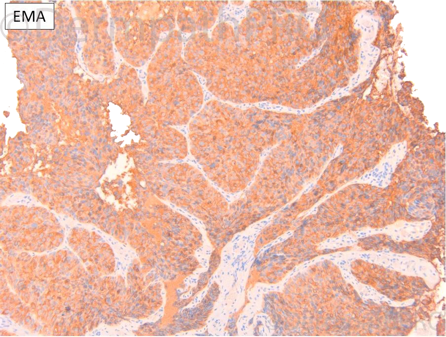
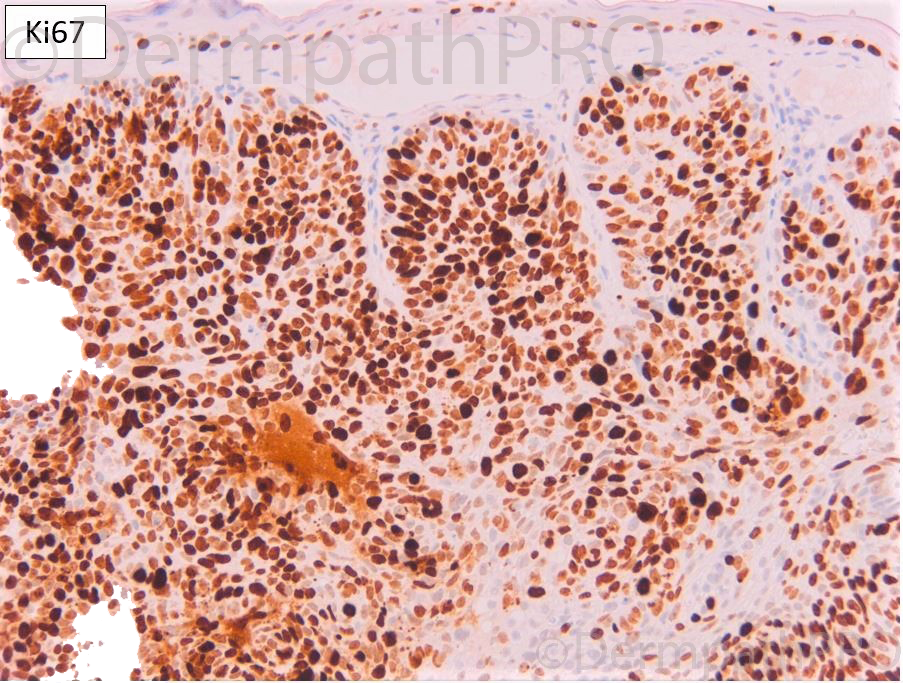
M65. Lower lid papilloma


  Report Record

User Feedback


Richard Logan

Posted

This ulcerated tumour appears biphasic.  There is a basaloid area on the left which suggests BCC.  To the right is a different population of cells which are more eosinophilic, showing a moderate degree of nuclear pleomorphism and a bubbly cytoplasm.  Given the site I suspect this is sebaceous carcinoma.

Share this comment


Link to comment
share_externally

daniellindsay

Posted

Agree, I was thinking sebaceous carcinoma. 

Share this comment


Link to comment
share_externally

Eman El-Nabarawy

Posted

My thought is sebaceous carcinoma too.

Share this comment


Link to comment
share_externally

Krishnakumar subramanian

Posted

Basaloid tumor with cells showing severe atypia. Cytoplasmic vacuoles are there in the tumor cells. I would rule out pooly differentiated SGC first even though SGC is rare in the lower lid

Share this comment


Link to comment
share_externally

Meenakshi Batrani

Posted

Agree with Sebaceous gland carcinoma. 

Share this comment


Link to comment
share_externally

On 25/06/2021 at 15:40, Richard Logan said:

This ulcerated tumour appears biphasic.  There is a basaloid area on the left which suggests BCC.  To the right is a different population of cells which are more eosinophilic, showing a moderate degree of nuclear pleomorphism and a bubbly cytoplasm.  Given the site I suspect this is sebaceous carcinoma.

Totally agree

Share this comment


Link to comment
share_externally

Krishnakumar subramanian

Posted

bases on IHC

The tumor on the right and left side is Ber EP 4 positive, EMA positive, High ki 67 and P53 positive. Synaptophysin positive

This suggests could be neuroendocrine type of BCC with sebaceous differentiation . Usually SGC is BEr EP4 negative and BCC is BErEP4 positive. SGC is EMA positive. P16 and p53 are expressed in periocular SGC

Expression of p16 and p53 in Intraepithelial Periocular Sebaceous Carcinoma. Ocul Oncol Pathol. 2015 Dec; 2(2): 71–75.

W. Robert Bell, Kamaljeet Singh, Anand Rajan KD, and Charles G. Eberhart*


also merkel cell carcinoma shows basaloid features, must we do further markers .

Merkel cell carcinoma frequently shows histologic features of basal cell carcinoma: a study of 30 cases.

Ball NJ, Tanhuanco-Kho G.J Cutan Pathol. 2007 Aug;34(8):612-9. doi: 10.1111/j.1600-0560.2006.00674.x.

Share this comment


Link to comment
share_externally

Dr. Richard Carr

Posted

For me morphologically this was a basaloid sebaceous carcinoma with evident vacuoles indenting the nucleus. No mucin or retraction to suggest bone fide BCC and BCC are always negative for EMA in the basaloid epithelium. In my experience dual BerEP4 and EMA is typical rather than unusual for sebaceous carcinoma. CK20 was negative. CD56 showed only focal positive staining and chromogranin was minimal. I have seen neuroendocrine differentiation in basaloid SCC and Bowen's previously, obviously it's very common in BCC but this is the first time I've seen such prominent neuroendocrine marker expression in a sebaceous carcinoma. That said it's unlikely I'd be running neuroendocrine markers routinely, the markers were done by a colleague.

Share this comment


Link to comment
share_externally

Krishnakumar subramanian

Posted

Thanks a lot sir, we would be more careful in interpreting markers

Share this comment


Link to comment
share_externally

Dr. Richard Carr

Posted

Forgot to say. p53 here is the highly aberrant diffuse strong (>75% moderate/strong) perhaps indicating a mutation that causes stabilisation. The p16 is probably wild type as it appears to be an exaggerated mosaic pattern and may be trying to compensate for the p53 issue. Of course we need studies of the genetics, epigenetics, RNA expression etc to confirms these speculations.

Share this comment


Link to comment
share_externally


Join the conversation

You can post now and register later. If you have an account, sign in now to post with your account.

Guest
Add a comment...

×   Pasted as rich text.   Paste as plain text instead

  Only 75 emoji are allowed.

×   Your link has been automatically embedded.   Display as a link instead

×   Your previous content has been restored.   Clear editor

×   You cannot paste images directly. Upload or insert images from URL.

Loading...

×
×
  • Create New...