Jump to content
In this section we have spot diagnoses posted on a daily basis since June 2010, now over 4000! You can review the archived cases and read the suggested diagnoses by users and the final comment by the contributors.
Case are uploaded each week day by 10 am UK time with the correct diagnosis will generally be posted at 8 pm UK time. Why not view the most recent spot diagnosis and proffer a diagnosis?

Case Number : Case 2782- 5 March 2021 Posted By: Richard Logan

Please read the clinical history and view the images by clicking on them before you proffer your diagnosis.
Submitted Date :
   (0 reviews)

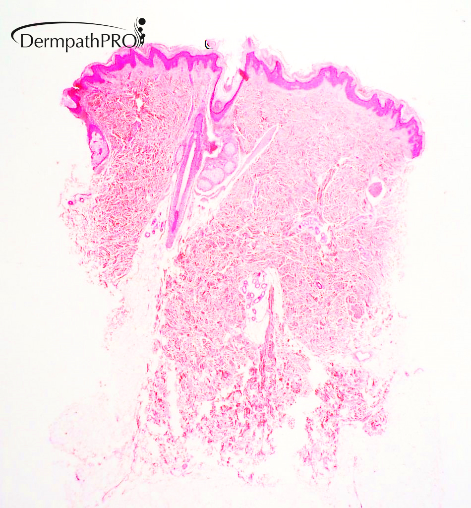
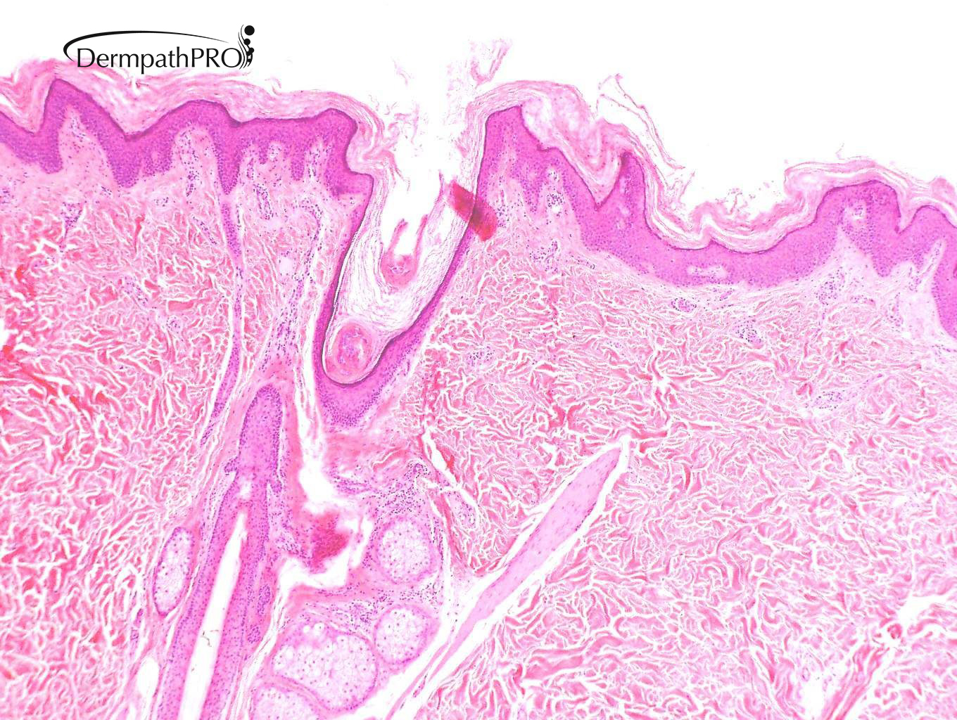
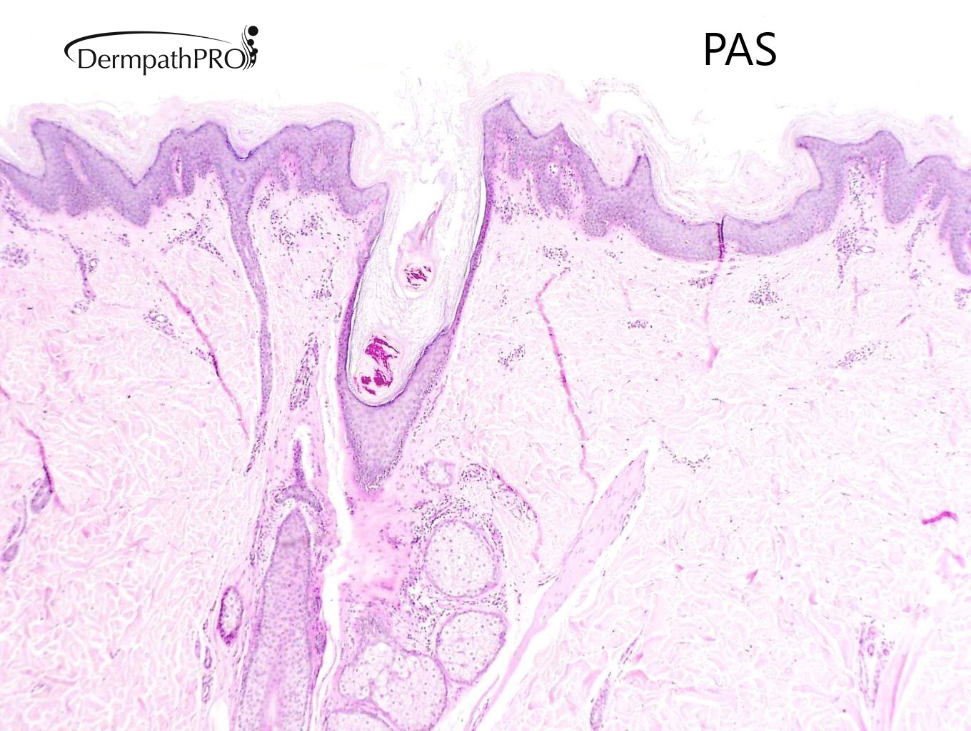
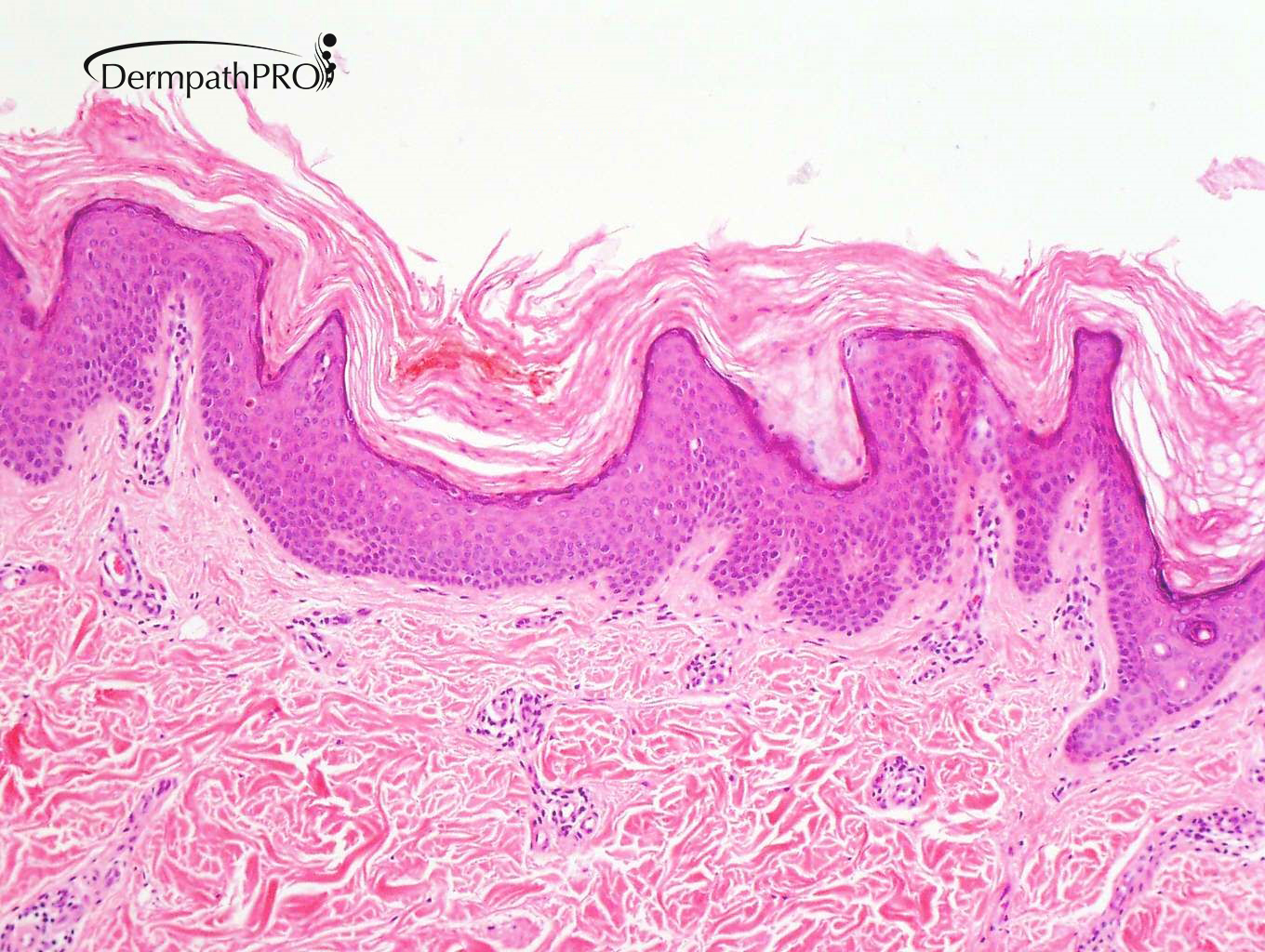
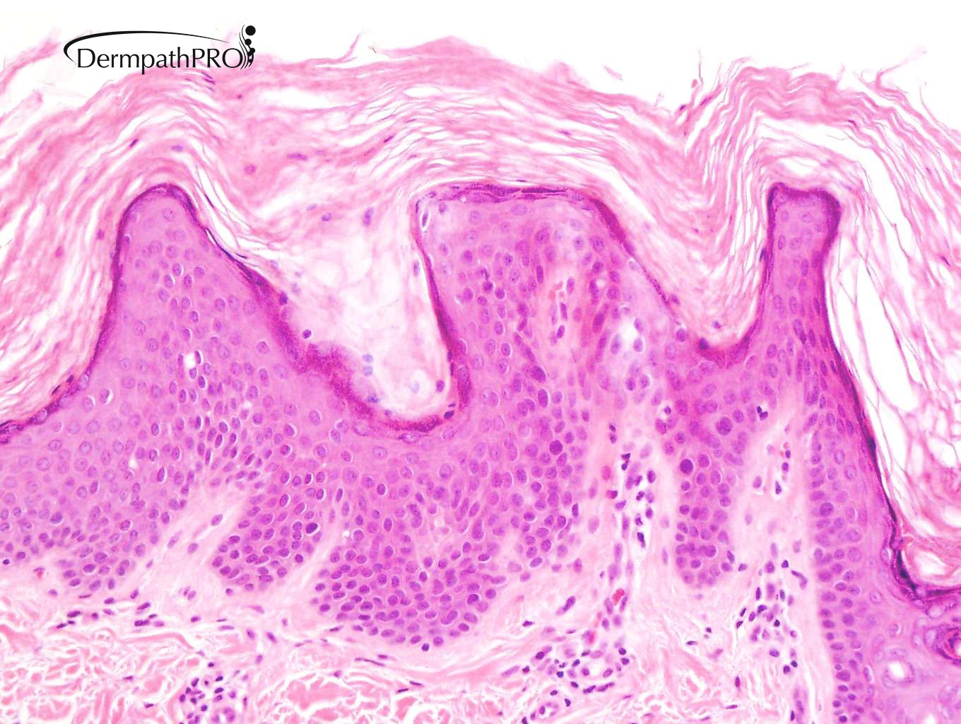
M 25 Previous history of Hodgkin's disease treated with chemotherapy and radiotherapy. Four year history of pigmented rash on trunk. Biopsy from trunk.


  • Like 1
  Report Record

User Feedback


Admin_Dermpath

Posted


posted By vincenzo

?Pityriasis Rubra Pilaris

Share this comment


Link to comment
share_externally

Admin_Dermpath

Posted

Posted By: Anil Patki

There's laminated hyperkeratosis, attenuated granular layer and follicular plugging with some pityrosporum spores in the follicle. PRP is a good suggestion by Vincenzo but I don't see any significant acanthosis. 

 This will need clinico-pathological correlation and the diagnosis will depend on clinical appearance of the lesions. 

 Since the lesions are pigmented, it's likely that it's acquired ichthyosis and pigmentation, both of which are paraneoplastic manifestations seen in patients with Hodgkin's lymphoma. 

 Investigations to rule out relapse of Hodgkin's may be needed in that case. 


Share this comment


Link to comment
share_externally

Alex-Ventura-Leon

Posted

Pigmented lesion on trunk made me think in Confluent and Reticulated Papillomatosis (CARP) 

Share this comment


Link to comment
share_externally

Richard Logan

Posted

7 hours ago, Alex-Ventura-Leon said:

Pigmented lesion on trunk made me think in Confluent and Reticulated Papillomatosis (CARP) 

Well done Alex, you are correct.  I will attach a clinical photo which shows the characteristic clinical appearance of confluent and reticulated papillomatosis seen in this case.  The histology shows the typical invagination of the epidermis by hyperkeratotic stratum corneum, and rather blunt-topped papillomatosis.  As Anil says, there are some follicular Pityrosporum yeasts but no ramifying hyphae as might be expected if this were Pityriasis versicolor.

DPP clinical 5.3.21.jpg

Share this comment


Link to comment
share_externally


Join the conversation

You can post now and register later. If you have an account, sign in now to post with your account.

Guest
Add a comment...

×   Pasted as rich text.   Paste as plain text instead

  Only 75 emoji are allowed.

×   Your link has been automatically embedded.   Display as a link instead

×   Your previous content has been restored.   Clear editor

×   You cannot paste images directly. Upload or insert images from URL.

Loading...

×
×
  • Create New...