Jump to content
In this section we have spot diagnoses posted on a daily basis since June 2010, now over 4000! You can review the archived cases and read the suggested diagnoses by users and the final comment by the contributors.
Case are uploaded each week day by 10 am UK time with the correct diagnosis will generally be posted at 8 pm UK time. Why not view the most recent spot diagnosis and proffer a diagnosis?

Case Number : Case 2787- 12 March 2021 Posted By: Richard Logan

Please read the clinical history and view the images by clicking on them before you proffer your diagnosis.
Submitted Date :
   (0 reviews)

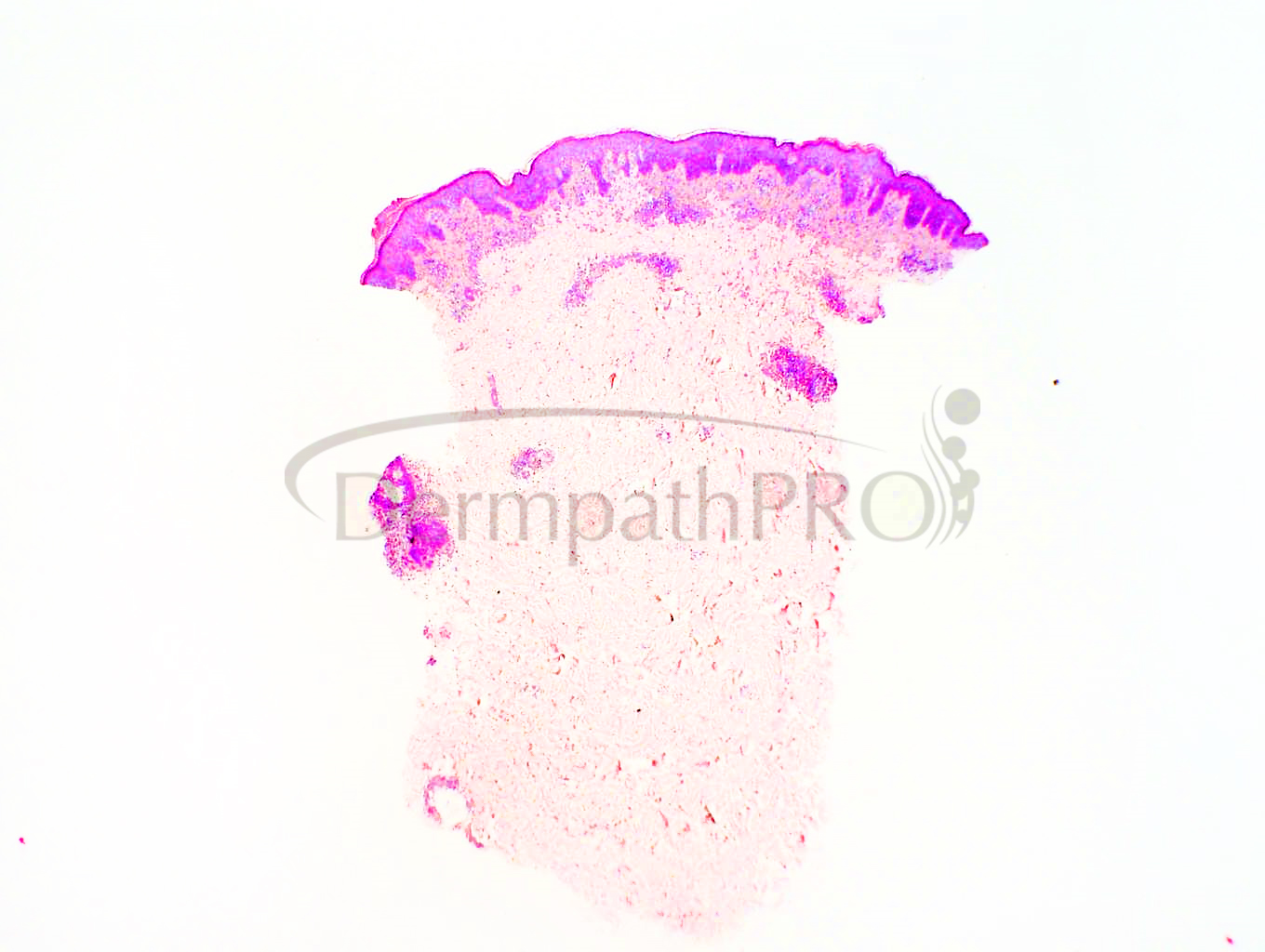
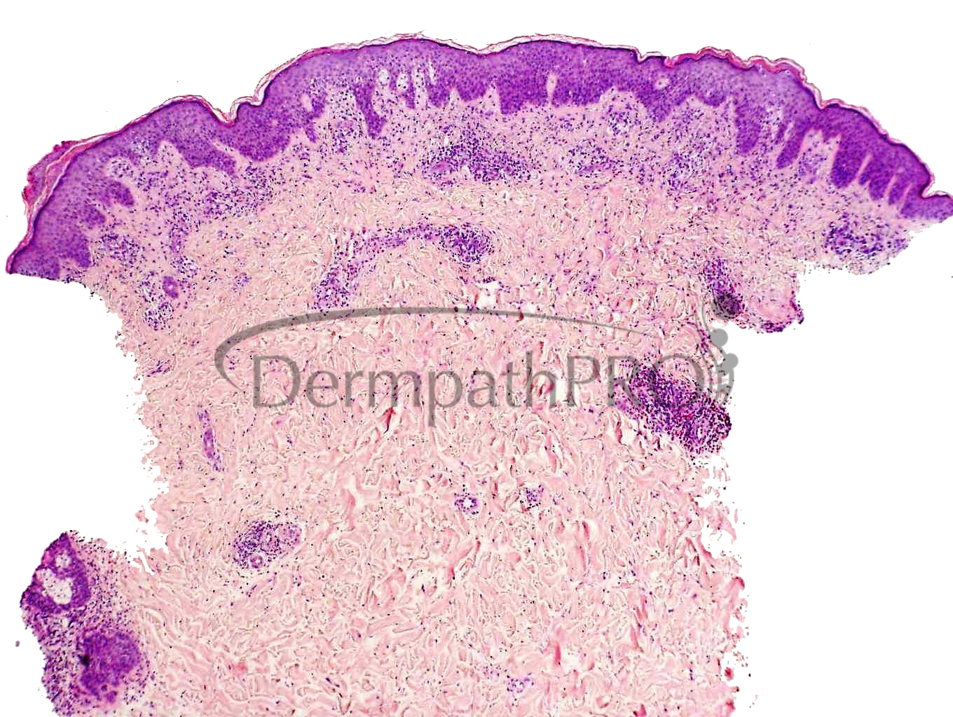
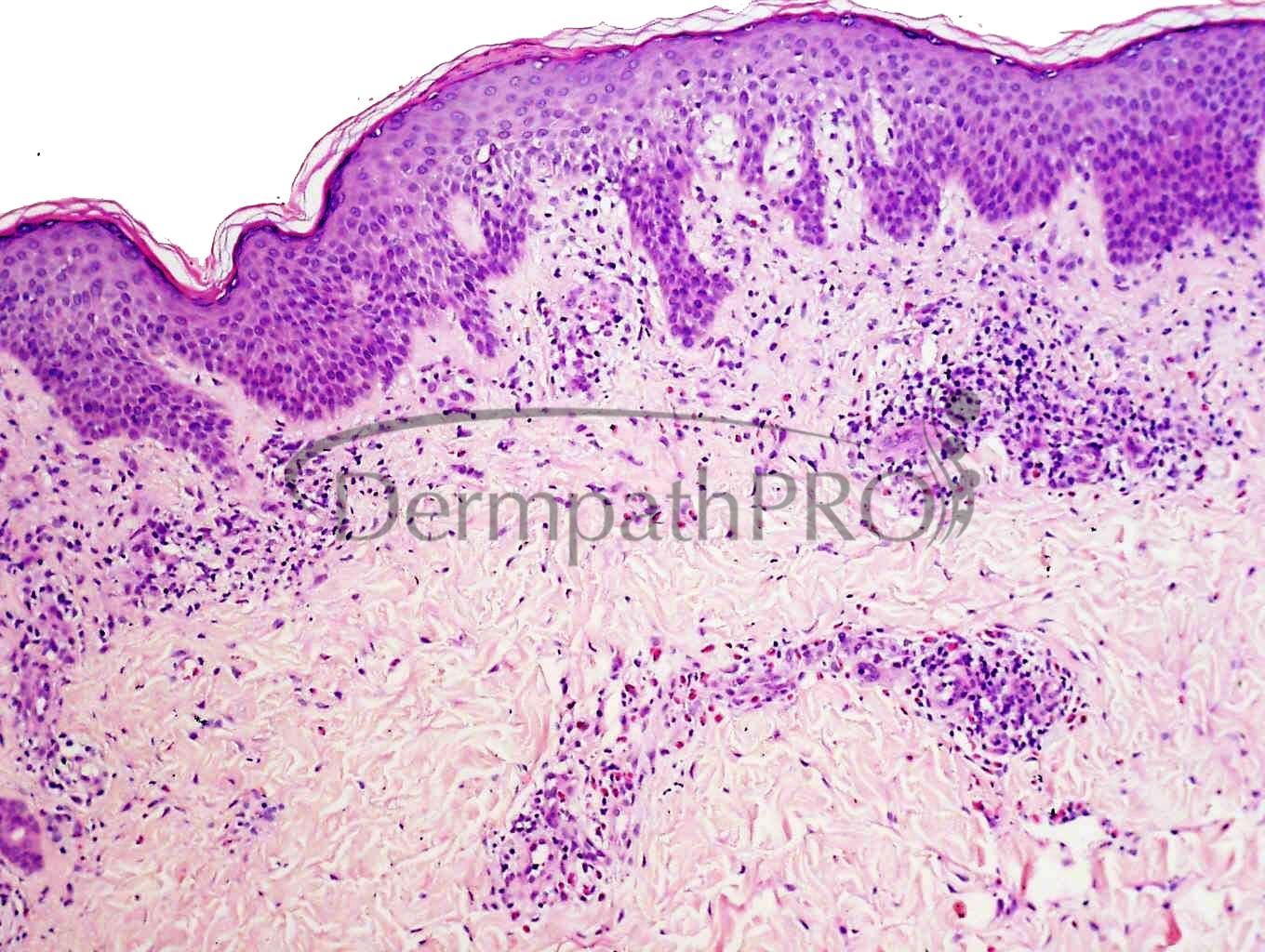
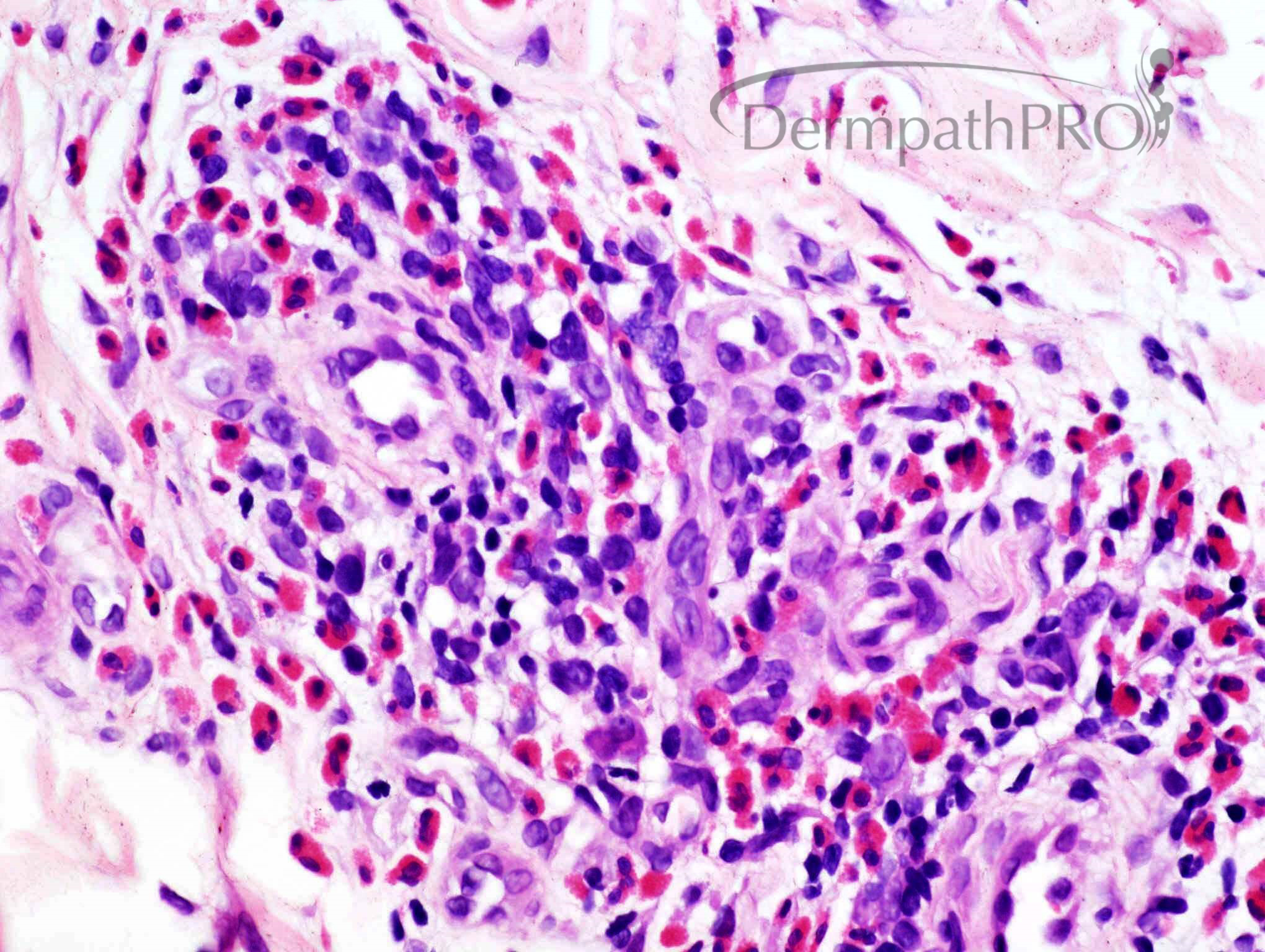
F 27 Biopsy from abdomen


  Report Record

User Feedback


Alex-Ventura-Leon

Posted

Hypersensitivity reaction pattern (Insect bite?, scabies? drug?)

Share this comment


Link to comment
share_externally

Saman Fatah

Posted

While this pattern of changes is not specific for a particular diagnosis, this is a biopsy from a young female and on abdomen hence one would consider Polymophic Eruption of Pregnancy (PEP) or PUPPP if the lady is pregnant especially primigravida. 

Usually such cases diagnosed clinically in the right context and if biopsy done perhaps a prebullous stage of pemphigoid gestationis was in the clinical differential and IMF needed to exclude or confirm the latter. 

Share this comment


Link to comment
share_externally

Richard Logan

Posted

8 hours ago, Saman Fatah said:

While this pattern of changes is not specific for a particular diagnosis, this is a biopsy from a young female and on abdomen hence one would consider Polymophic Eruption of Pregnancy (PEP) or PUPPP if the lady is pregnant especially primigravida. 

Usually such cases diagnosed clinically in the right context and if biopsy done perhaps a prebullous stage of pemphigoid gestationis was in the clinical differential and IMF needed to exclude or confirm the latter. 

Well done Saman - this was a case of polymorphic eruption of pregnancy.  I deliberately withheld the crucial information that she was pregnant.  As you say most of these cases are diagnosed clinically without the need for a biopsy.

There is an eosinophil-rich dermal infiltrate with mild spongiosis.  The images shown don't really allow for a close inspection of the epidermal cytology, but there weren't any eosinophils in the epidermis, so strictly speaking these slides do not demonstrate eosinophilic spongiosis.

Share this comment


Link to comment
share_externally


Join the conversation

You can post now and register later. If you have an account, sign in now to post with your account.

Guest
Add a comment...

×   Pasted as rich text.   Paste as plain text instead

  Only 75 emoji are allowed.

×   Your link has been automatically embedded.   Display as a link instead

×   Your previous content has been restored.   Clear editor

×   You cannot paste images directly. Upload or insert images from URL.

Loading...

×
×
  • Create New...