Jump to content
In this section we have spot diagnoses posted on a daily basis since June 2010, now over 4000! You can review the archived cases and read the suggested diagnoses by users and the final comment by the contributors.
Case are uploaded each week day by 10 am UK time with the correct diagnosis will generally be posted at 8 pm UK time. Why not view the most recent spot diagnosis and proffer a diagnosis?

Case Number : Case 2827- 7 May 2021 Posted By: Dr. Richard Carr

Please read the clinical history and view the images by clicking on them before you proffer your diagnosis.
Submitted Date :
   (0 reviews)

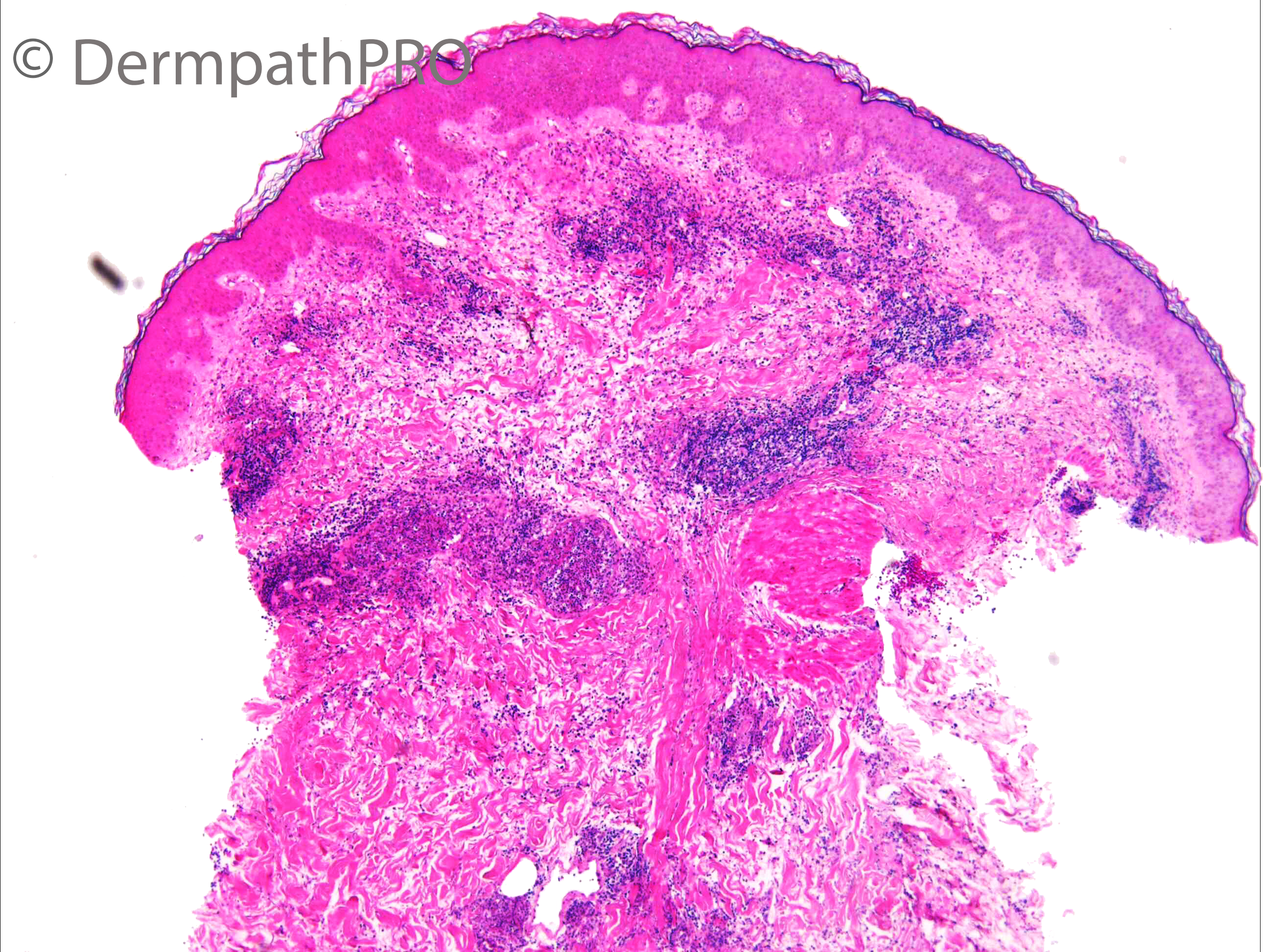
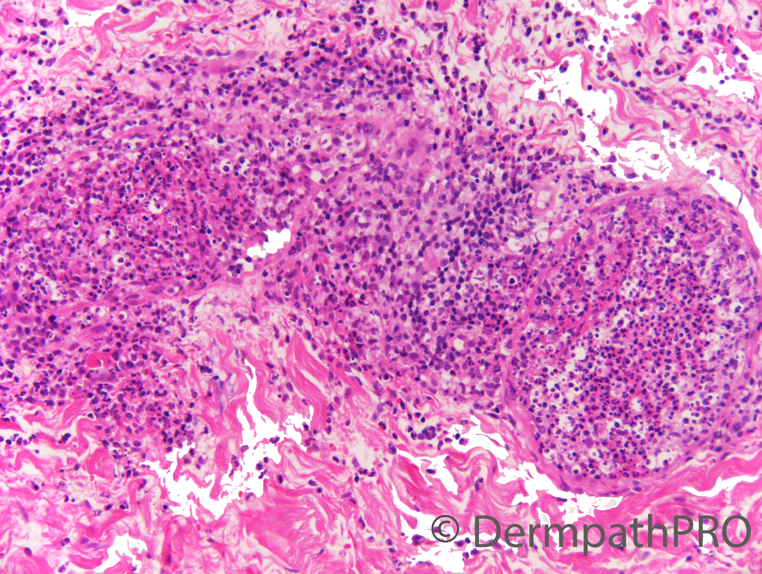
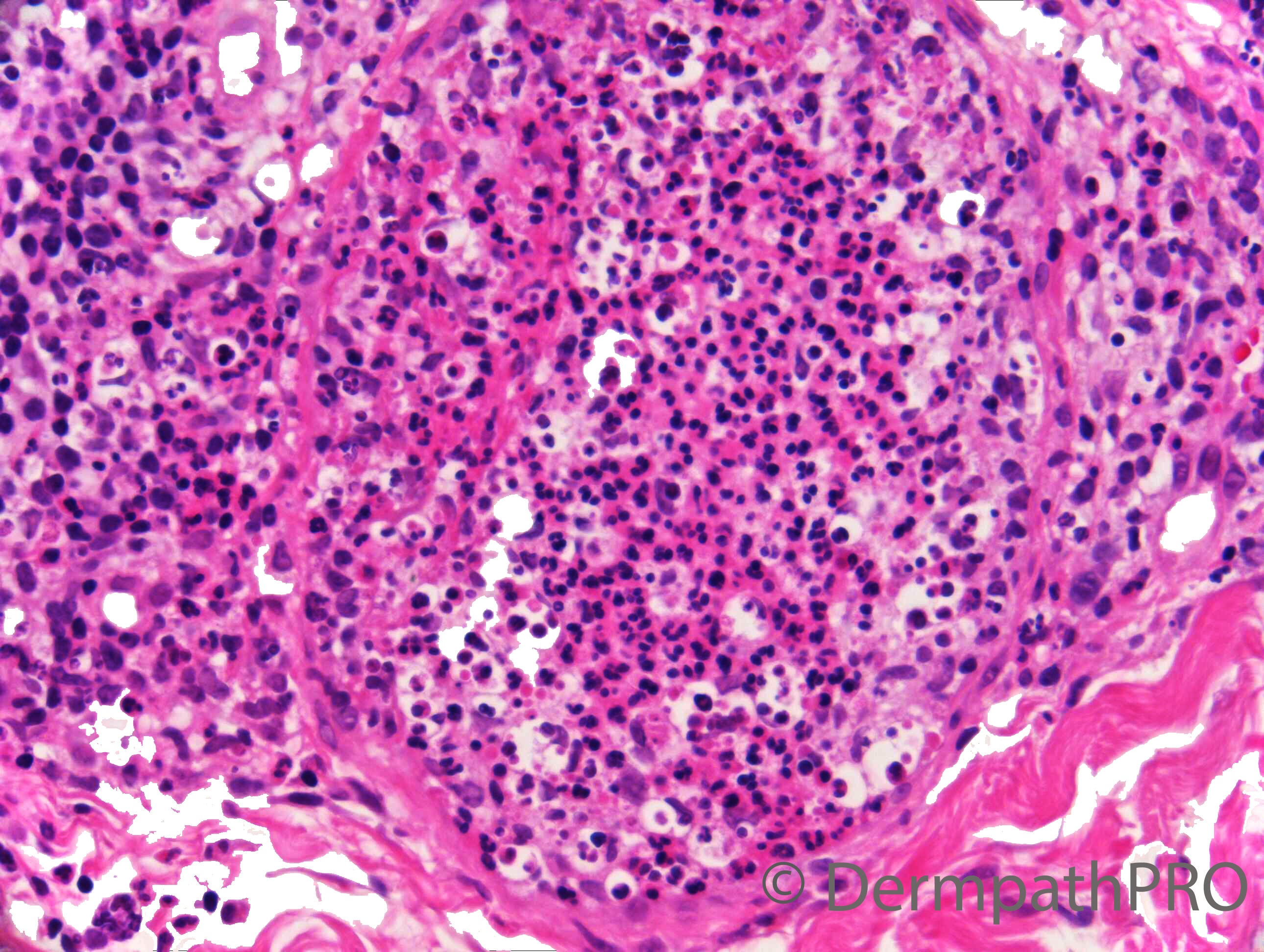
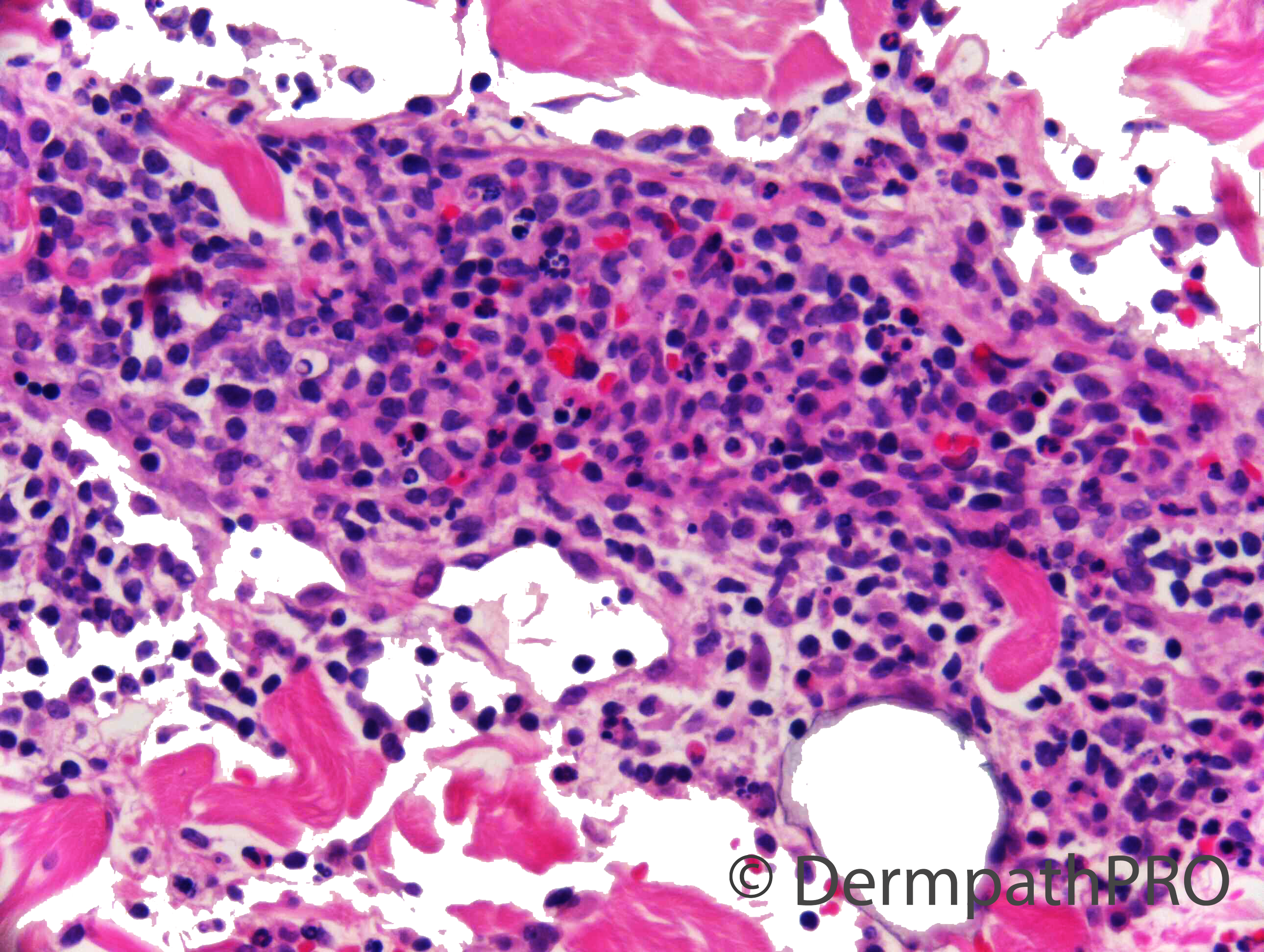
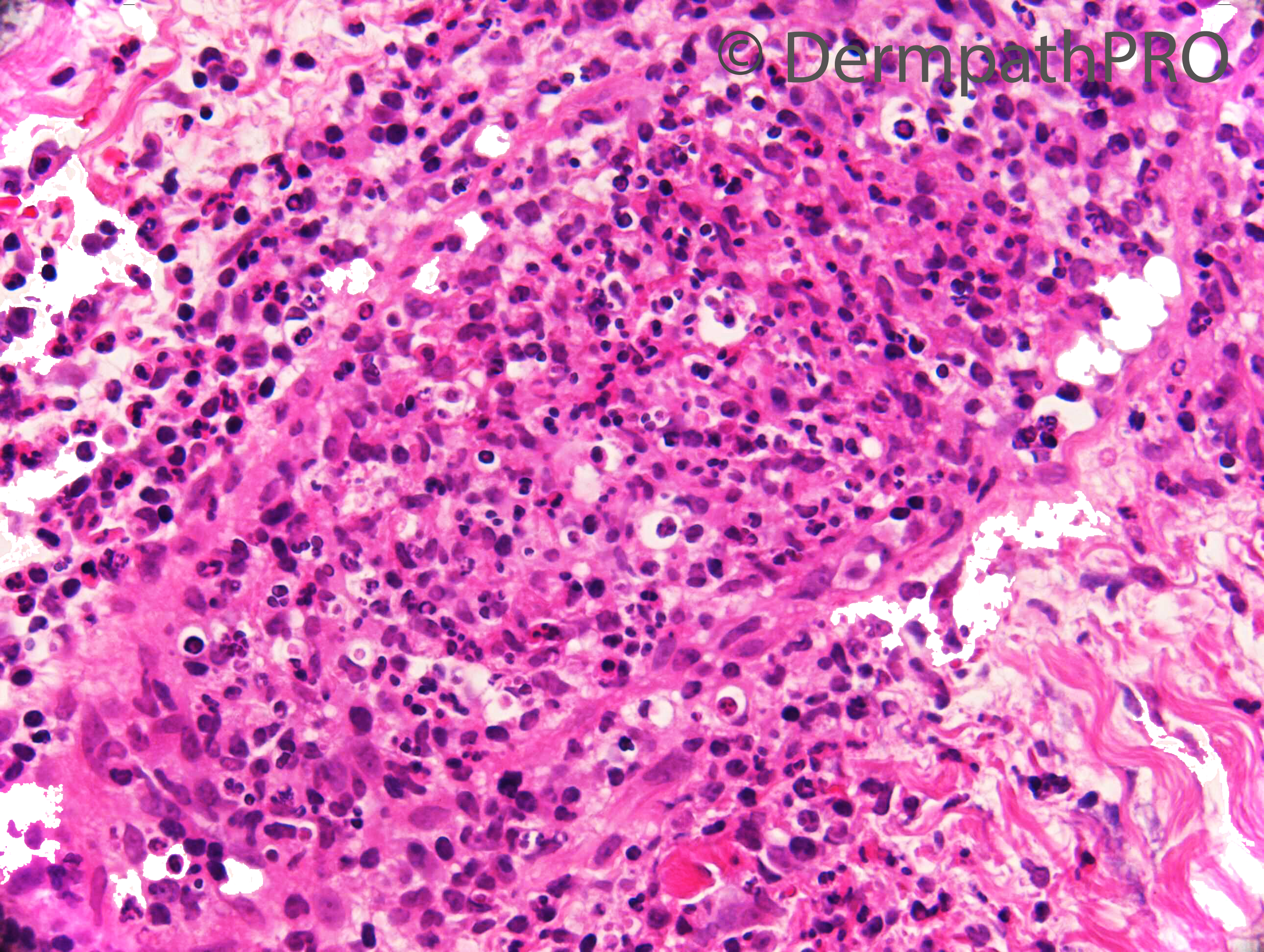
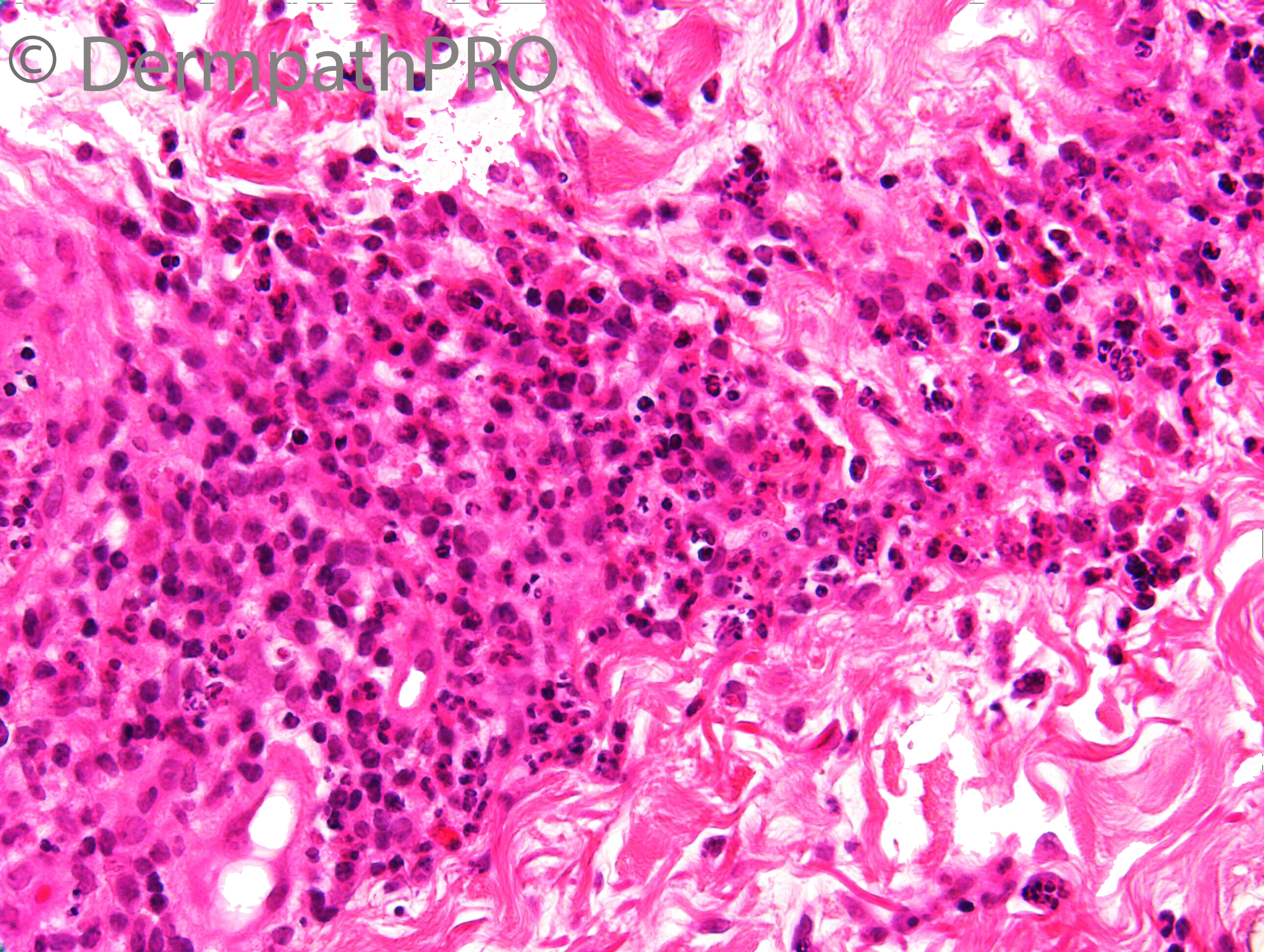
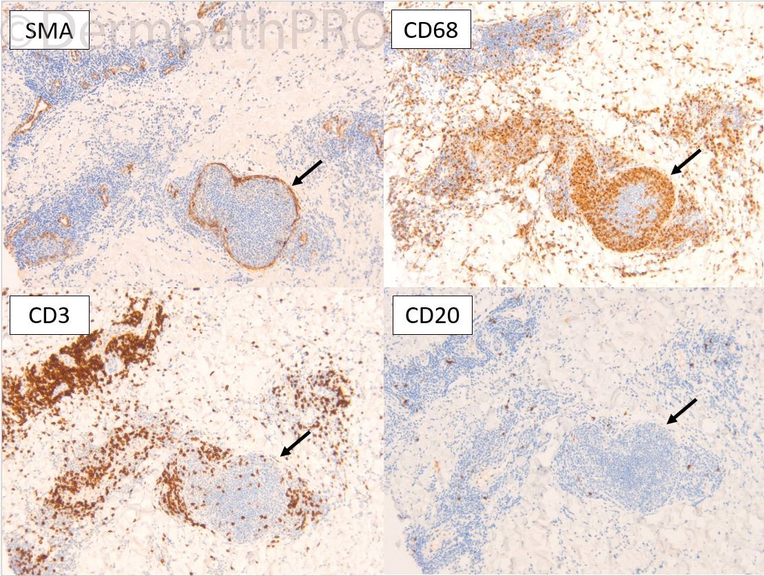
F75. Right hip. 4/12 hx of red / purple discolouration extending down right leg from right buttock. ?Contact reaction to prosthesis right hip replacement 2 years ago. ?Vasculitis. Does not have phenotype of shingles


  Report Record

User Feedback


daniellindsay

Posted

Superficial and deep relatively well circumscribed dermal infiltrate of neutrophil polmorphs, associated with more immature appearing cells. I can see any obvious vasculitic component but is difficult to see vessels clearly. I would consider histiocytoid Sweet's syndrome. Also need to correlate with microbiology to exclude infective process, although long history since surgery. The features are not typical of a chronic peri-prosthetic reaction. 

Share this comment


Link to comment
share_externally

Richard Logan

Posted

I agree that the histology is suggestive of Sweet's syndrome.  However, the localised and persistent nature of the lesion is odd for that diagnosis and I too would be wondering about an infective process.

Share this comment


Link to comment
share_externally

Krishnakumar subramanian

Posted

leuko cytoclastic vasculitis histology with somewhat  ? pseudo-cryptococcal artefact, can we have a PAS stain

Share this comment


Link to comment
share_externally

Cem Leblebici

Posted

Intravascular histiocytosis

Share this comment


Link to comment
share_externally

Saman Fatah

Posted

Infection requires exclusion first through appropriate stains and tissue cultures. If sterile nature of the infiltrate is established, the histiocytoid/cryptococcoid neutrophilic diseases is in the DDx and requires clinical correlation. Agree with Richard such localisation will be unusual clinically. 

The list of ....oid is getting bigger. 
 

It will be interesting to see the results of other premature granulocytic markers especially MPO (if performed) and any background haematological malignancy. 

Share this comment


Link to comment
share_externally

Dr. Richard Carr

Posted

Thanks for all the great discussion comments and suggestions. I'd like to have shared the clinicals but it's becoming difficult to do. The clinical distribution was very confined around the hip and buttock - large red blotches (so let's shelve CML and infection on that basis). Her prosthesis was ceramic and not metal. I neglected to say she has a 20 year history of polymyalgia rheumatica and had a raised CRP and PV. She initially was treated with dermovate but also was developing joint pain, intermittent chest cramps and lethargy. Her oral prednisolone was increased to 15mm OD and the skin eruption resolved completely. I favoured a diagnosis of Sweet's-like (Sweet-oid!) intravascular histiocytosis related to her PMR and previous joint replacement. I did not do any "bug" stains actually as I thought it was an "immunological" reaction. I guess this is a slightly unique case but I think you all did well.

Share this comment


Link to comment
share_externally


Join the conversation

You can post now and register later. If you have an account, sign in now to post with your account.

Guest
Add a comment...

×   Pasted as rich text.   Paste as plain text instead

  Only 75 emoji are allowed.

×   Your link has been automatically embedded.   Display as a link instead

×   Your previous content has been restored.   Clear editor

×   You cannot paste images directly. Upload or insert images from URL.

Loading...

×
×
  • Create New...