Jump to content
In this section we have spot diagnoses posted on a daily basis since June 2010, now over 4000! You can review the archived cases and read the suggested diagnoses by users and the final comment by the contributors.
Case are uploaded each week day by 10 am UK time with the correct diagnosis will generally be posted at 8 pm UK time. Why not view the most recent spot diagnosis and proffer a diagnosis?

Case Number : Case 2836- 20 May 2021 Posted By: Saleem Taibjee

Please read the clinical history and view the images by clicking on them before you proffer your diagnosis.
Submitted Date :
   (0 reviews)

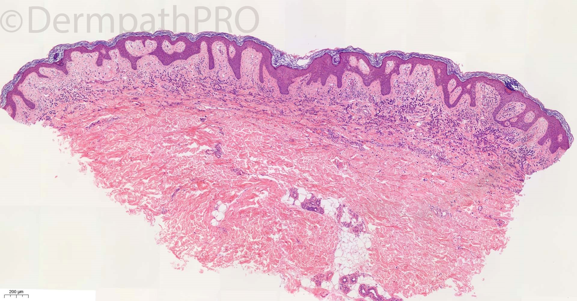
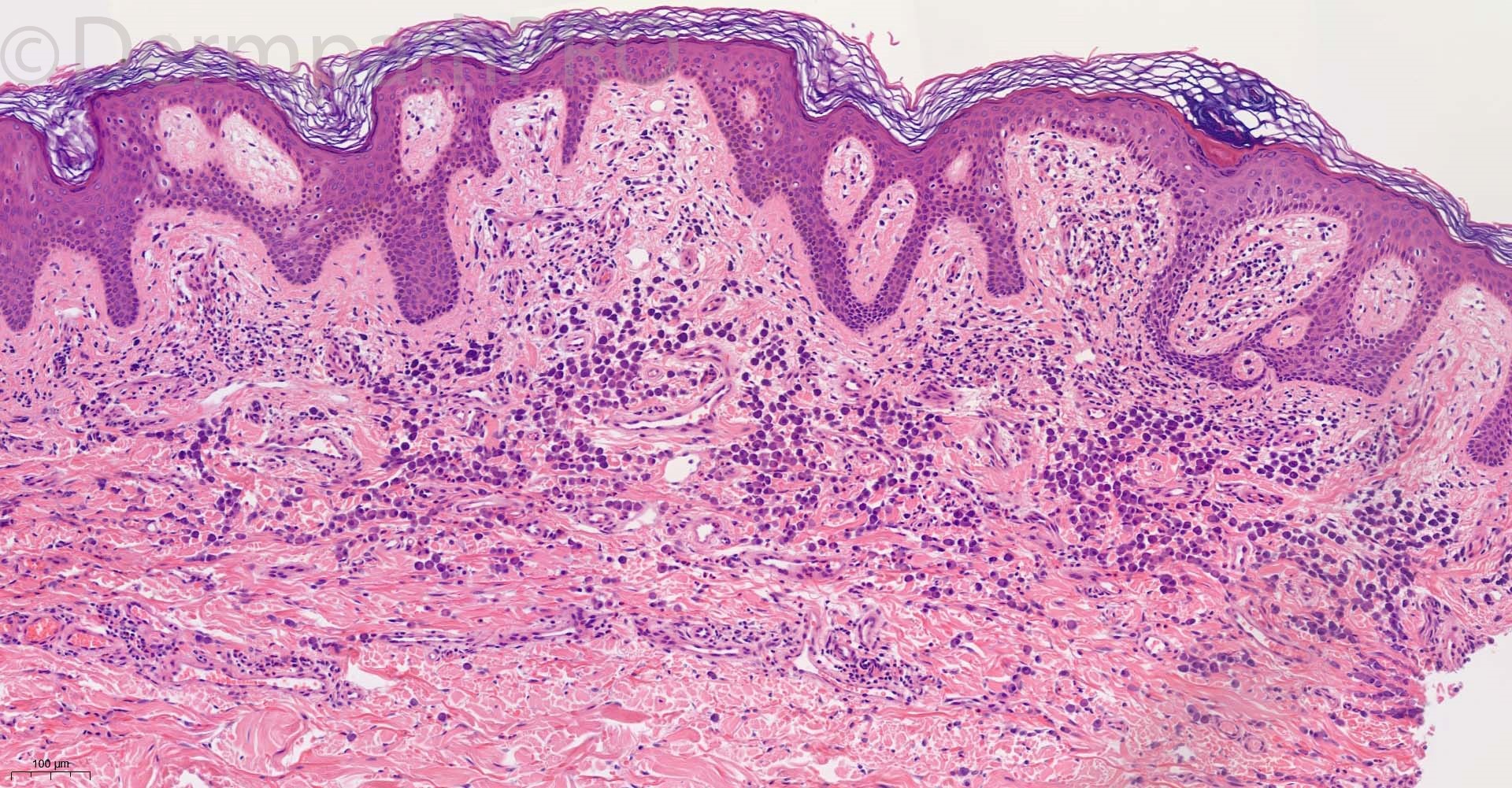
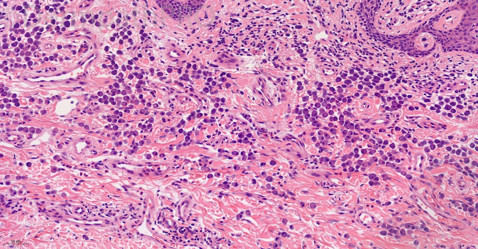
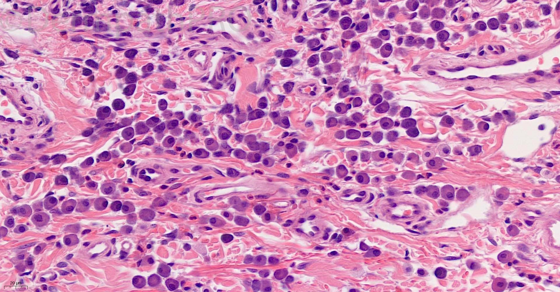
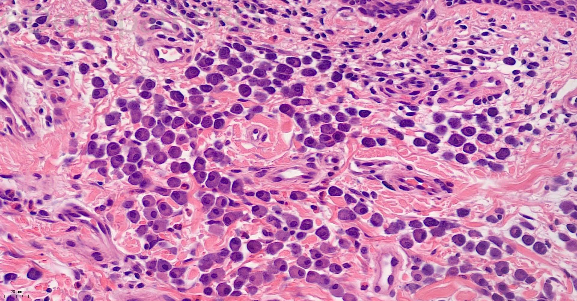
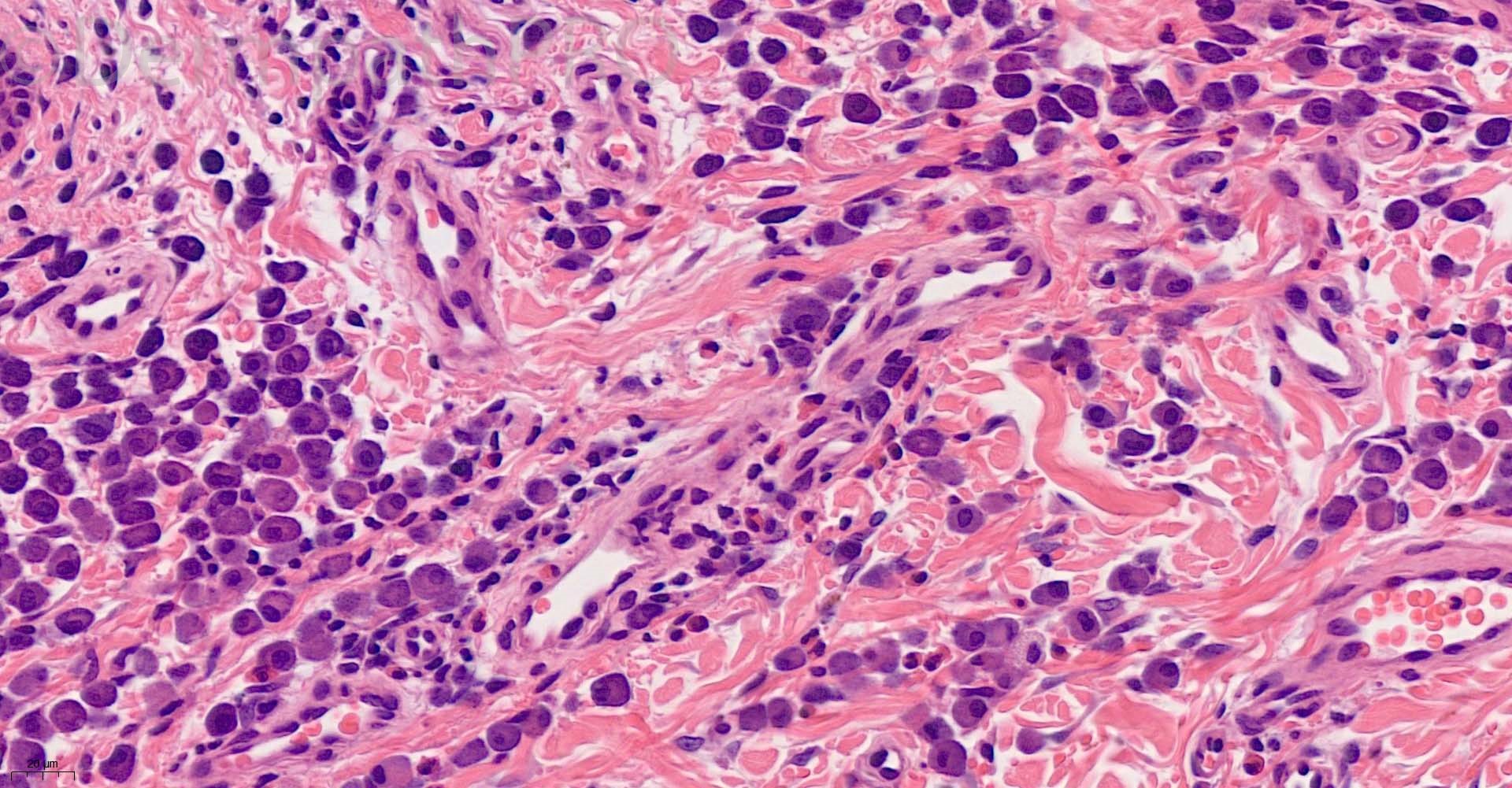
67M erythematous rash with the shiny surface? lichen planus?Grover’s disease


  Report Record

User Feedback


Richard Logan

Posted

I agree that some of the cells look a little like fried eggs, but too many of them have an eccentric nucleus with a perinuclear halo so I think they are plasma cells.  I am sure immuonhistochemistry would sort that out.

Given that I think these are plasma cells then of course syphilis and plasmacytoma come into the differential diagnosis.  Of the two, given the age of the patient, and the clinical description which doesn't really suggest syphilis, I am inclined towards plasmacytoma,

Share this comment


Link to comment
share_externally

Saleem Taibjee

Posted

This is already proving to be an interesting discussion. Here is a closer up image of the cells, to promote this further.

13895_40.0xb crop.jpg

Share this comment


Link to comment
share_externally

Richard Logan

Posted

I'm sticking to the plasma cell theory.  I think some form of plasma cell dyscrasia would have to be considered, as it sounds as though this is clinically a more widespread problem than a solitary plasmacytoma.

Share this comment


Link to comment
share_externally

Krishnakumar subramanian

Posted

dermal perivascular plasma cells

first is serology to rule out syphilis

second get IHC for clonal plasma cell disease and then rule pout IgG4 inflammation and also get serum igg4

Share this comment


Link to comment
share_externally

Dr. Mona Abdel-Halim

Posted

Cutaneous plasmacytosis with extracellular crystal deposition. Will check clonality of plasma cells.

Share this comment


Link to comment
share_externally

vincenzo

Posted

Agree with Colleagues there are too features heading to plasma cells...but there is a basophilic granularity of cytoplasm. For me it’s a mastocytosis. 

Share this comment


Link to comment
share_externally

Saleem Taibjee

Posted

To take this further... The original images shown were snapshots taken of the digitised slides. The slides are digitised at our lab (Poundbury Cancer Institute), and I routinely report cases digitally arising from this lab. 

By contrast, below is the same slide viewed on an Olympus BX53 microscope, with images taken on the Olympus SC50 camera (with Olympus cellSens Entry software).


HE_10x.jpg

HE_60x.jpg

HE_2x.jpg

Share this comment


Link to comment
share_externally

Richard Logan

Posted

There is an obvious granularity to the cytoplasm, but is this an optical artefact because of the equipment being used which adds an extra complexity to the interpretation of digitised slides?  The overall cellular morphology remains plasmacytoid.

Share this comment


Link to comment
share_externally

daniellindsay

Posted

Very challenging to distinguish, could this be dual plasma cell and mast cell pathology? IHC needed. 

Share this comment


Link to comment
share_externally

Saleem Taibjee

Posted

Thanks for all the comments.

The immunohistochemistry has demonstrated positive staining for CD117, and negative staining for CD20, CD79a, CD138, Kappa, Lambda, myeloperoxidase.

Hence this recent case was signed out as mastocytosis. I did advise haematology referral, checking serum tryptase, etc, to assess for any possible systemic involvement.

But as you have all picked up on, there are some intriguing aspects.

Firstly, this was a rare case where I felt that the digital view was a bit misleading, in so far as the cytoplasmic granules were definitely much more subtle as compared to the microscope view. This is one of those occasional cases illustrating pitfalls of digital pathology, where the glass can be superior. Don't get me wrong, I am a full convert/preacher of the benefits of digital pathology, but just to remember that there are such occasional cases where despite the improvements in scanning resolution, glass can be superior for morphology.

Secondly, there is no doubt that many of the cells display a peri-nuclear halo / hof, which we usually associate with plasma cells.

I am yet to find a reference to a similar phenomenon in mast cells, and would be grateful if any of you come across anything in the literature. I did consult our haematologists, and they too suggested to rule out a dual / bilineage plasma cell / mast cell pathology given the close resemblance to plasma cells. I think the immunopanel has now excluded this. I intend to review past cases of mastocytosis to check whether this morphology is evident in any previous cases.

BW

Saleem

Share this comment


Link to comment
share_externally

Richard Logan

Posted

Well done Vincenzo - a lone voice crying in the wilderness!

Immunohistochemistry trumps cytology!

Share this comment


Link to comment
share_externally

Richard Logan

Posted

Perhaps there's a paper in this for you Saleem?

Share this comment


Link to comment
share_externally

Saleem Taibjee

Posted

Yes, well done Vincenzo. And yes, we are planning a short letter to one of the journals! We might even mention that the case was posted on the internet and generated some interesting discussion as evidence of the difficulty/confusing aspects of the case.

Share this comment


Link to comment
share_externally

vincenzo

Posted

Thanks you all. I’m a lucky to frequent this web site and learning really a lot.  

Share this comment


Link to comment
share_externally

Richard Logan

Posted

How does the final histological diagnosis tie in with the clinical description of "rash with shiny surface"?   Shiny surface is normally suggestive of lichen planus rather than urticaria pigmentosa.  Perhaps the rather "chunky" and dense nature of the infiltrate is mimicking lichen planus clinically?

Share this comment


Link to comment
share_externally

Richard Logan

Posted

I will look forward to hearing if you find any more cases of mastocytosis with a plasmacytoid cytology.  If you do, then the concept of mast cell cytology will have to be broadened.

Share this comment


Link to comment
share_externally

vincenzo

Posted

17 hours ago, Richard Logan said:

How does the final histological diagnosis tie in with the clinical description of "rash with shiny surface"?   Shiny surface is normally suggestive of lichen planus rather than urticaria pigmentosa.  Perhaps the rather "chunky" and dense nature of the infiltrate is mimicking lichen planus clinically?

Not sure; maybe the thick band nature of the mast cells infiltrate, I thought so too. 

Share this comment


Link to comment
share_externally

Saleem Taibjee

Posted

Good question...

Yes, I'm guessing it must be a consequence of pathology with a band-like infiltrate, but must admit I've never really considered this before.

This is speculation, but presumably an optical consequence - perhaps the band in effect creates an optical interface with a sufficiently different refractive index to tissue above (epidermis) and below (reticular dermis), such that this promotes reflection rather than transmission of incident light rays when the light approaches the tissue interface beyond the 'critical angle'. I am trying to remember from A-level physics 30 years ago! As I begin to read, I start to recall Snell's law ... see https://www.optics4kids.org/what-is-optics/refraction/snell-s-law,-reflection,-and-refraction

We might then imagine why any sufficiently dense band-like infiltrate may appear shiny e.g. lichen nitidus, lichenoid MF, sometimes lichen sclerosis.

Another logical consequence would be why the apparent shiny nature of lichenoid rashes is often accentuated by looking at the rash from an angle (increasing the angle of incidence to promote this effect).

There is probably a paper to write on this theme too, if not already done so!

Share this comment


Link to comment
share_externally

Richard Logan

Posted

Do you have access to any clinical photographs of the case in question?

Share this comment


Link to comment
share_externally

Saleem Taibjee

Posted

sadly not, the case is not from a local source

Share this comment


Link to comment
share_externally

tyelaine

Posted

Thanks for sharing this interesting case, especially with regards to images from digital pathology scanned slides. Seems there are limitations with scanning for fine granules, were the images scanned in 40x? Must take note of returning to glass slides in strange cases. On first glance I did thought the cells were plasma cells, but they did look different from the usual ones with more cytoplasm (more centrally located nucleus). Paranuclear hof represents the Golgi apparatus, and seems from older EM studies of mast cells, they do also have presence of Golgi apparatus - can this explain the plasmacytoid phenomenon?

Found in an old paper about human mast cells, mentioning 'The Golgi complex was moderately prominent, consisting of flattened lamellae, vesicles, and occasional vacuoles'.

image.thumb.png.61cd297cab189834c44faf8e5b20a66b.png

Dobbins WO 3rd, Tomasini JT, Rollins EL. Electron and light microscopic identification of the mast cell of the gastrointestinal tract. Gastroenterology. 1969 Feb;56(2):268-79. PMID: 5764594.

Share this comment


Link to comment
share_externally


Join the conversation

You can post now and register later. If you have an account, sign in now to post with your account.

Guest
Add a comment...

×   Pasted as rich text.   Paste as plain text instead

  Only 75 emoji are allowed.

×   Your link has been automatically embedded.   Display as a link instead

×   Your previous content has been restored.   Clear editor

×   You cannot paste images directly. Upload or insert images from URL.

Loading...

×
×
  • Create New...