Jump to content
In this section we have spot diagnoses posted on a daily basis since June 2010, now over 4000! You can review the archived cases and read the suggested diagnoses by users and the final comment by the contributors.
Case are uploaded each week day by 10 am UK time with the correct diagnosis will generally be posted at 8 pm UK time. Why not view the most recent spot diagnosis and proffer a diagnosis?

Case Number : Case 2837- 21 May 2021 Posted By: Dr. Richard Carr

Please read the clinical history and view the images by clicking on them before you proffer your diagnosis.
Submitted Date :
   (0 reviews)

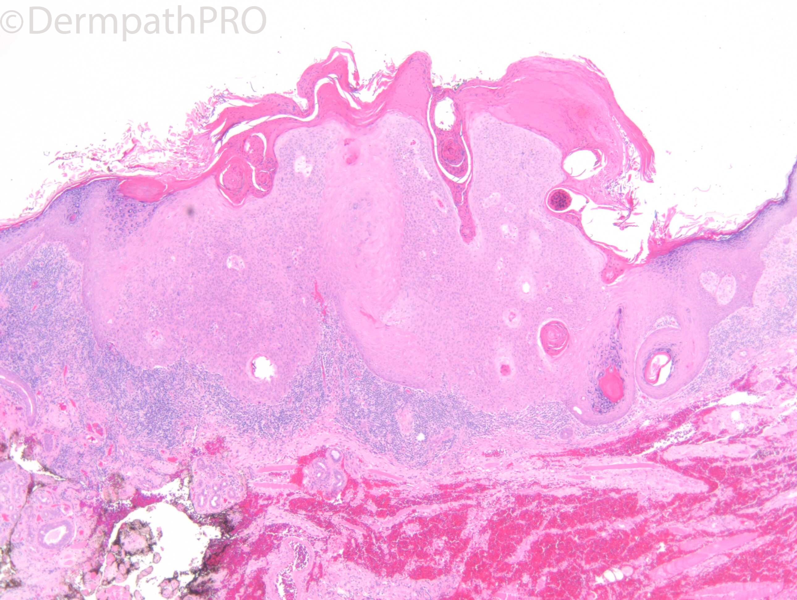
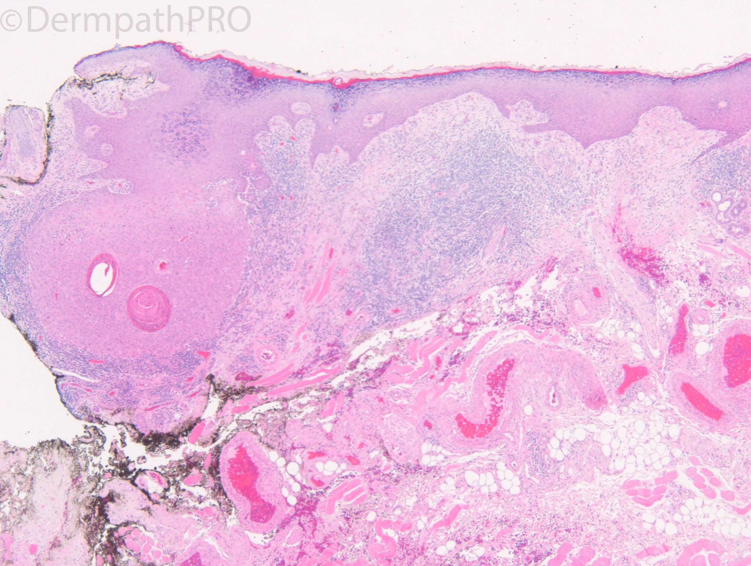
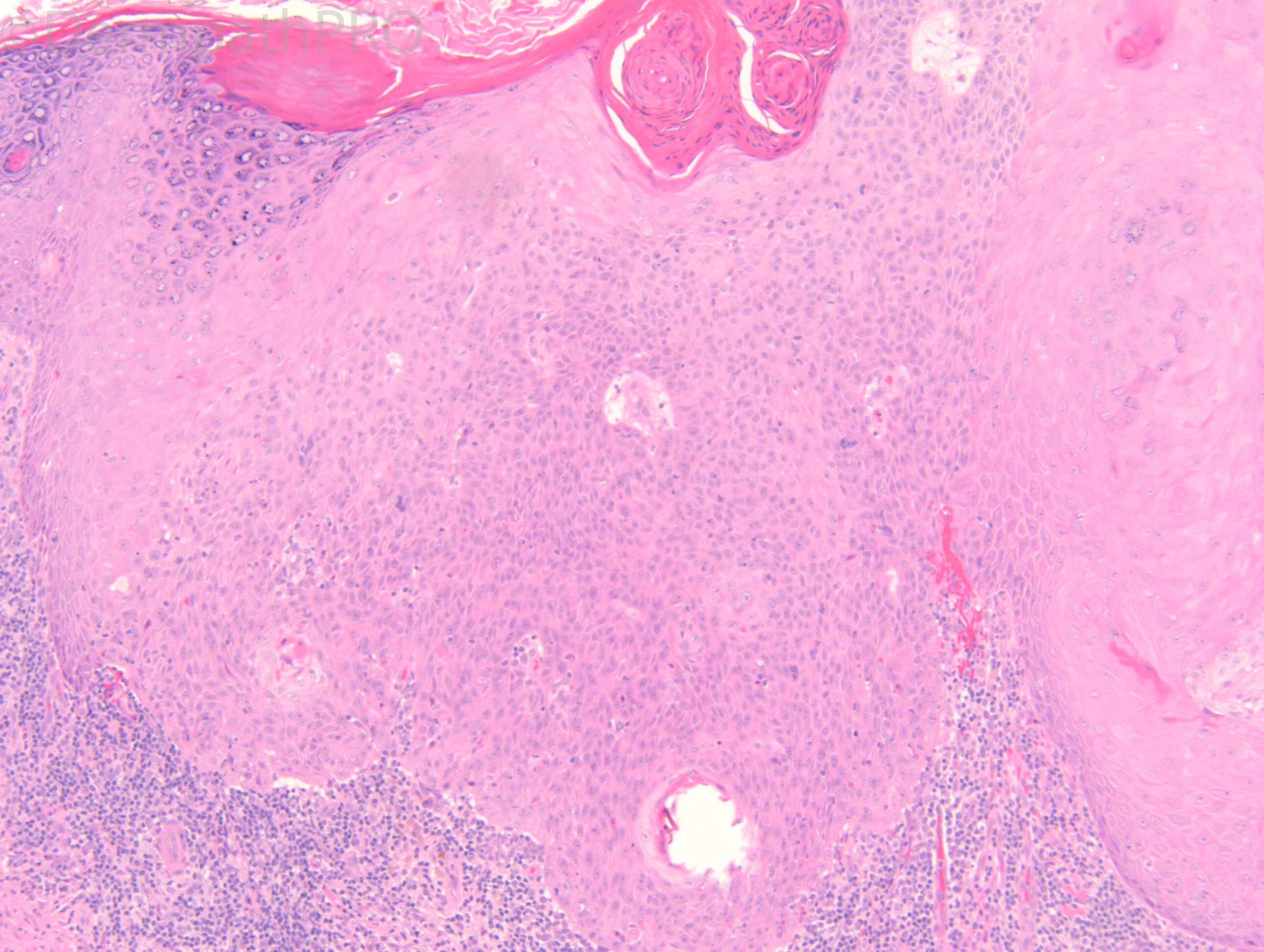
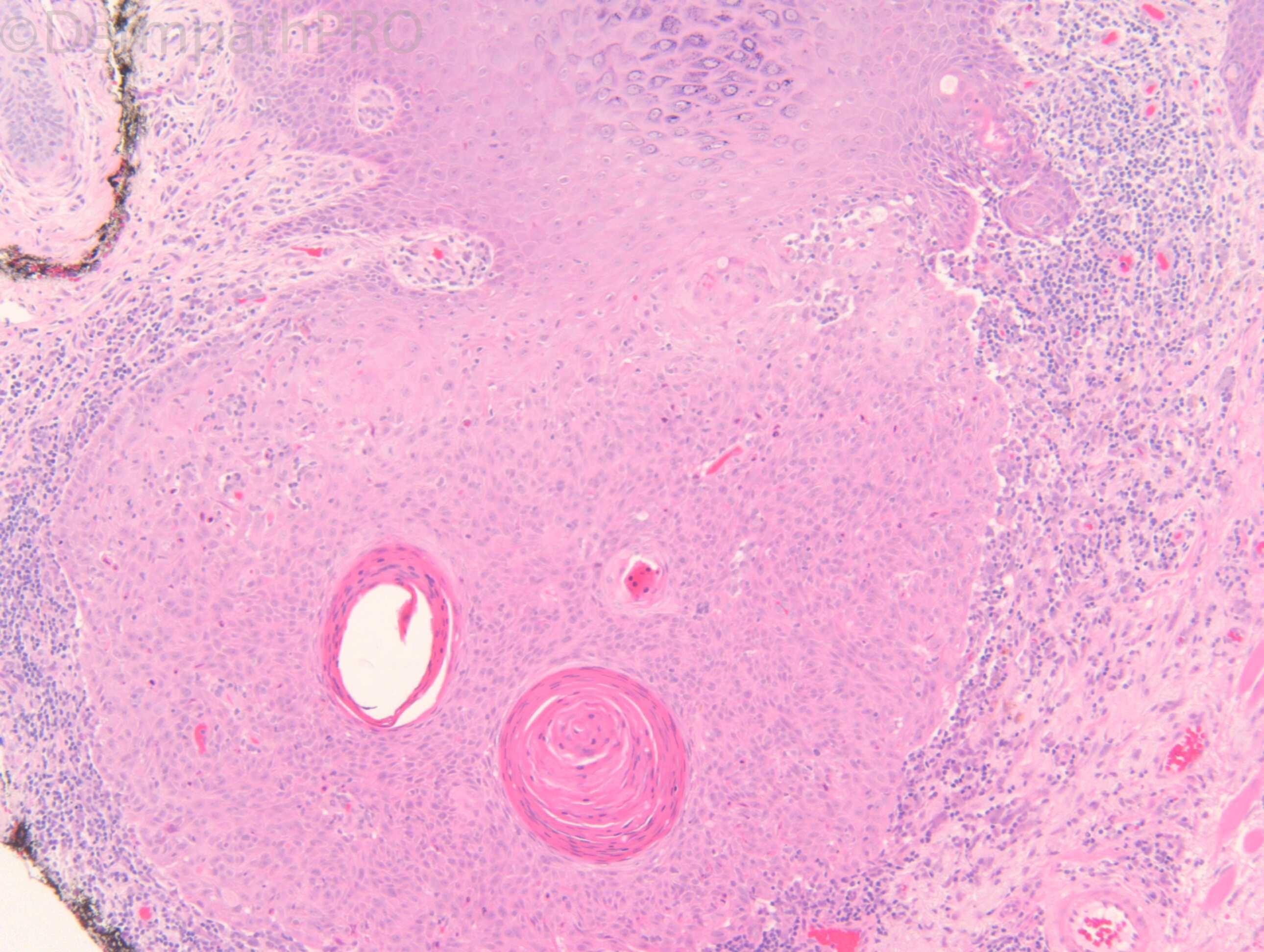
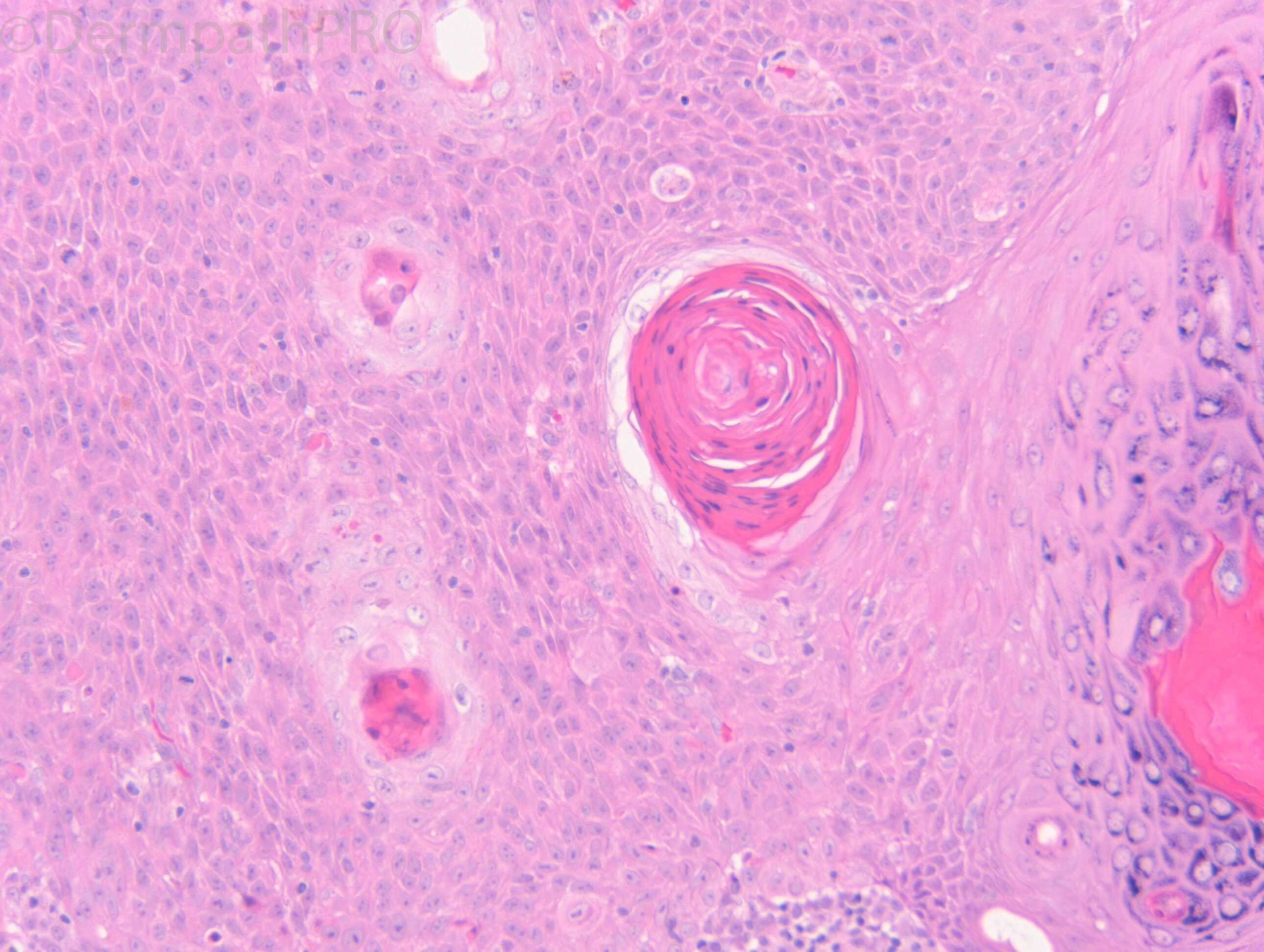
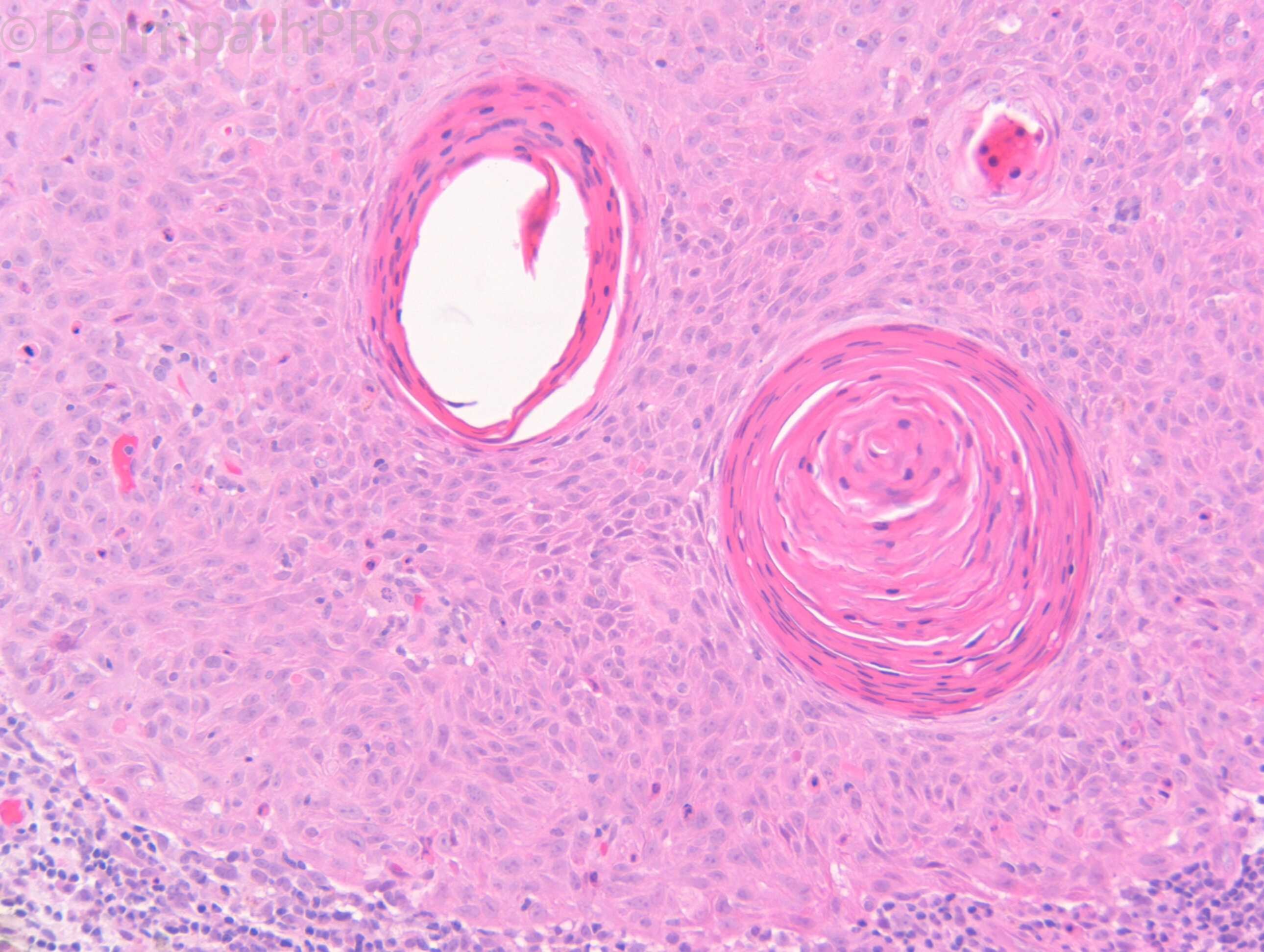
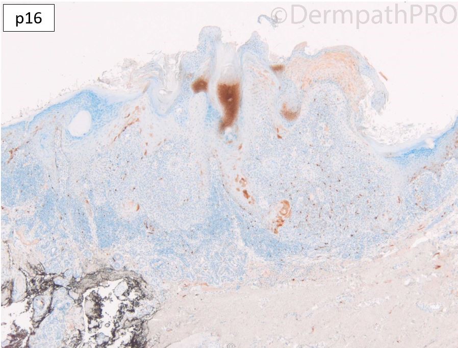
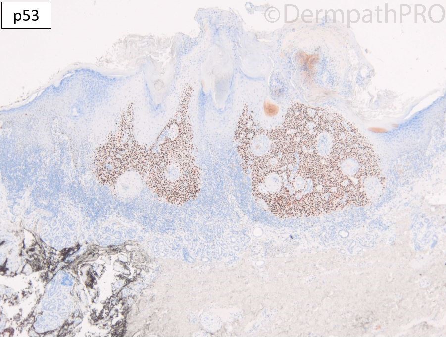
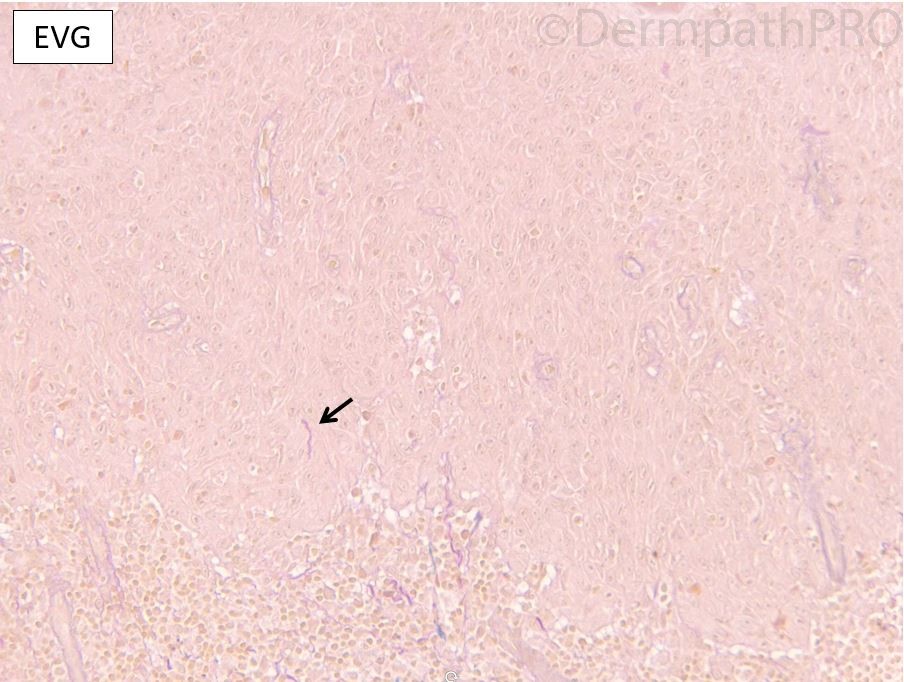
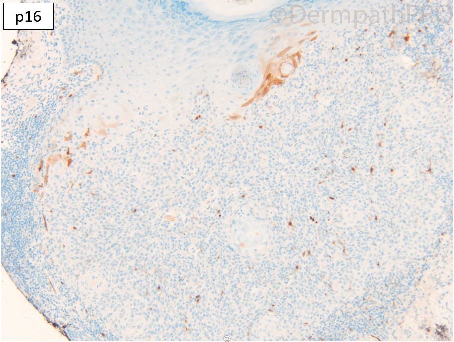
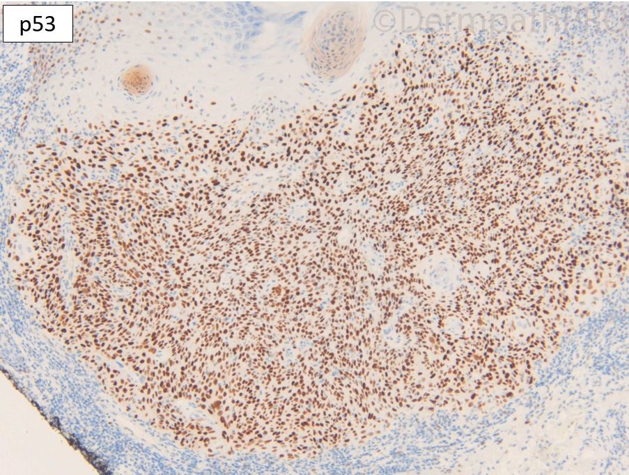
M85. Medial canthus. >1 year history, 7 x 6mm nodule, keratotic centre. ?SCC


  Report Record

User Feedback


Krishnakumar subramanian

Posted

sir i would think of keratotic squamous cell carcinoma, considering the age.

i would also consider seborrheic keratosis

Share this comment


Link to comment
share_externally

yuriy shatovsky

Posted

Inverted follicular keratosis .


Share this comment


Link to comment
share_externally

Meenakshi Batrani

Posted

I am favouring benign. Irritated SK/ IFK.

Share this comment


Link to comment
share_externally

vincenzo

Posted

Hypertrophic Actinic Keratosis. 

Share this comment


Link to comment
share_externally

Dr. Richard Carr

Posted

There is helpful IHC on this case. I'll ask for it to  be posted.


Share this comment


Link to comment
share_externally

vincenzo

Posted

Zonal pattern with Em/Eo and zonal pattern with p53. So it could really be a hypertrophic/Bowenoid Actinic Keratosis. 

Share this comment


Link to comment
share_externally

Dr. Richard Carr

Posted

Okay thanks. I think the resemblance to seborrhoeic keratosis (an infundibular tumour) here is more than a coincidence. As you know I believe most SCC show follicular infundibular-tricholemmal differentiation. In this case we have a tumour that is more toward the pure infundibular pattern. Conventional actinic keratosis are not as circumscript and unlike AK where you get orthokeratosis from the infundibular orifices in follicular AK/SCC you get parakeratosis which is the real feature that should concern you for in situ carcinoma here. EVG shows minimal entrapment of doubtful signficance although strictly speaking this may be due to early invasion. But based on small size and circumscript borders I'd report this lesion as having minimal risk for metastasis and we would not recommend routine follow-up for this lesion in isolation. p16 is a null (staining reactive melanocytes and mosaic in the reactive infundibular and epidermal areas) and p53 is aberrantly up-regulated (perhaps in compensation from the disturbed p16 pathway). I've been reviewing the literature extensively and it is known the p16/RB1 pathway is aberrant in most SCC it just seems not to have been given any diagnostic significance until now. So to summarise I'd call this a folliclar AK/SCC with minimal metastatic potential. I hope you are happy to keep seeing these types of cases because for me the p16/p53 panel is now routine in challenging SPL's including subtle dysplasia, clonal lesions, odd SEBK/IFK- and KA-like lesions.

Share this comment


Link to comment
share_externally

Krishnakumar subramanian

Posted

sir, thanks, bit complicated, anyway shall read these points care fully 

Share this comment


Link to comment
share_externally

tyelaine

Posted

Richard, thanks for the great case. Solves puzzles in the daily cases we encounter - like in this case, a very small follicular SCC. I am attempting these stains too for strange SPL and learning how to use them.

Share this comment


Link to comment
share_externally

msofopoulos

Posted

On 28/05/2021 at 10:45, Dr. Richard Carr said:

because for me the p16/p53 panel is now routine in challenging SPL's

Is there any bibliography on how you interpret these stains? Nathan Harvey's paper [Pathology (June 2013) 45(4), pp. 402–407] on p16 somehow clarifies p16 interpretation but how about p53?

Thanks


Share this comment


Link to comment
share_externally

Dr. Mona Abdel-Halim

Posted

On 28/05/2021 at 09:45, Dr. Richard Carr said:

Okay thanks. I think the resemblance to seborrhoeic keratosis (an infundibular tumour) here is more than a coincidence. As you know I believe most SCC show follicular infundibular-tricholemmal differentiation. In this case we have a tumour that is more toward the pure infundibular pattern. Conventional actinic keratosis are not as circumscript and unlike AK where you get orthokeratosis from the infundibular orifices in follicular AK/SCC you get parakeratosis which is the real feature that should concern you for in situ carcinoma here. EVG shows minimal entrapment of doubtful signficance although strictly speaking this may be due to early invasion. But based on small size and circumscript borders I'd report this lesion as having minimal risk for metastasis and we would not recommend routine follow-up for this lesion in isolation. p16 is a null (staining reactive melanocytes and mosaic in the reactive infundibular and epidermal areas) and p53 is aberrantly up-regulated (perhaps in compensation from the disturbed p16 pathway). I've been reviewing the literature extensively and it is known the p16/RB1 pathway is aberrant in most SCC it just seems not to have been given any diagnostic significance until now. So to summarise I'd call this a folliclar AK/SCC with minimal metastatic potential. I hope you are happy to keep seeing these types of cases because for me the p16/p53 panel is now routine in challenging SPL's including subtle dysplasia, clonal lesions, odd SEBK/IFK- and KA-like lesions.

Thank You Dr Carr. Looking forward to reading ur published IHC work for these lesions..

Share this comment


Link to comment
share_externally

Dr. Richard Carr

Posted

On 30/05/2021 at 16:21, msofopoulos said:

Is there any bibliography on how you interpret these stains? Nathan Harvey's paper [Pathology (June 2013) 45(4), pp. 402–407] on p16 somehow clarifies p16 interpretation but how about p53?

Thanks


I'm currently writing it up so hopefully you'll have a definitive reference in 2022. But basically the commonest abnormality in malignant tumours of sun-exposed skin is a complete loss of IHC staining for p16 (including ~70% of naevoid melanomas). This was known in SCC but appears to not have been used diagnostically. BAK can also show a null frequently (for p16 or p53). The strong diffuse pattern of p16 can be associated with high risk HPV associated Bowenoid lesions in non-sun-exposed skin. However the fascinating thing is that KA show striking mosaic pattern in the proliferative phase (and never null), similar to Spitz naevi, so it leads me to believe typical KA are probably benign lesions. Any KA-like SPL that has a complete knock-out of p16 or p53 by IHC should be viewed with extreme caution and removed completely. You may see striking up-regulation of p53 when p16 is highly aberrant, presumably a compensatory increase, but a diffuse strong p53 can also be a clue to a mutation which stabilises the protein. In KA and other benign SPL p53 shows a graded pattern i.e. strongest in the basal cells and progressively weaker towards the centre differentiated cells. Rarely do benign lesions show >50% moderate/strong staining for p53. 

Share this comment


Link to comment
share_externally

msofopoulos

Posted

On 31/05/2021 at 20:14, Dr. Richard Carr said:

I'm currently writing it up so hopefully you'll have a definitive reference in 2022

Thanks! it would be extremely helpful!

Share this comment


Link to comment
share_externally


Join the conversation

You can post now and register later. If you have an account, sign in now to post with your account.

Guest
Add a comment...

×   Pasted as rich text.   Paste as plain text instead

  Only 75 emoji are allowed.

×   Your link has been automatically embedded.   Display as a link instead

×   Your previous content has been restored.   Clear editor

×   You cannot paste images directly. Upload or insert images from URL.

Loading...

×
×
  • Create New...