Jump to content
In this section we have spot diagnoses posted on a daily basis since June 2010, now over 4000! You can review the archived cases and read the suggested diagnoses by users and the final comment by the contributors.
Case are uploaded each week day by 10 am UK time with the correct diagnosis will generally be posted at 8 pm UK time. Why not view the most recent spot diagnosis and proffer a diagnosis?

Case Number : Case 2842- 28 May 2021 Posted By: Dr. Richard Carr

Please read the clinical history and view the images by clicking on them before you proffer your diagnosis.
Submitted Date :
   (0 reviews)

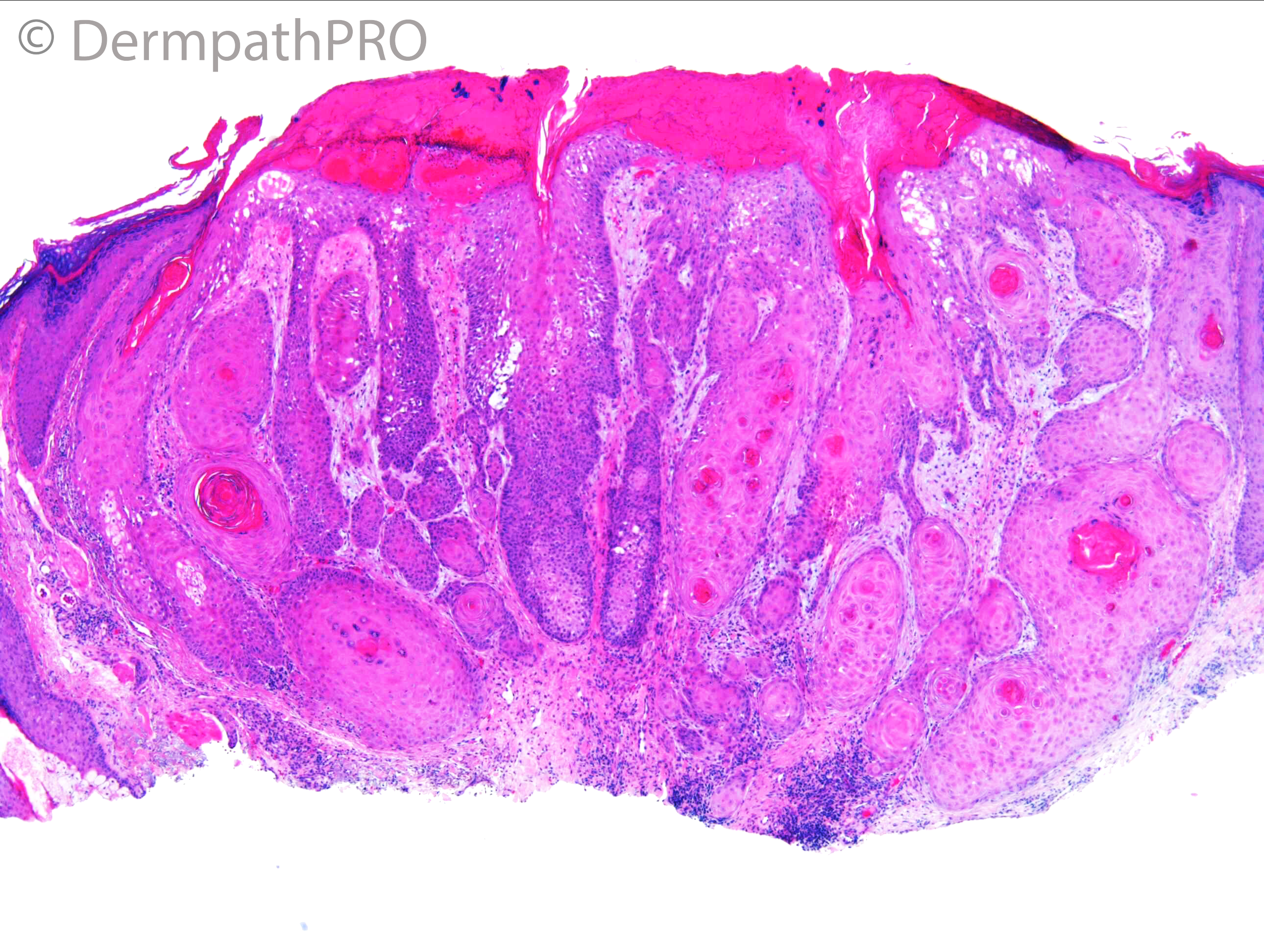
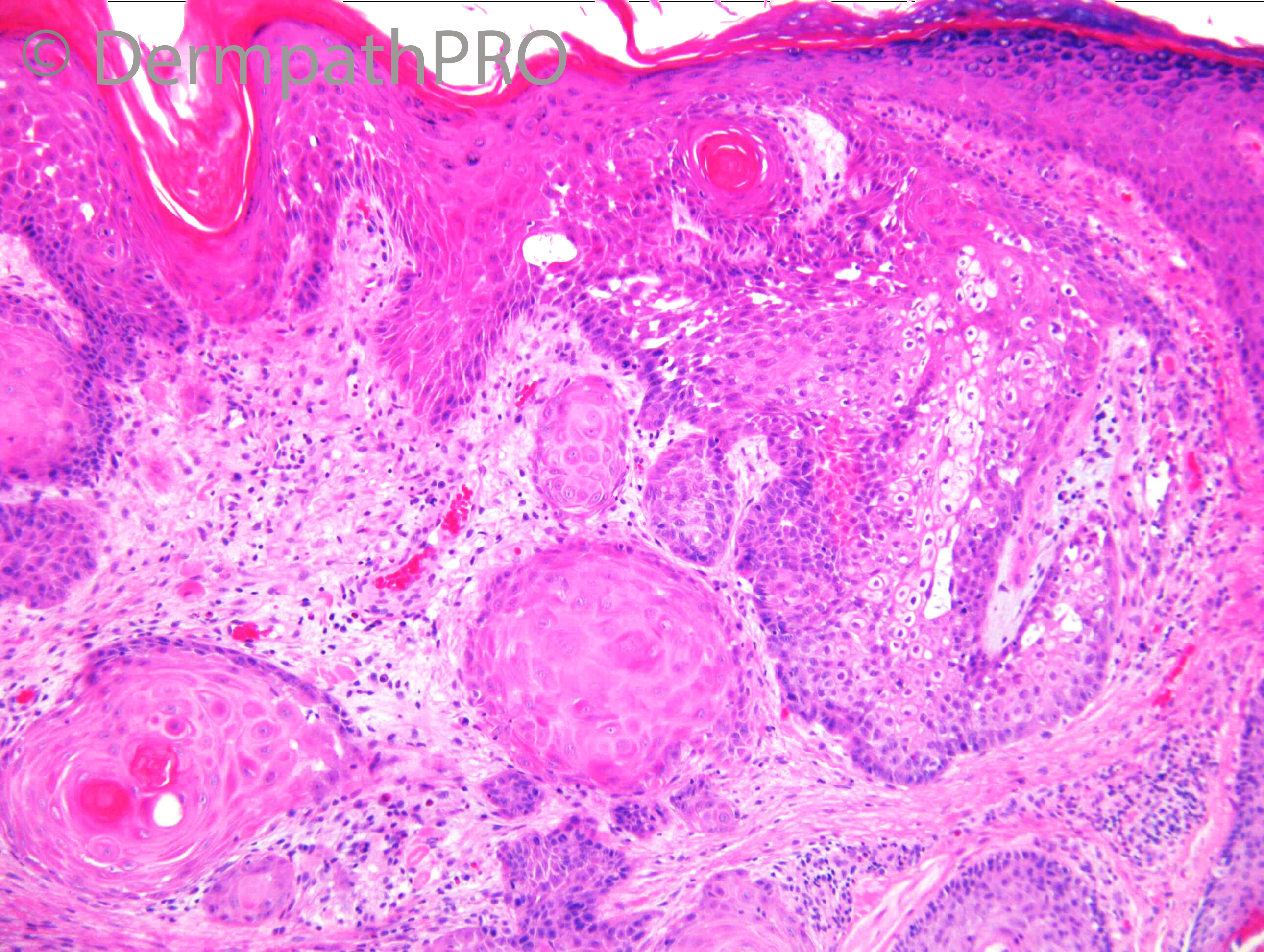
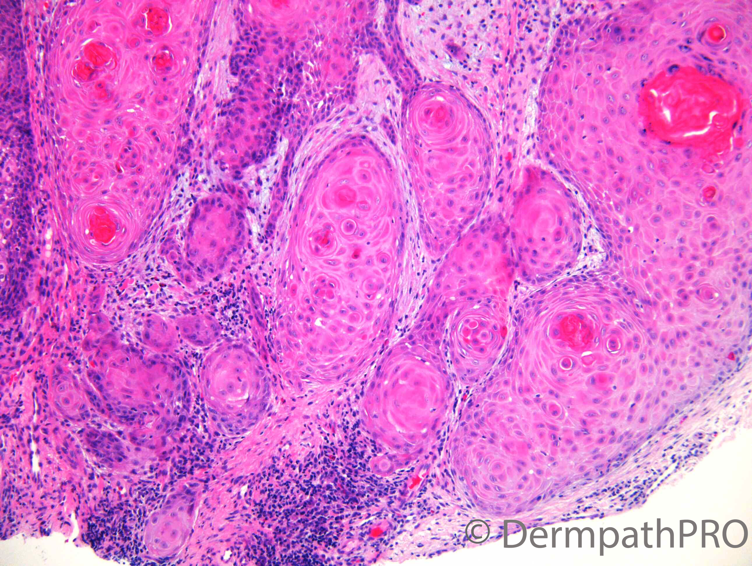
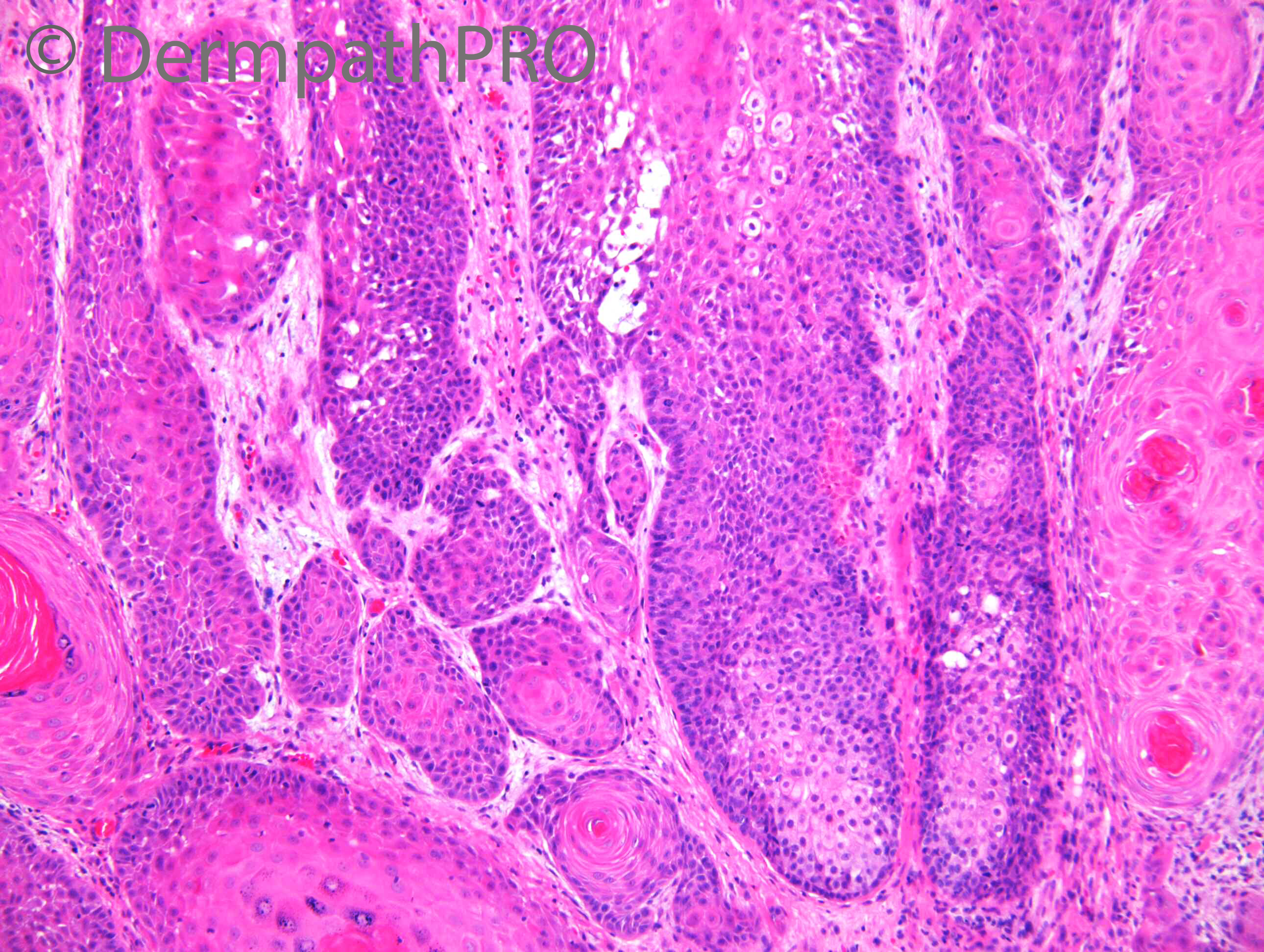
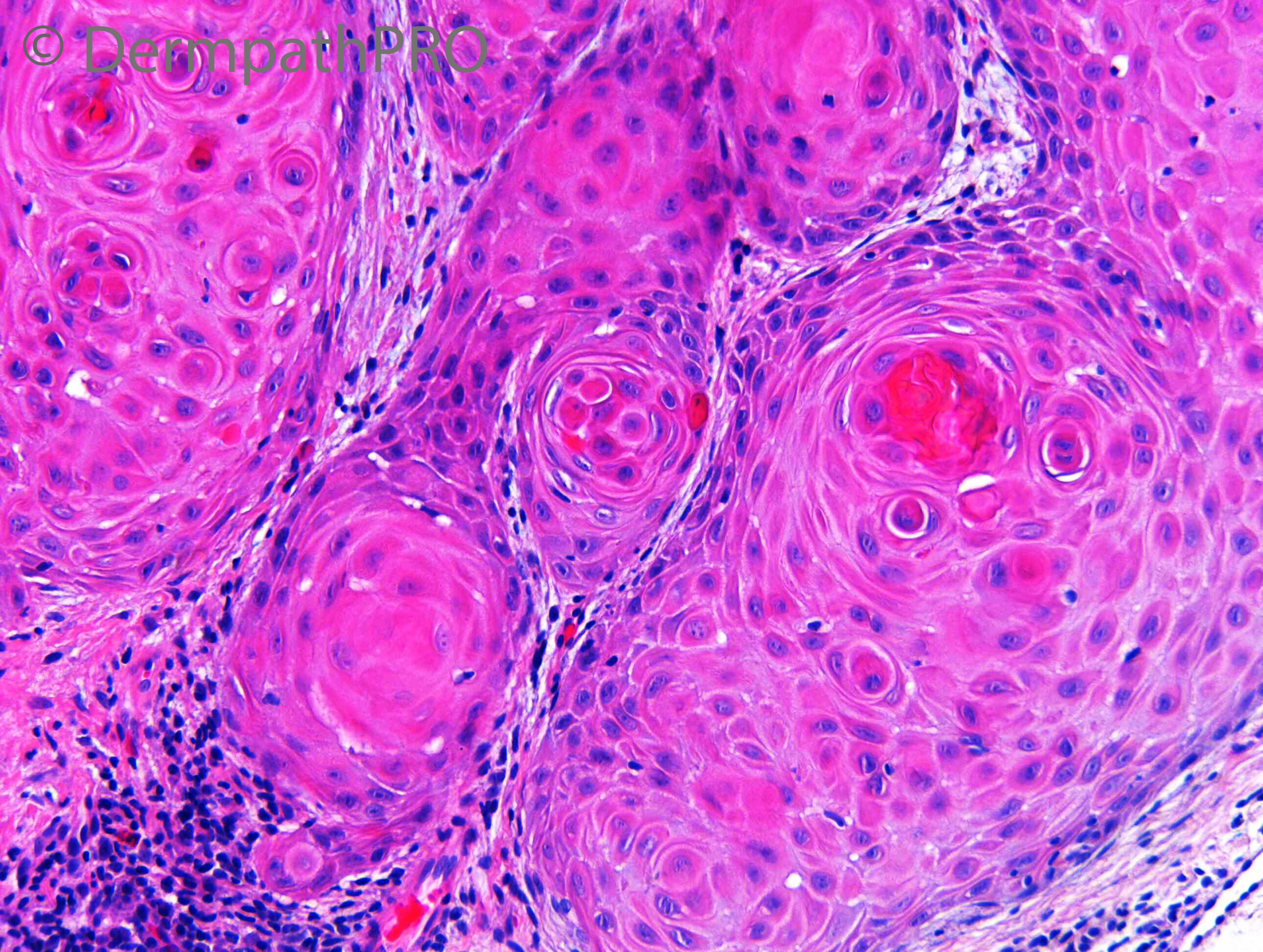
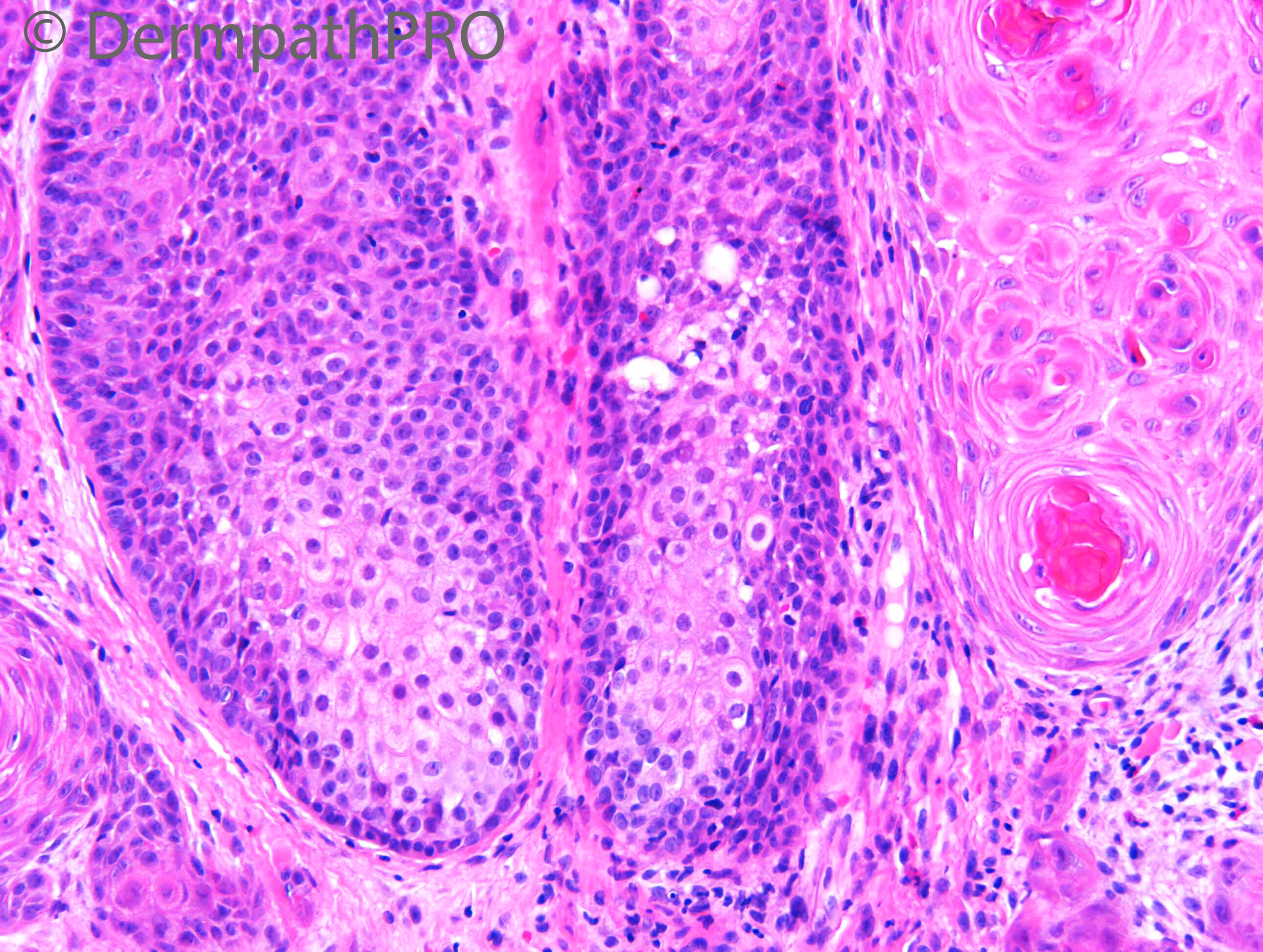
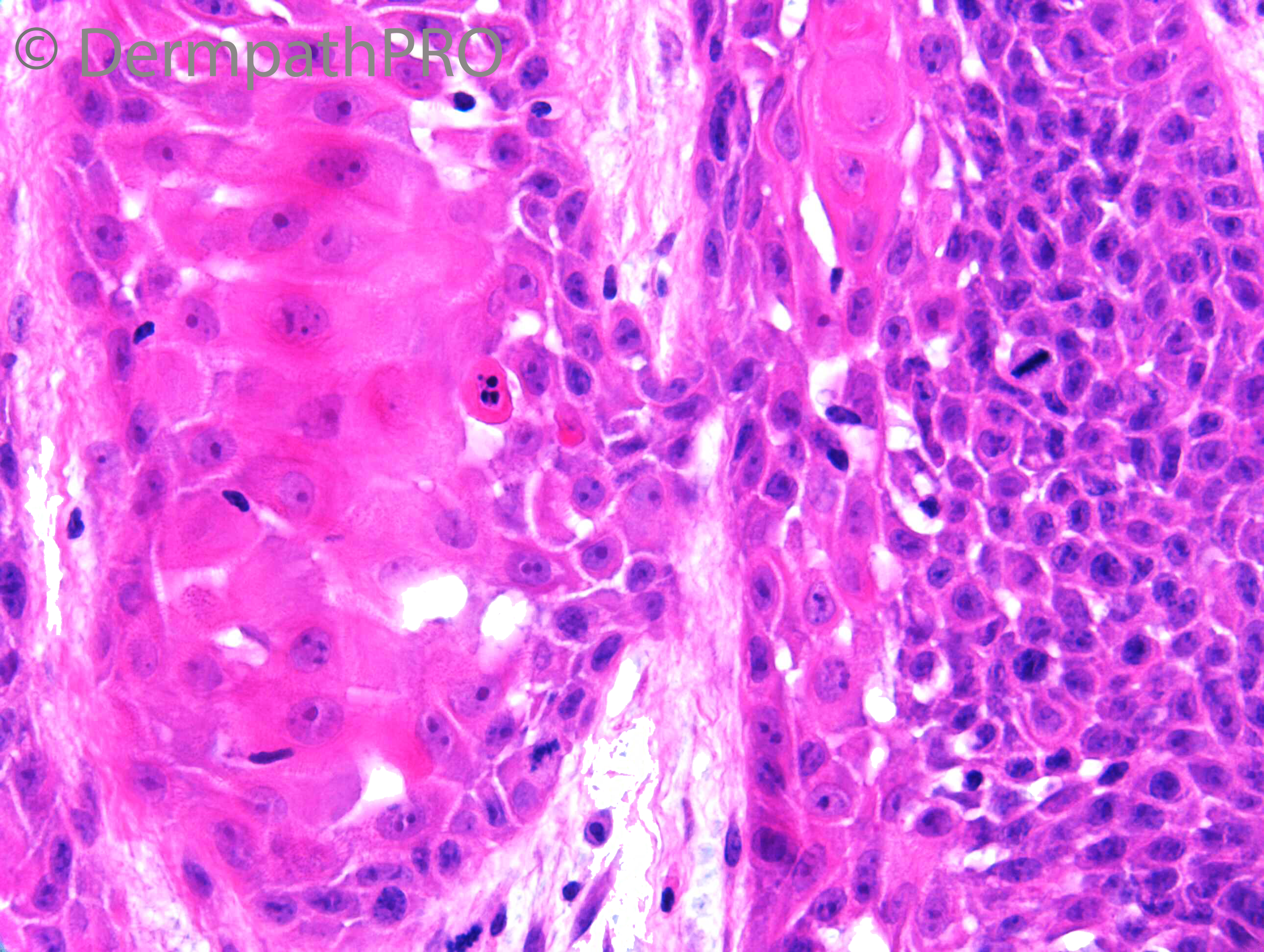
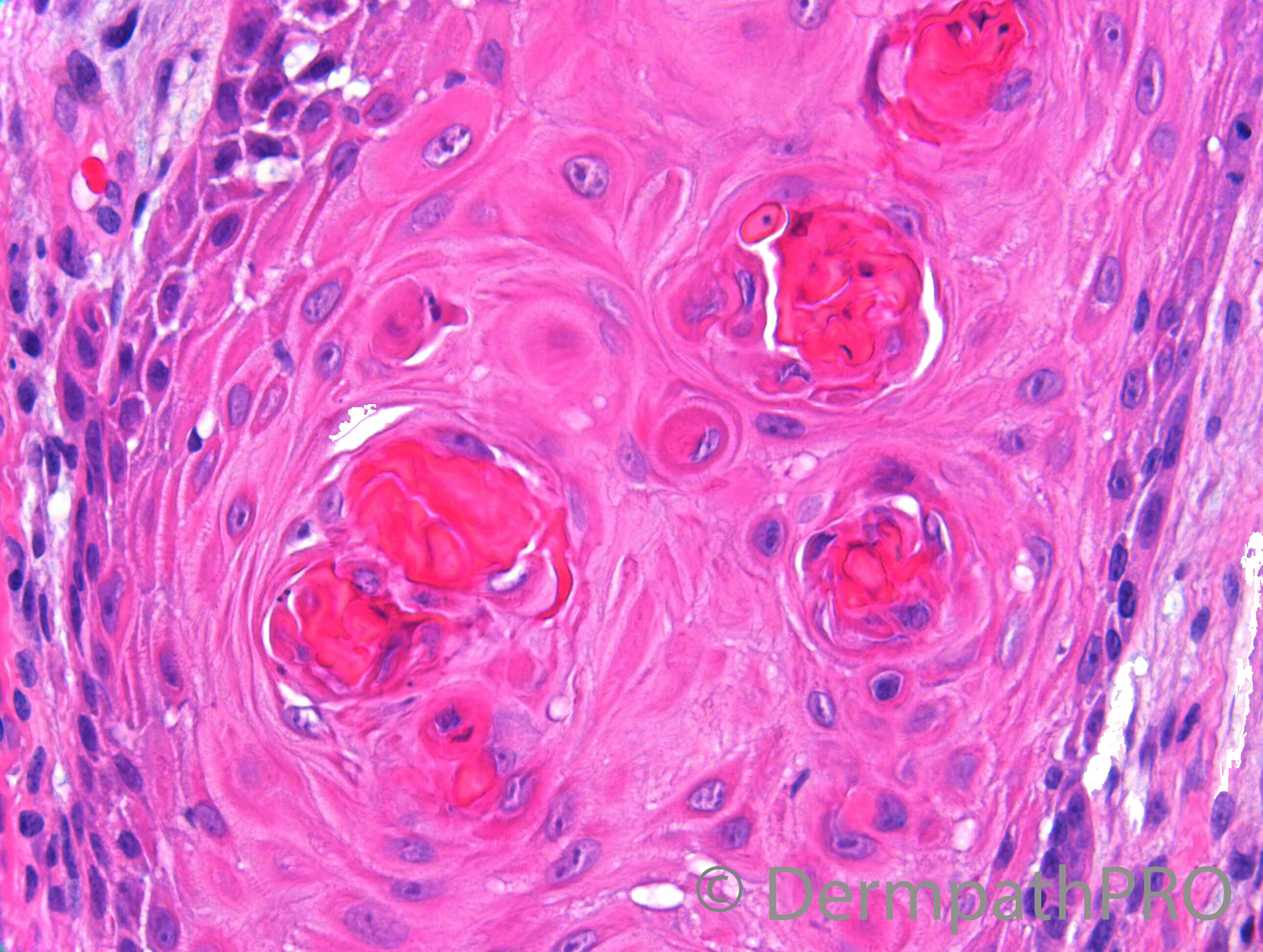
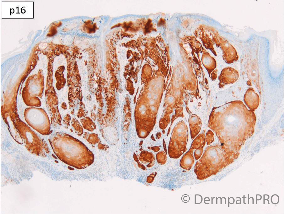
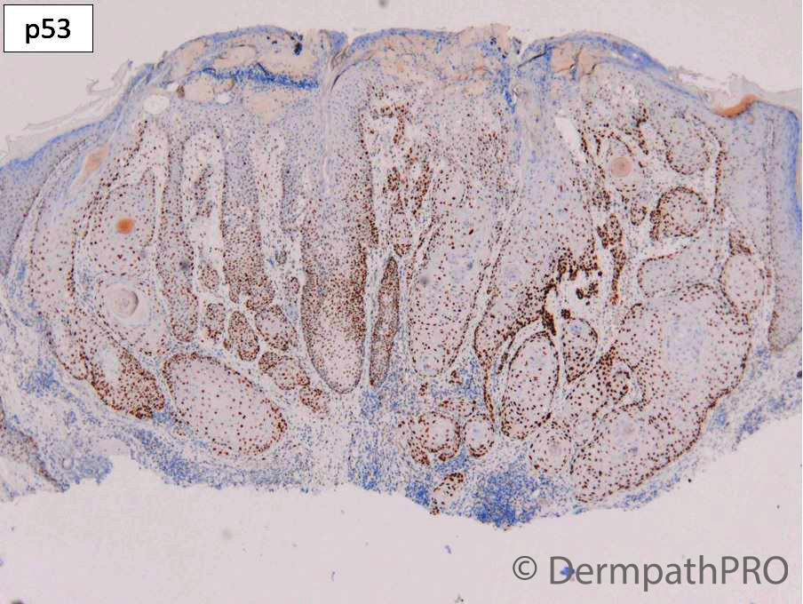
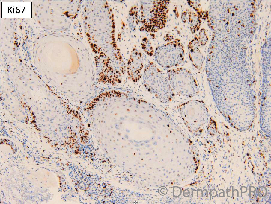
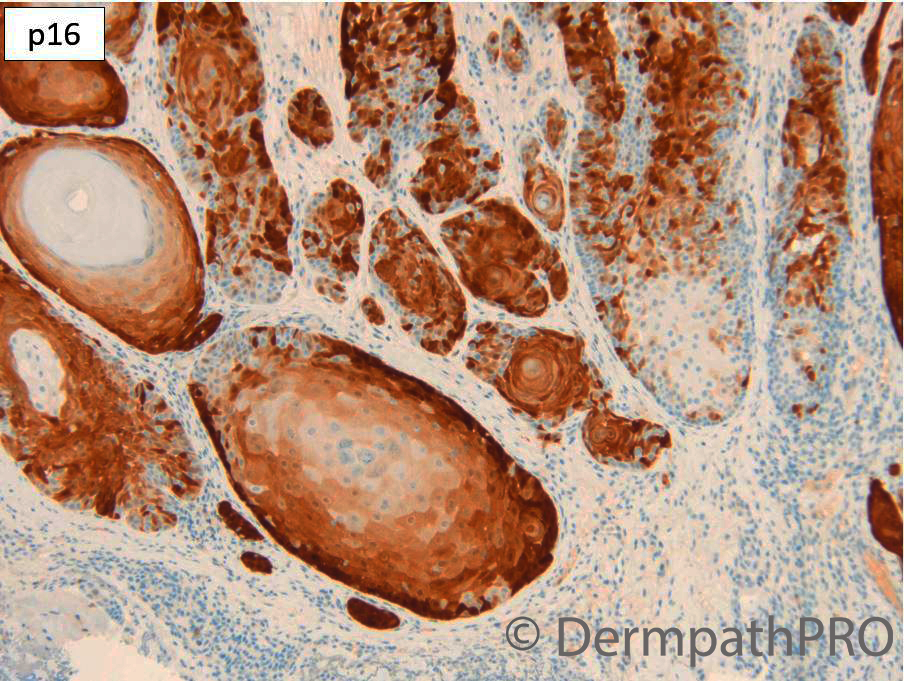
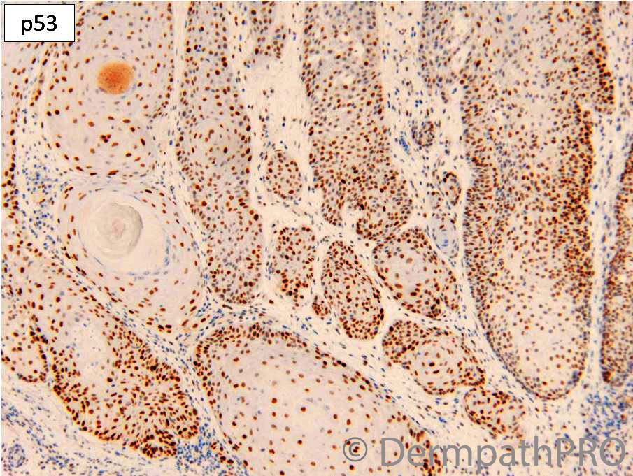
M65. Has had cholangiocarcinoma and urothelial carcinoma.


  Report Record

User Feedback


Krishnakumar subramanian

Posted

Sir, No comments, you are ultimate, i will try and learn

Share this comment


Link to comment
share_externally

Krishnakumar subramanian

Posted

P53 is increased, P16 nuclear and cytoplasmic positive- invasive follicular squamous cell carcinoma

Share this comment


Link to comment
share_externally

vincenzo

Posted

p53 > but p16 isn’t null as expected in fSCC, so it’s difficult , for me, making a diagnosis based on these markers. Not sure, but...how about a proliferating pilar tumor?

Share this comment


Link to comment
share_externally

Dr. Richard Carr

Posted

I'll wait for a few more comments

Share this comment


Link to comment
share_externally

Dr. Richard Carr

Posted

Those brave soles who did comment did not note the sebaceous differentiation or relative lack of significant cellular plemorphism or the rather organoid architecture. The patient has Muir Torre syndrome (MTS) and has had many sebaceous adenomas. I concluded this was sebaceous adenoma with striking squamous differentiation (probably from the sebaceous duct) i.e. a seboacanthoma. I could not find much on google but according to wikipedia! these types of lesions may be specific to MTS. p16 is up-regulated mosaic (wild type) and p53 shows up-regulated wild type (nice gradation in staining from stronger peripheral to weak and negative centrally - as discussed last week). The lesion had been seen by two other pathologists who had favoured a well differentiated squamous cell carcinoma

Share this comment


Link to comment
share_externally

vincenzo

Posted

Yes. Now I’m seeing the nice sebaceous diff. Thanks, Richard. I was off the track, with my guess!

Share this comment


Link to comment
share_externally

Krishnakumar subramanian

Posted

yes we are able to appreciate the sebaceous component

Share this comment


Link to comment
share_externally


Join the conversation

You can post now and register later. If you have an account, sign in now to post with your account.

Guest
Add a comment...

×   Pasted as rich text.   Paste as plain text instead

  Only 75 emoji are allowed.

×   Your link has been automatically embedded.   Display as a link instead

×   Your previous content has been restored.   Clear editor

×   You cannot paste images directly. Upload or insert images from URL.

Loading...

×
×
  • Create New...