Jump to content
In this section we have spot diagnoses posted on a daily basis since June 2010, now over 4000! You can review the archived cases and read the suggested diagnoses by users and the final comment by the contributors.
Case are uploaded each week day by 10 am UK time with the correct diagnosis will generally be posted at 8 pm UK time. Why not view the most recent spot diagnosis and proffer a diagnosis?

Case Number : Case 2966 - 18 November 2021 Posted By: Saleem Taibjee

Please read the clinical history and view the images by clicking on them before you proffer your diagnosis.
Submitted Date :
   (0 reviews)

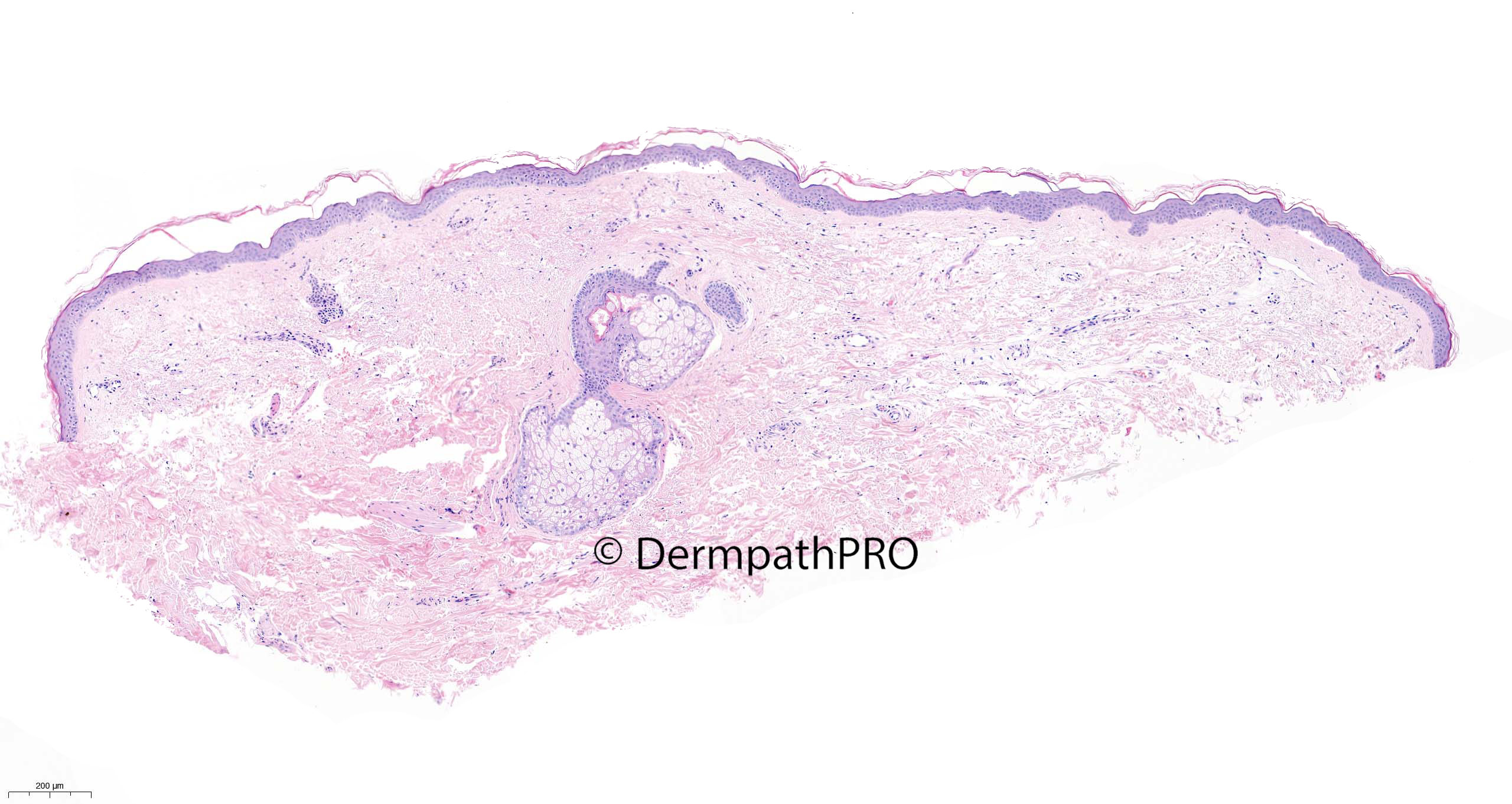
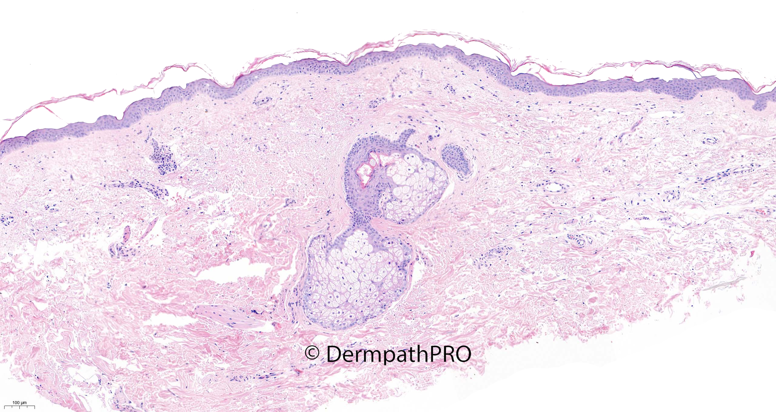
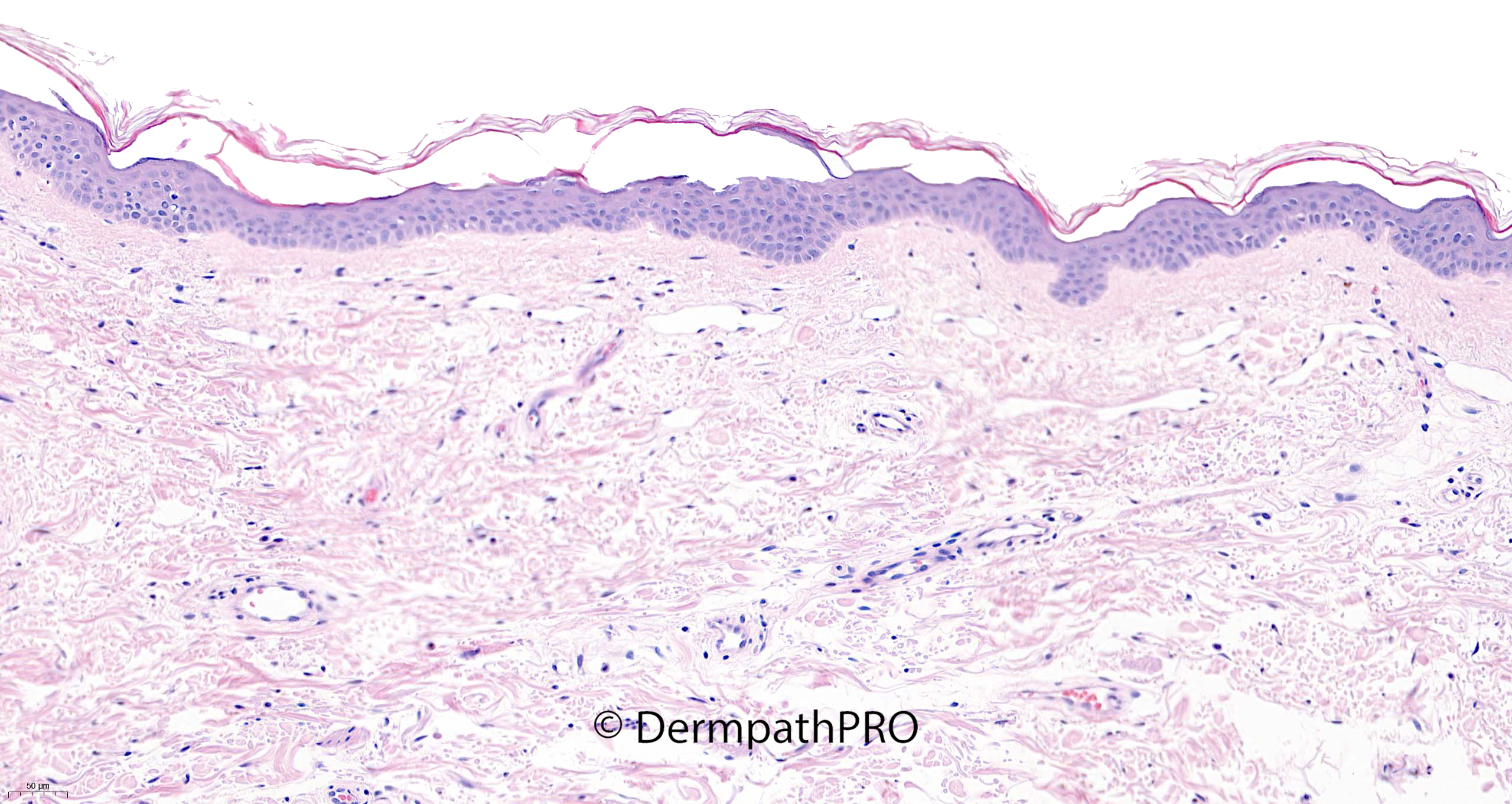
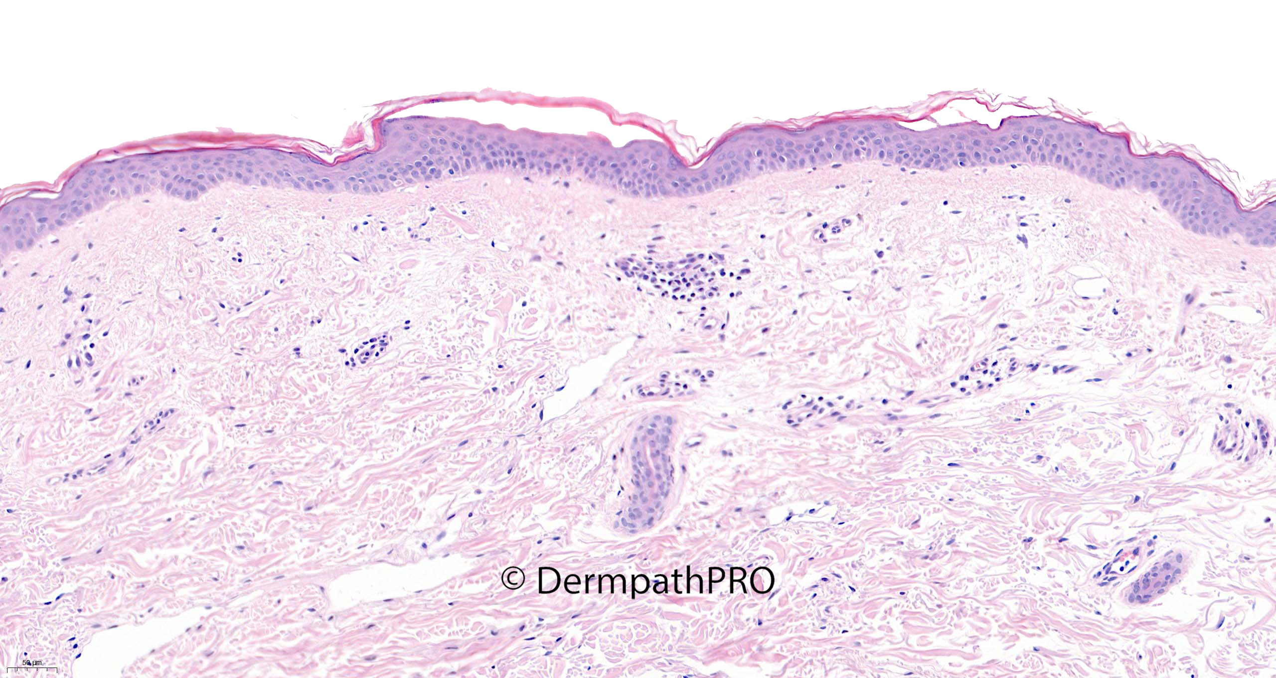
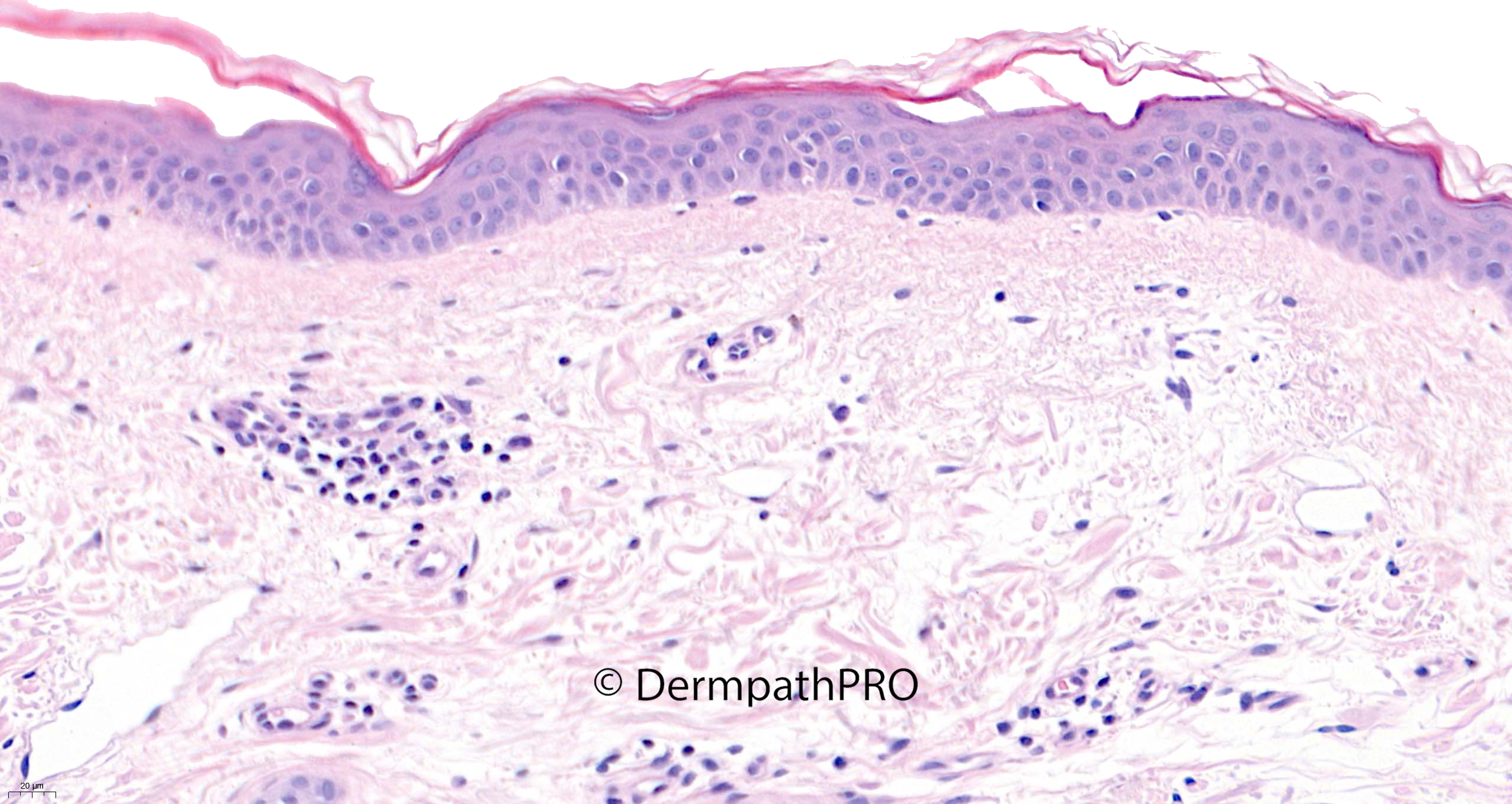
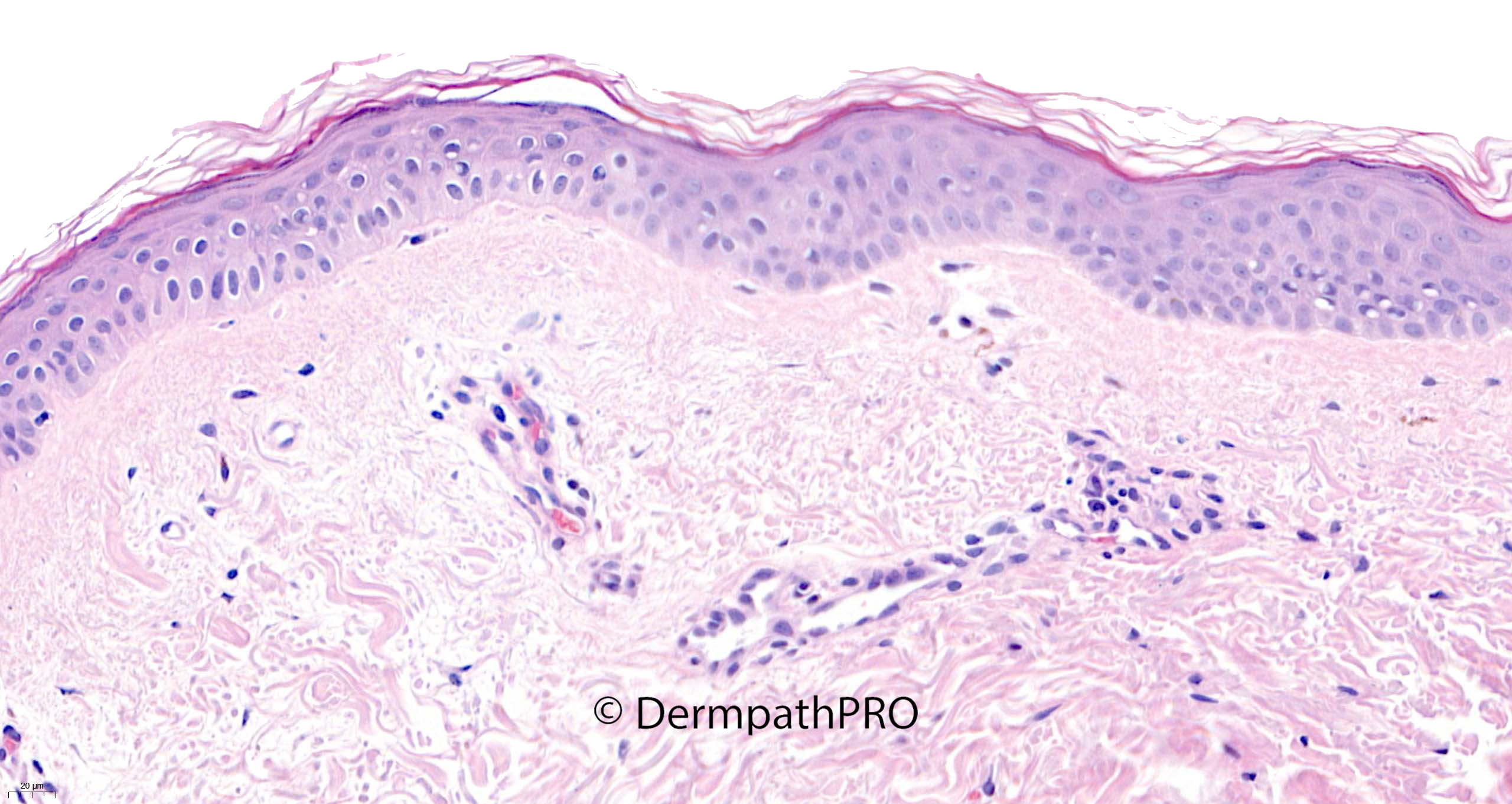
56F, punch biopsy right side of neck. White macules with slight atrophy on face, neck and body


  Report Record

User Feedback


Anil Patki

Posted (edited)

Pseudoxanthoma elasticum (PXE) and PXE- like papillary dermal elastolysis are the main differentials here. Clinicopathological correlation after staining for elastic fibres and von Kossa stain to detect  calcification of degraded elastic fibres will help in arrival at the diagnosis. 

Edited by Anil Patki

Share this comment


Link to comment
share_externally

Meenakshi Batrani

Posted

Would consider PXE-like papillary dermal elastolysis or white fibrous papulosis of neck- both related disorders. Elastic stains and CPC needed. 

Share this comment


Link to comment
share_externally

Cem Leblebici

Posted

I think there are no melanocytes in epidermis. First, I would like to perform SOX10. 

Share this comment


Link to comment
share_externally

Krishnakumar subramanian

Posted

I thought of LSA with evidence of actinic damage, but widespread LSA is rare, 

new thing i learnt

Share this comment


Link to comment
share_externally

Richard Logan

Posted

I agree with Anil and Meenakshi.  I think this is PXE-like papillary dermal elastolysis.  The gender and age of the patient are typical.

Share this comment


Link to comment
share_externally

Without clinical features my first spot had been TMEP mastocytosis, but…waiting for final answer. 

Share this comment


Link to comment
share_externally

Richard Logan

Posted

51 minutes ago, daniellindsay said:

I agree with possible TMEP. Need IHC

TMEP doesn't fit with the clinical details.

Share this comment


Link to comment
share_externally

Saleem Taibjee

Posted

I was being a bit deceitful. I did not mention that the clinician queried vitiligo versus lichen sclerosus. 

As Cem astutely points out, there are no melanocytes in the basal layer. This is further confirmed by Melan-A (see below). Hence I did think this is very likely to be vitiligo, another case we can add to the differential diagnosis of 'invisible dermatoses'. Of course, this is usually a clinical diagnosis and rarely biopsied.

I have checked back on the slide, and I do feel that the elastic is within normal limits in this case. So I don't think this is PXE-like papillary dermal elastolysis or other disorder of elastic fibres (although good suggestions), and similarly I cannot see any increase in mast cells.

BW
Saleem

60676_20.0x Melan-A labelled.jpg

Share this comment


Link to comment
share_externally

Richard Logan

Posted (edited)

I accept the loss of melanocytes, but how do you explain the "slight atrophy"?

Edited by Richard Logan
Grammar.

Share this comment


Link to comment
share_externally

Saleem Taibjee

Posted

I can't readily explain the atrophy noted clinically. But I am not really seeing any histological counterpart (the epidermis looks of normal thickness). I suppose topical steroid could be a factor if a history of prolonged usage. I think it may be a red herring.

Share this comment


Link to comment
share_externally


Join the conversation

You can post now and register later. If you have an account, sign in now to post with your account.

Guest
Add a comment...

×   Pasted as rich text.   Paste as plain text instead

  Only 75 emoji are allowed.

×   Your link has been automatically embedded.   Display as a link instead

×   Your previous content has been restored.   Clear editor

×   You cannot paste images directly. Upload or insert images from URL.

Loading...

×
×
  • Create New...