Jump to content
In this section we have spot diagnoses posted on a daily basis since June 2010, now over 4000! You can review the archived cases and read the suggested diagnoses by users and the final comment by the contributors.
Case are uploaded each week day by 10 am UK time with the correct diagnosis will generally be posted at 8 pm UK time. Why not view the most recent spot diagnosis and proffer a diagnosis?

Case Number : Case 2971 - 25 November 2021 Posted By: Saleem Taibjee

Please read the clinical history and view the images by clicking on them before you proffer your diagnosis.
Submitted Date :
   (0 reviews)

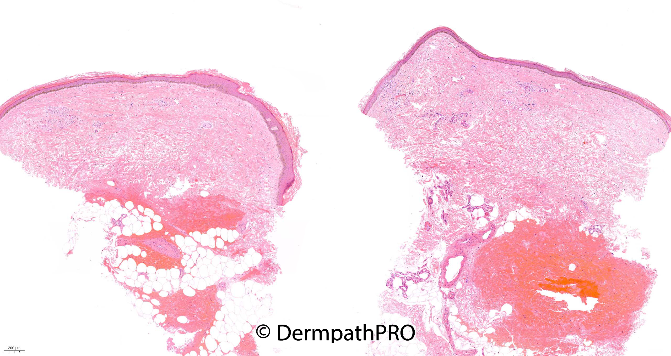
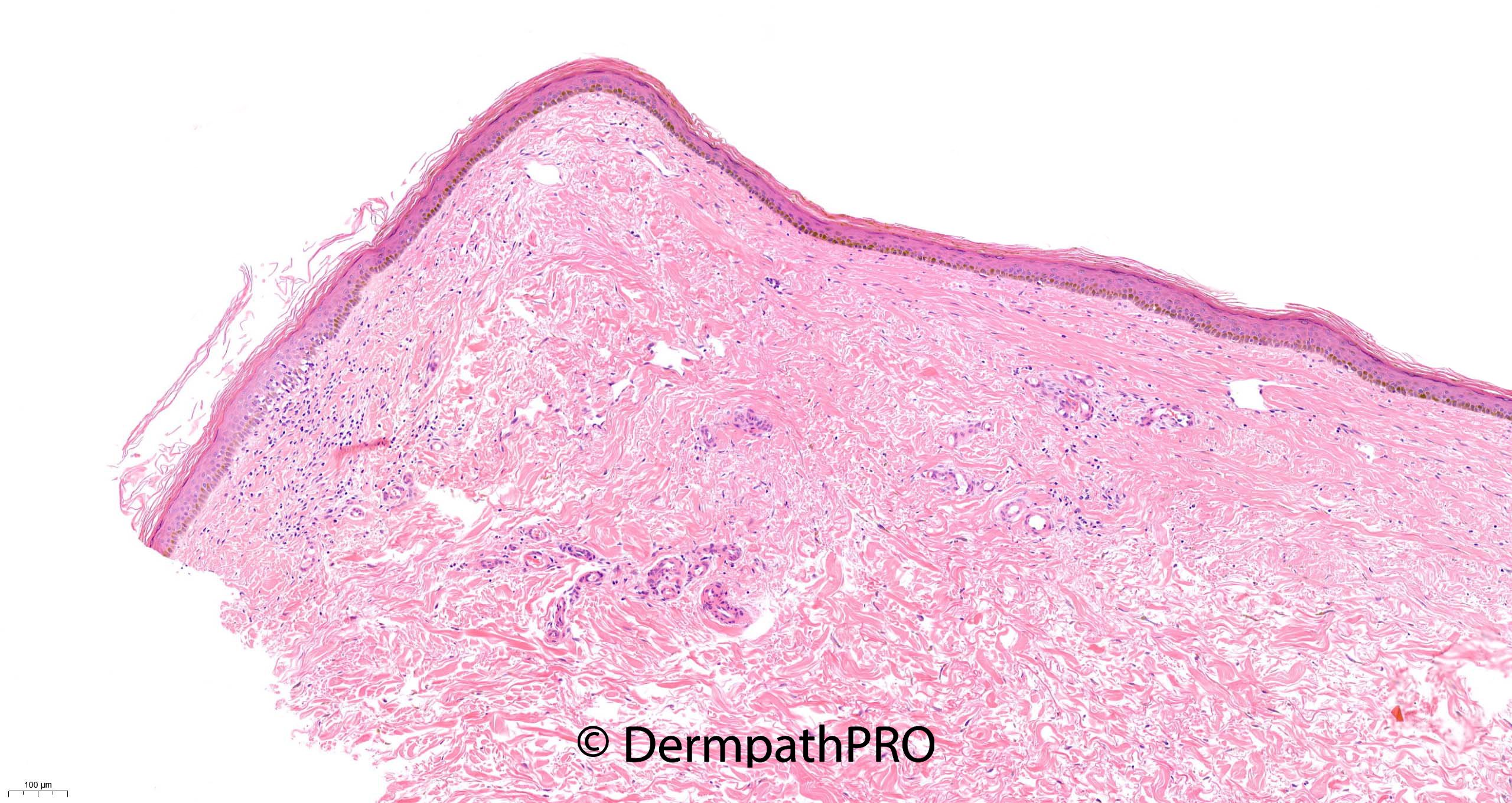
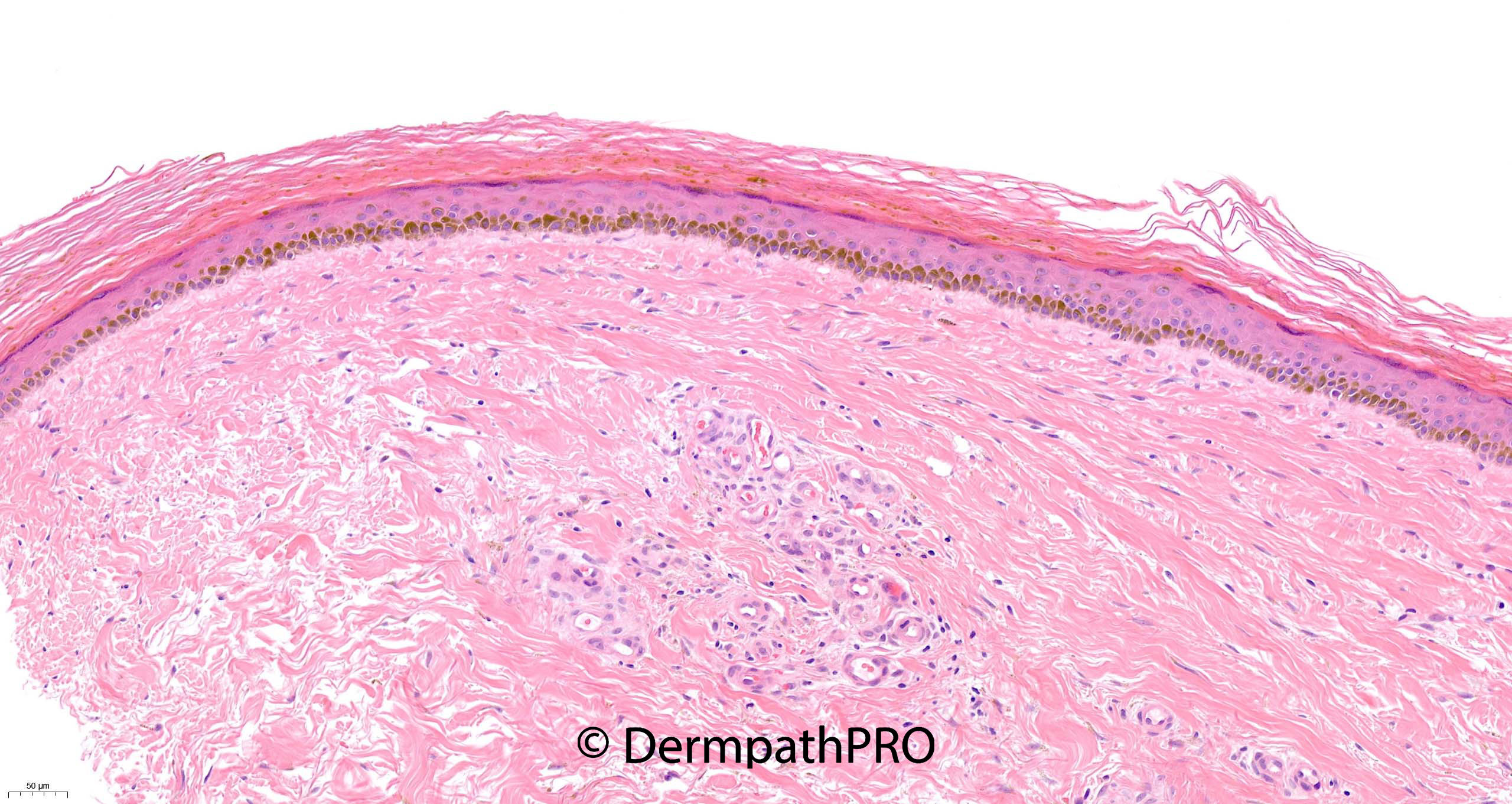
62F punch biopsy right lower leg ?skin spots ?acanthosis nigricans ?necrobiosis lipoidica


  • Thanks 1
  Report Record

User Feedback


daniellindsay

Posted (edited)

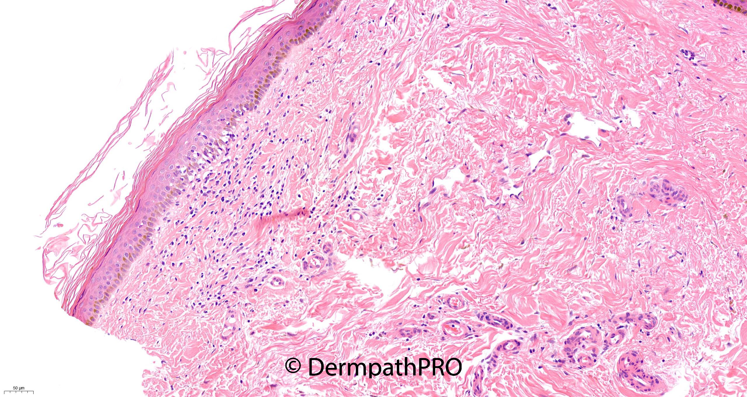
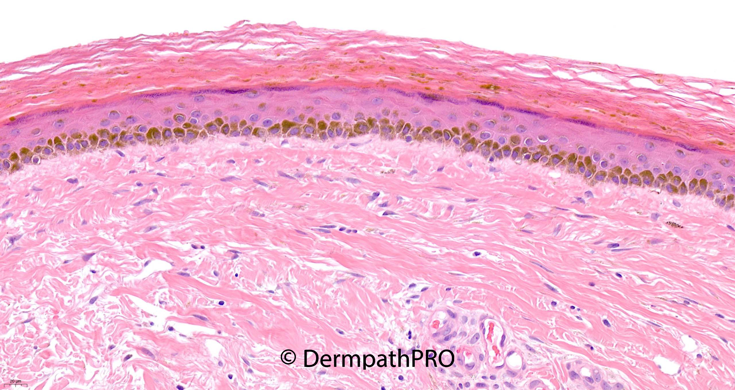
There is mild lichenoid inflammation with pigment incontinence. ?lichen nitidus - but doesnt fit very well clinically

Edited by daniellindsay

Share this comment


Link to comment
share_externally

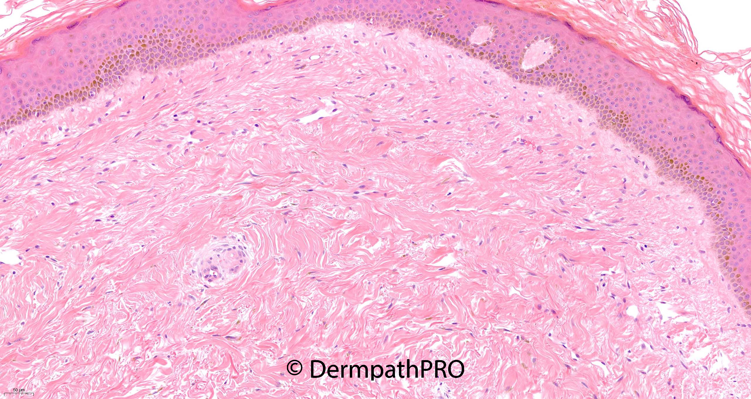
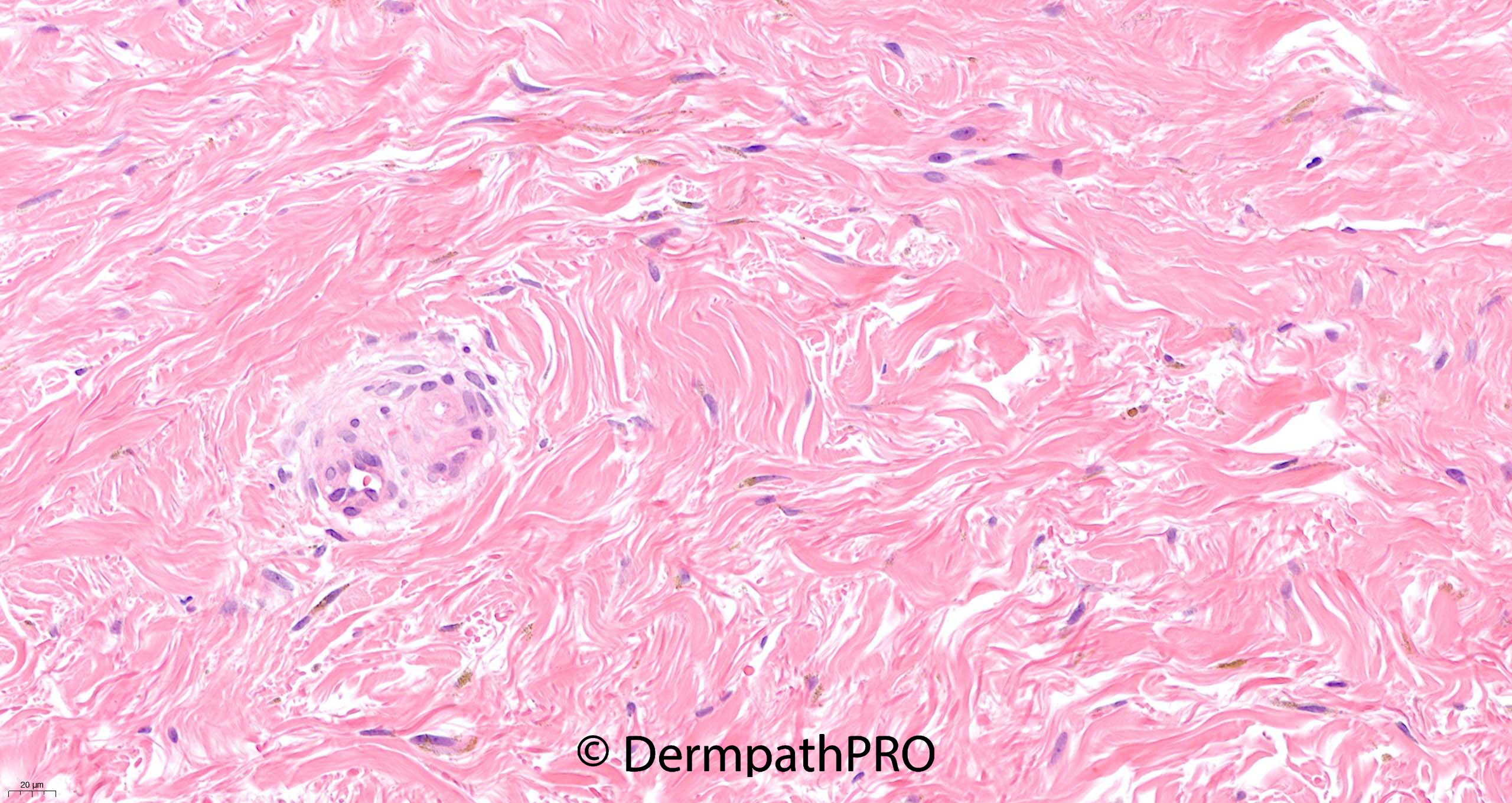
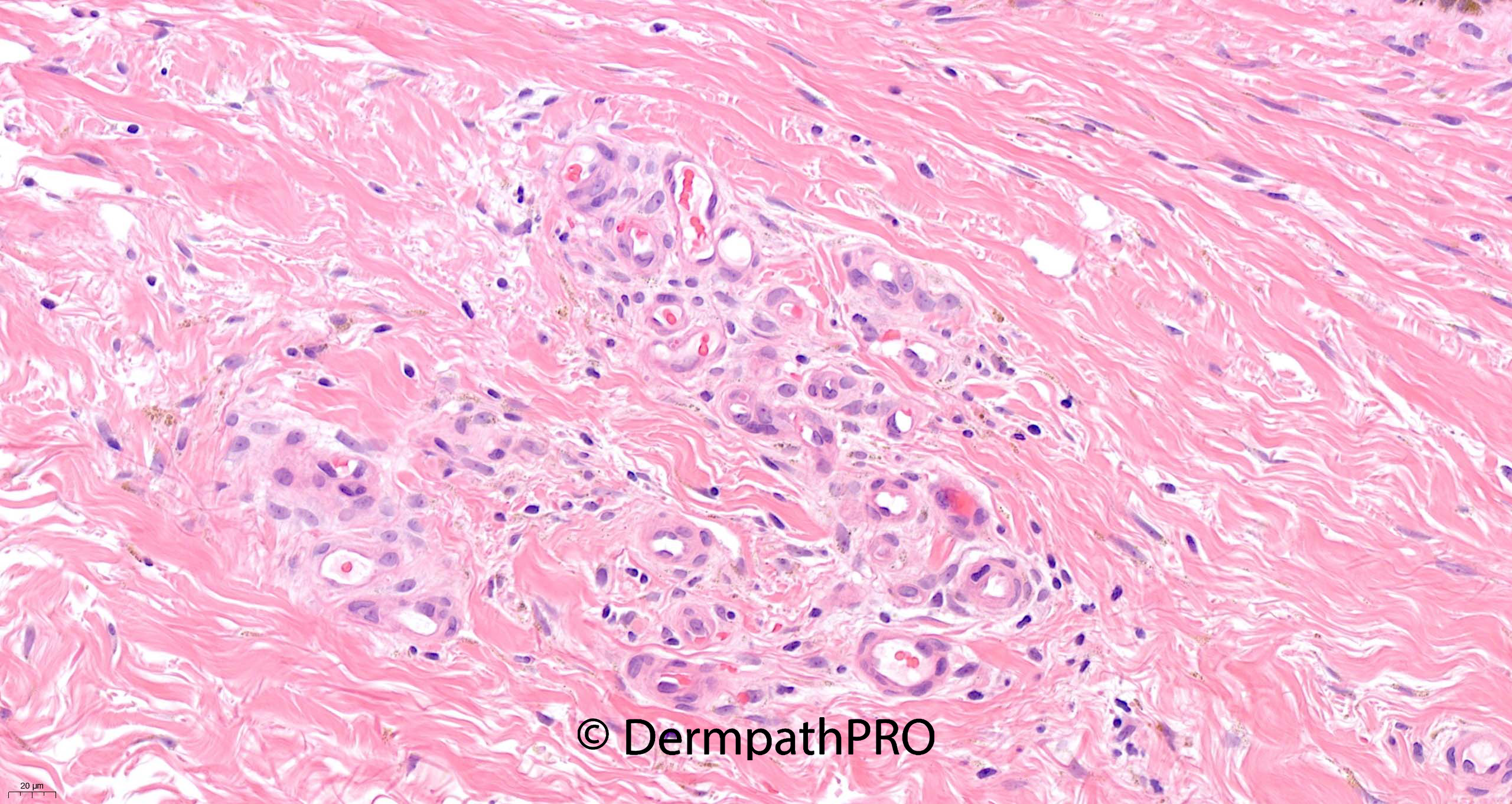
Epidermal atrophy, dermal sclerosis and neovascularization are the features suggesting stasis dermatitis/ lipodermatosclerosis. Panniculus is not seen in the given pictures. A mild lichenoid reaction is seen at a place which does not seem to be significant in the overall scheme of things.

Share this comment


Link to comment
share_externally

any chance of haemochromatosis??

from Weedon's :Thinning of epidermis; increased melanin in basal layer; increased yellow–brown granules of hemosiderin in dermis, especially around vessels and sweat glands basement membrane

Share this comment


Link to comment
share_externally

?hemosiderin or melanin in dermis. Difficult for me. A subtle hobnail hemangioma???

Share this comment


Link to comment
share_externally

Richard Logan

Posted

There is a small focus of lichenoid inflammation.  Much of the epidermis is effaced with loss of the rete ridge pattern, suggesting a previous lichenoid inflammatory process.  The epidermis is heavily pigmented which could be racial, and there is subtle pigmentation in the dermis and around sweat glands.  I am wondering about a lichenoid condition in a patient who has been on minocycline, hydroxychloroquine or the like.

Share this comment


Link to comment
share_externally

Saleem Taibjee

Posted

I favoured diabetic dermopathy (skin spots/pigmented pretibial patches), but did also raise the possibility of drug-induced pigmentation.

Now I will summarise from Weedon's textbook:

It states that these are the most common cutaneous finding in diabetes mellitus (and yet we rarely ever see biopsies of this condition). The clinical description is flat-topped, dull-red papules that are round or oval, discrete or grouped, and situated mainly on the pretibial areas. As the lesions evolve they develop a thin scale and finally become variably atrophic and hyperpigmented, varying from 0.5 cm in diameter up to large patches covering much of the pretibial skin.


Early lesions may show oedema of the papillary dermis and a mild perivascular lymphocytic infiltrate with some extravasation of red blood cells. There may be mild epidermal spongiosis and focal parakeratosis. Hyaline microangiopathy is invariably present. In atrophic lesions there is neovascularization of the papillary dermis, a sparse perivascular infiltrate of lymphocytes, and small amounts of hemosiderin, mostly in macrophages. Attention has been drawn to the presence of a few perivascular plasma cells in this condition, but plasma cells are almost invariably present whenever there is hemosiderin deposition in the skin. In a recent case studied by the author, hemosiderin was also present in the epidermis, between basal cells and along the basement membrane, a finding not previously recorded.

In a post-mortem study https://pubmed.ncbi.nlm.nih.gov/21762390/

"only 4 out of 14 skin biopsies showed moderate to severe wall thickening of arterioles or medium-sized arteries on periodic acid Schiff (PAS) stains. Only mild basement membrane thickening was noted in 11 of 14 which was highlighted by the PAS stain. Pigmented material was identified within the dermis of 13 cases. In 10 of the cases, the material was positive for Perl's iron stain. Ten cases had material staining positive for Fontana-Masson in the dermis. Nine cases had markedly increased epidermal melanin. The findings suggest that hemosiderin deposition in conjunction with the deposition of melanin contribute to the clinical features of diabetic dermopathy."

I think we do have some of these various (non-specific) features in this case.

I show the Masson-Fontana and Perls below.

I think the Masson-Fontana is the most interesting in highlighting conspicuous melanin within the stratum corneum. The Perls was cutting out, but does show some sparse haemosiderin within the dermis.

BW, Saleem

56544_20.0x MF labelled.jpg

56544_40.0x MFb labelled.jpg

56544_40.0x Perls labelled.jpg

Share this comment


Link to comment
share_externally


Join the conversation

You can post now and register later. If you have an account, sign in now to post with your account.

Guest
Add a comment...

×   Pasted as rich text.   Paste as plain text instead

  Only 75 emoji are allowed.

×   Your link has been automatically embedded.   Display as a link instead

×   Your previous content has been restored.   Clear editor

×   You cannot paste images directly. Upload or insert images from URL.

Loading...

×
×
  • Create New...