Jump to content
In this section we have spot diagnoses posted on a daily basis since June 2010, now over 4000! You can review the archived cases and read the suggested diagnoses by users and the final comment by the contributors.
Case are uploaded each week day by 10 am UK time with the correct diagnosis will generally be posted at 8 pm UK time. Why not view the most recent spot diagnosis and proffer a diagnosis?

Case Number : Case 2936 - 07 October 2021 Posted By: Saleem Taibjee

Please read the clinical history and view the images by clicking on them before you proffer your diagnosis.
Submitted Date :
   (0 reviews)

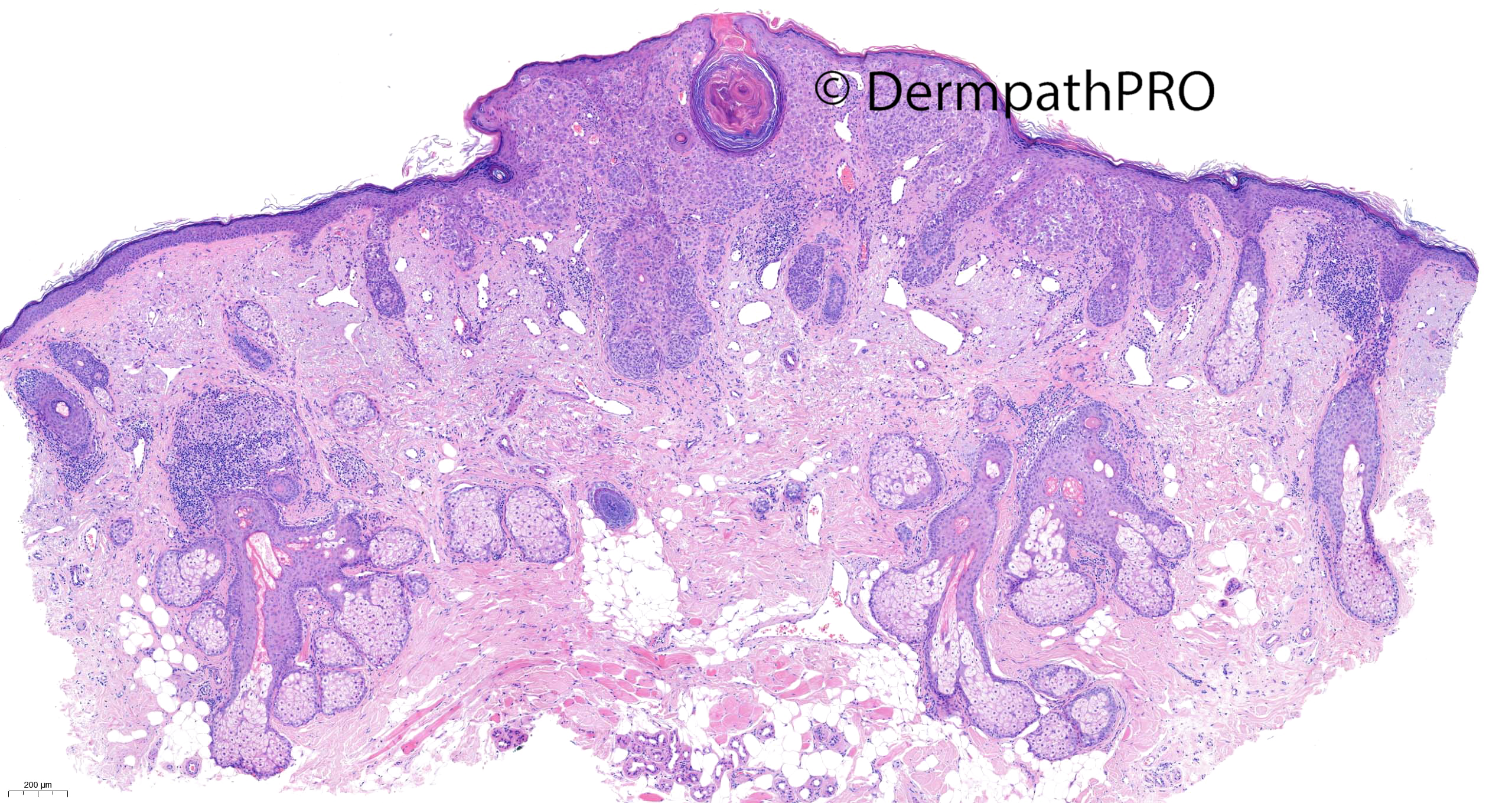
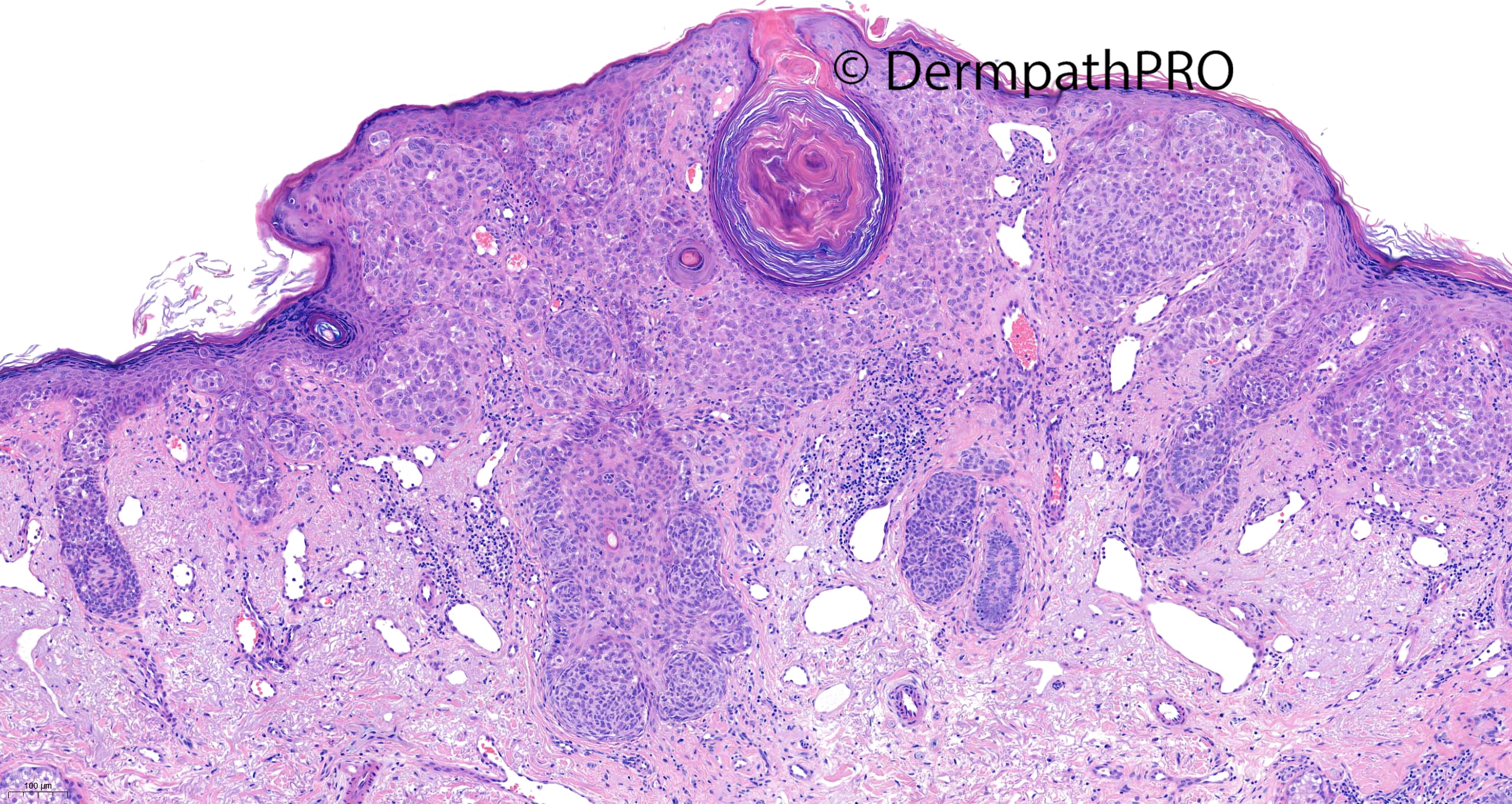
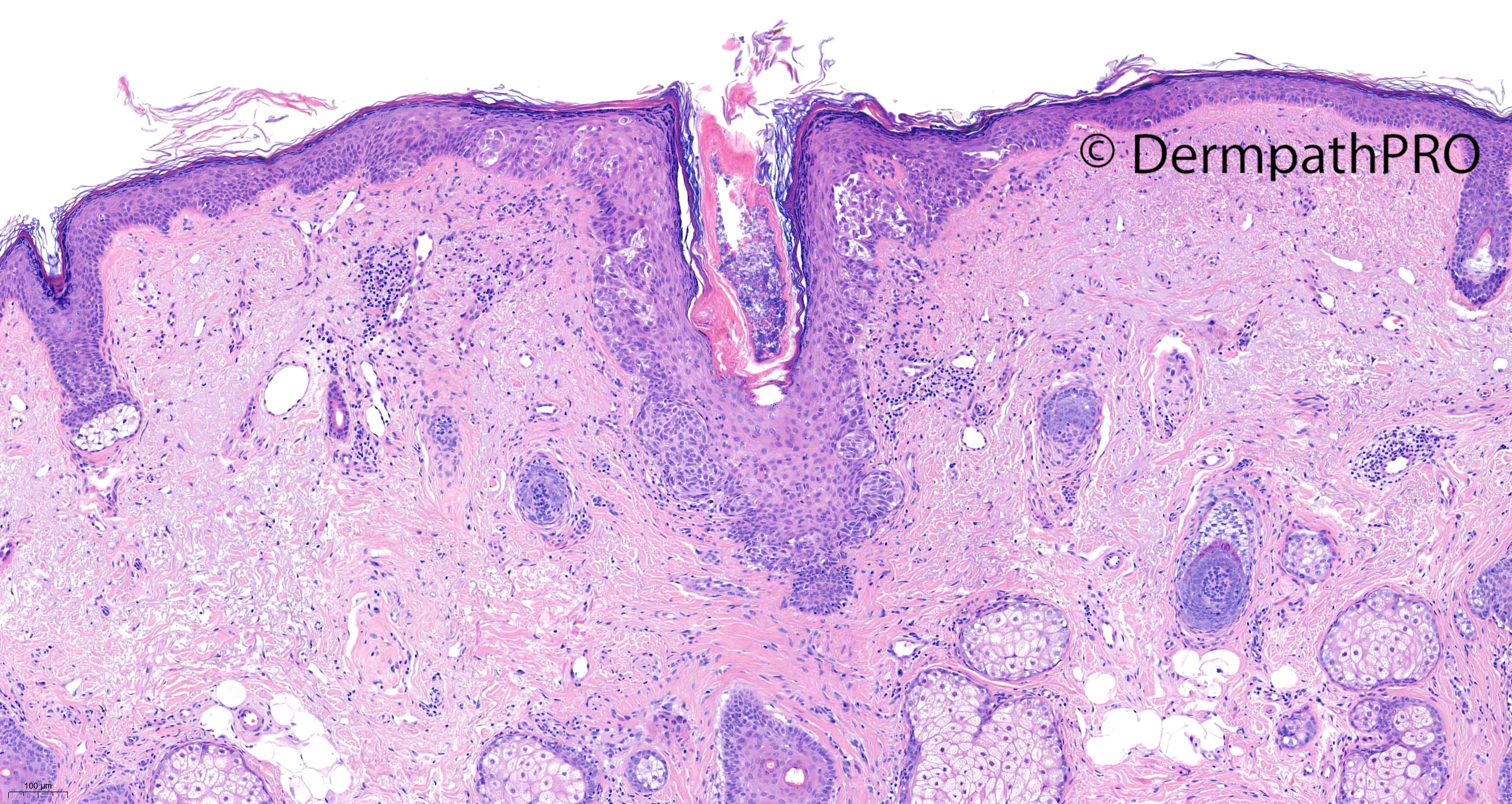
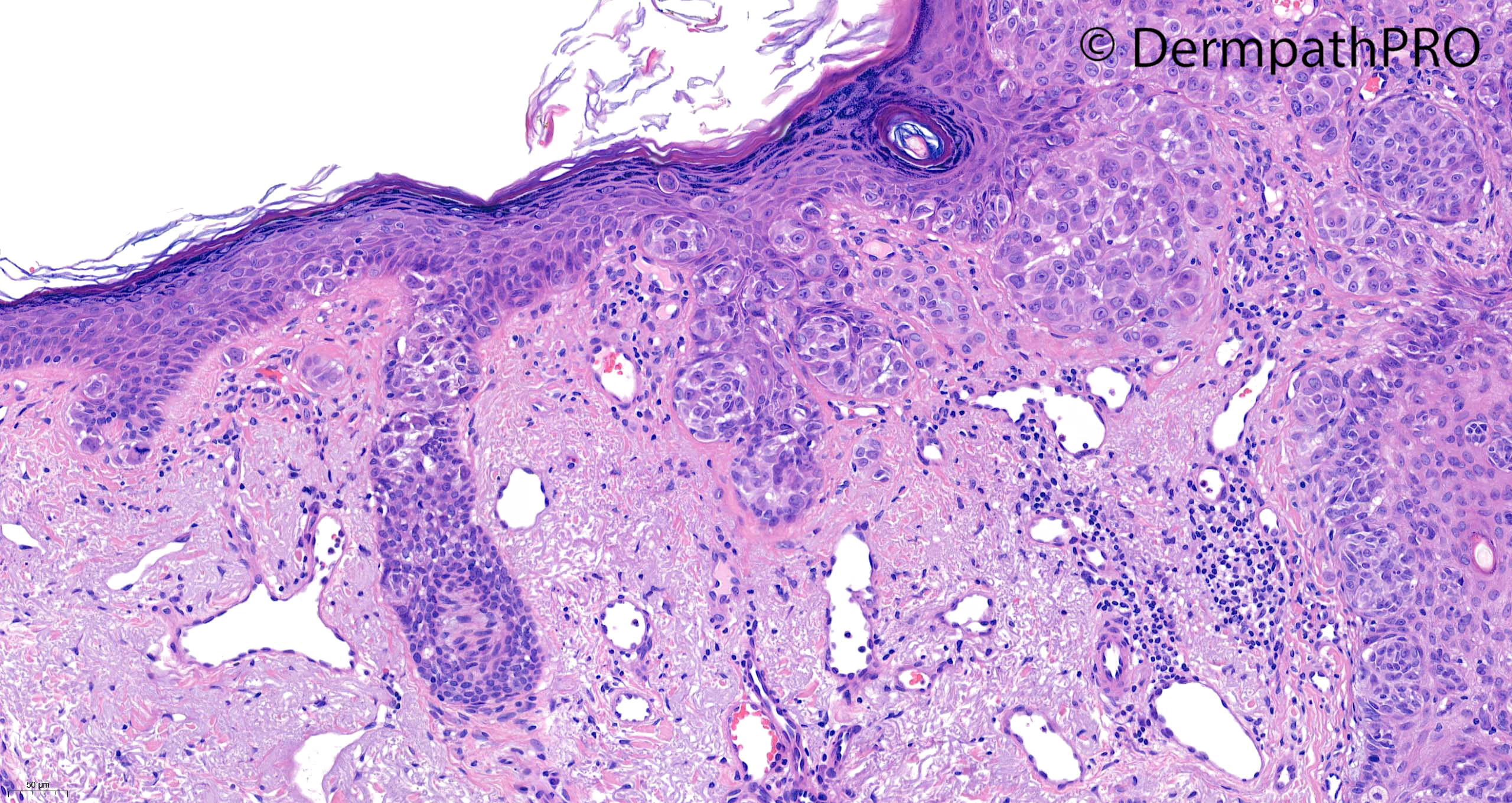
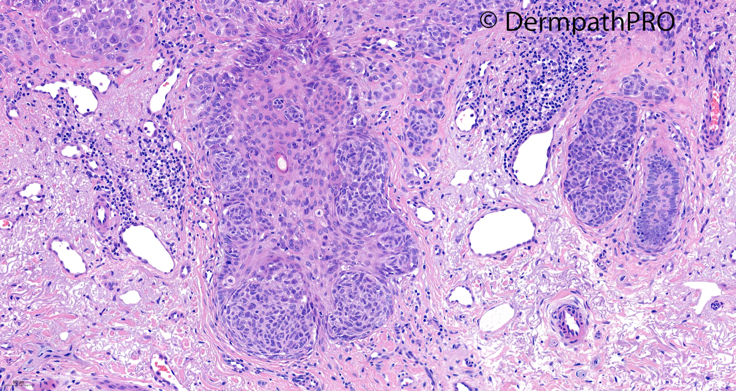
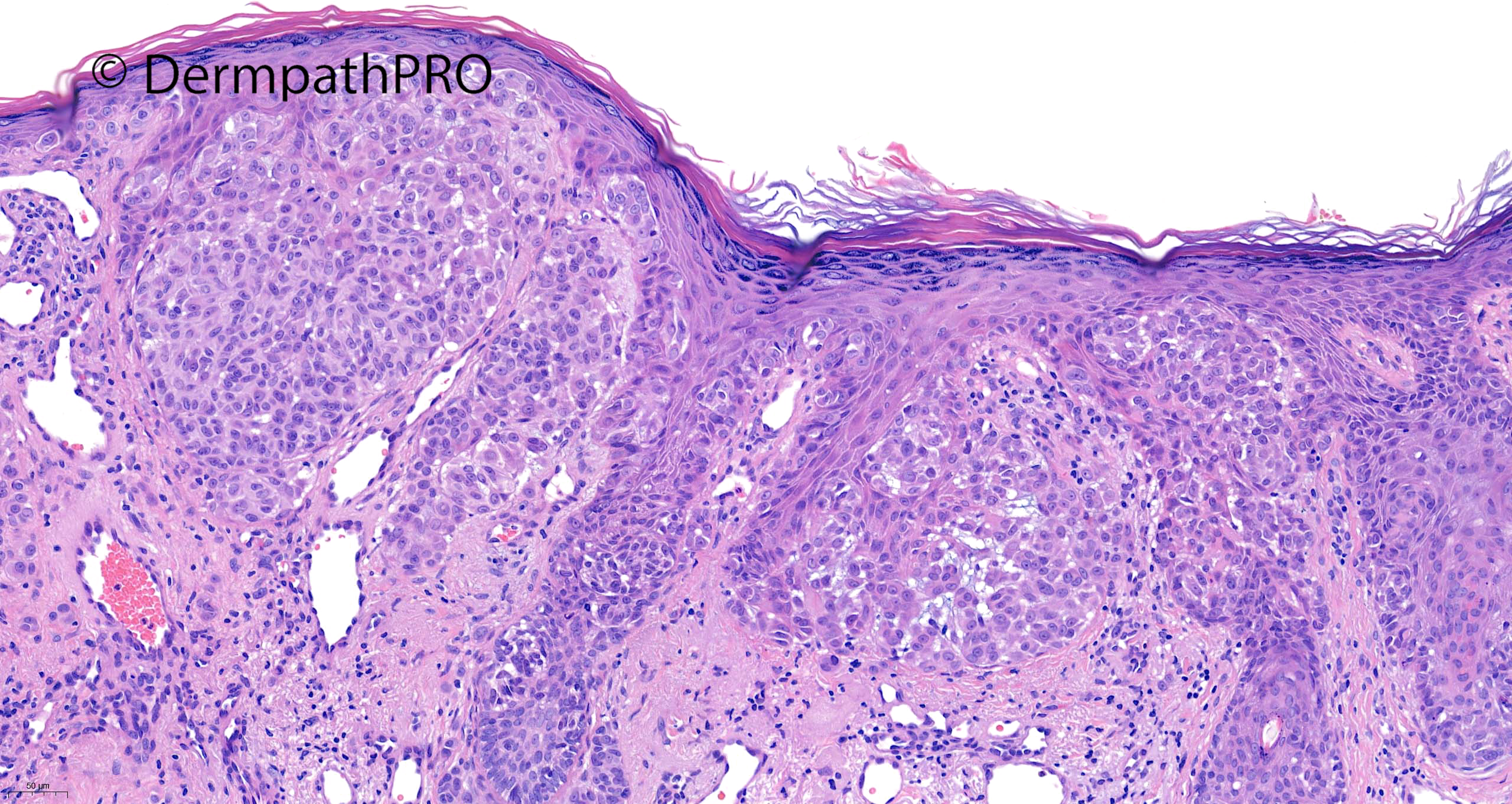
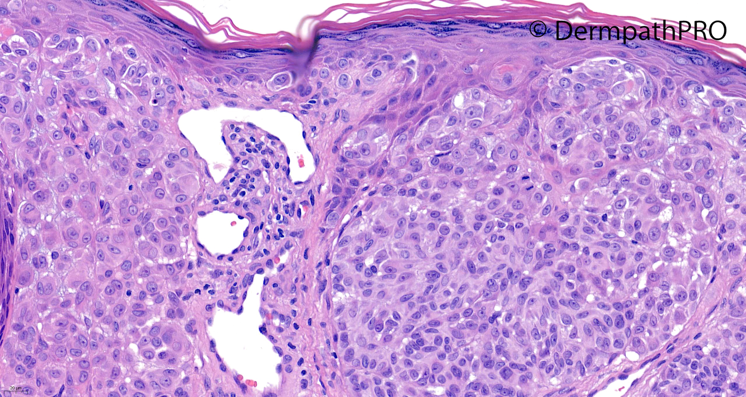
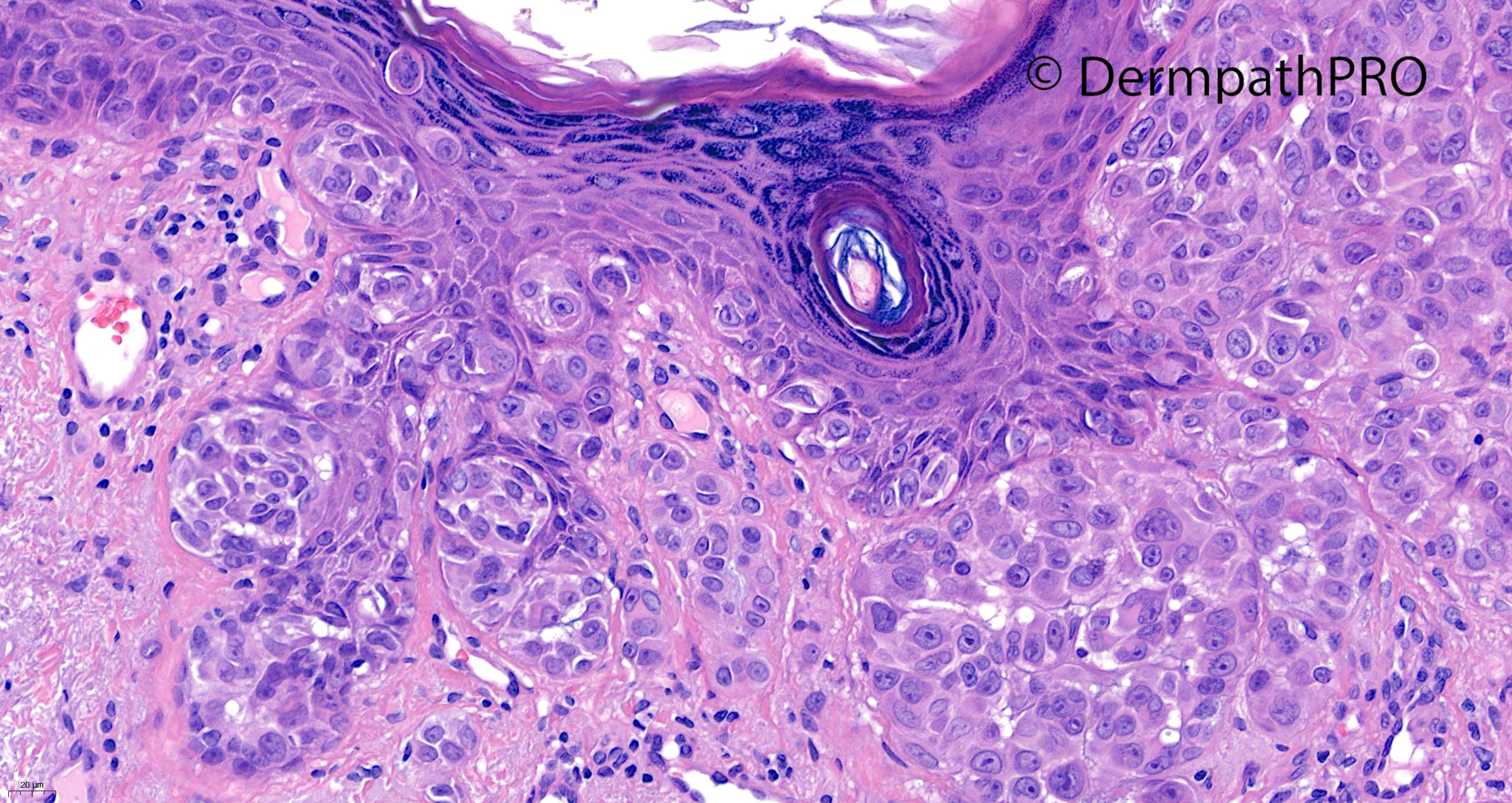
62F punch excision on nose ?BCC (previous history of melanoma on the back several years ago)


  Report Record

User Feedback


Richard Logan

Posted

I think this is a second primary melanoma.  Looks as though there is some superficial invasion.

Share this comment


Link to comment
share_externally

Agree with Richard. This is a radial stage, of a superficial spreading type melanoma, instead of a lentigo maligna type, as expected in this location. It’s initially invasive, but not with a penetrating intradermal nodule. 

Share this comment


Link to comment
share_externally

Krishnakumar subramanian

Posted

agree superficial spreading melanoma

Share this comment


Link to comment
share_externally

it is MM as it is spreads down the hair follicles 

Share this comment


Link to comment
share_externally

Saleem Taibjee

Posted

So it seems that there is a clear consensus from the respondents on melanoma....

However, although there are some worrying features here, I was not sure there is enough for outright melanoma. It is only a 3mm lesion!

I preferred to sit on the fence here, and a MELTUMP/STUMP designation, but advising a conservative re-excision given the close margins.

Of further interest is the immunohistochemistry...

This indicated expression of ROS-1 and lended weight to a possible underlying ROS-1 fusion as a driver, and BRAFV600E negative, and thus true Spitz lineage. For what it's worth, PRAME was negative, although I would not trust it in this situation. p16 appears to be preserved, Ki67 low.  See below.

It would be interesting to know if the immunos would alter the opinions of any of the respondents? For me at least, melanocytic pathology is becoming an increasingly grey area!

BW

Saleem

44342_10.0x ROS-1 labelled.jpg

44342_10.0x BRAFV600E labelled.jpg

44342_5.0x PRAME labelled.jpg

44342_10.0x p16 labelled.jpg

44342_10.0x Ki-67 labelled.jpg


Share this comment


Link to comment
share_externally

msofopoulos

Posted

ROS-1 fusion makes it a Spitzoid lesion. Given it retains p16 would you classify this as a simple Spitz Nevus or an Atypical Spitz Tumor?

Share this comment


Link to comment
share_externally

Meenakshi Batrani

Posted

18 hours ago, msofopoulos said:

ROS-1 fusion makes it a Spitzoid lesion. Given it retains p16 would you classify this as a simple Spitz Nevus or an Atypical Spitz Tumor?

Agree. I have the same thought

Share this comment


Link to comment
share_externally

Saleem Taibjee

Posted

  • I classified as Atypical Spitz Tumour. I would not rely upon p16. Certainly it is more worrying if there is loss (and might imply p16 deletion at the molecular level), but there are still worrying features in this case, including the demographic.

Share this comment


Link to comment
share_externally


Join the conversation

You can post now and register later. If you have an account, sign in now to post with your account.

Guest
Add a comment...

×   Pasted as rich text.   Paste as plain text instead

  Only 75 emoji are allowed.

×   Your link has been automatically embedded.   Display as a link instead

×   Your previous content has been restored.   Clear editor

×   You cannot paste images directly. Upload or insert images from URL.

Loading...

×
×
  • Create New...