Jump to content
In this section we have spot diagnoses posted on a daily basis since June 2010, now over 4000! You can review the archived cases and read the suggested diagnoses by users and the final comment by the contributors.
Case are uploaded each week day by 10 am UK time with the correct diagnosis will generally be posted at 8 pm UK time. Why not view the most recent spot diagnosis and proffer a diagnosis?

Case Number : Case 2937 - 08 October 2021 Posted By: Dr. Richard Carr

Please read the clinical history and view the images by clicking on them before you proffer your diagnosis.
Submitted Date :
   (0 reviews)

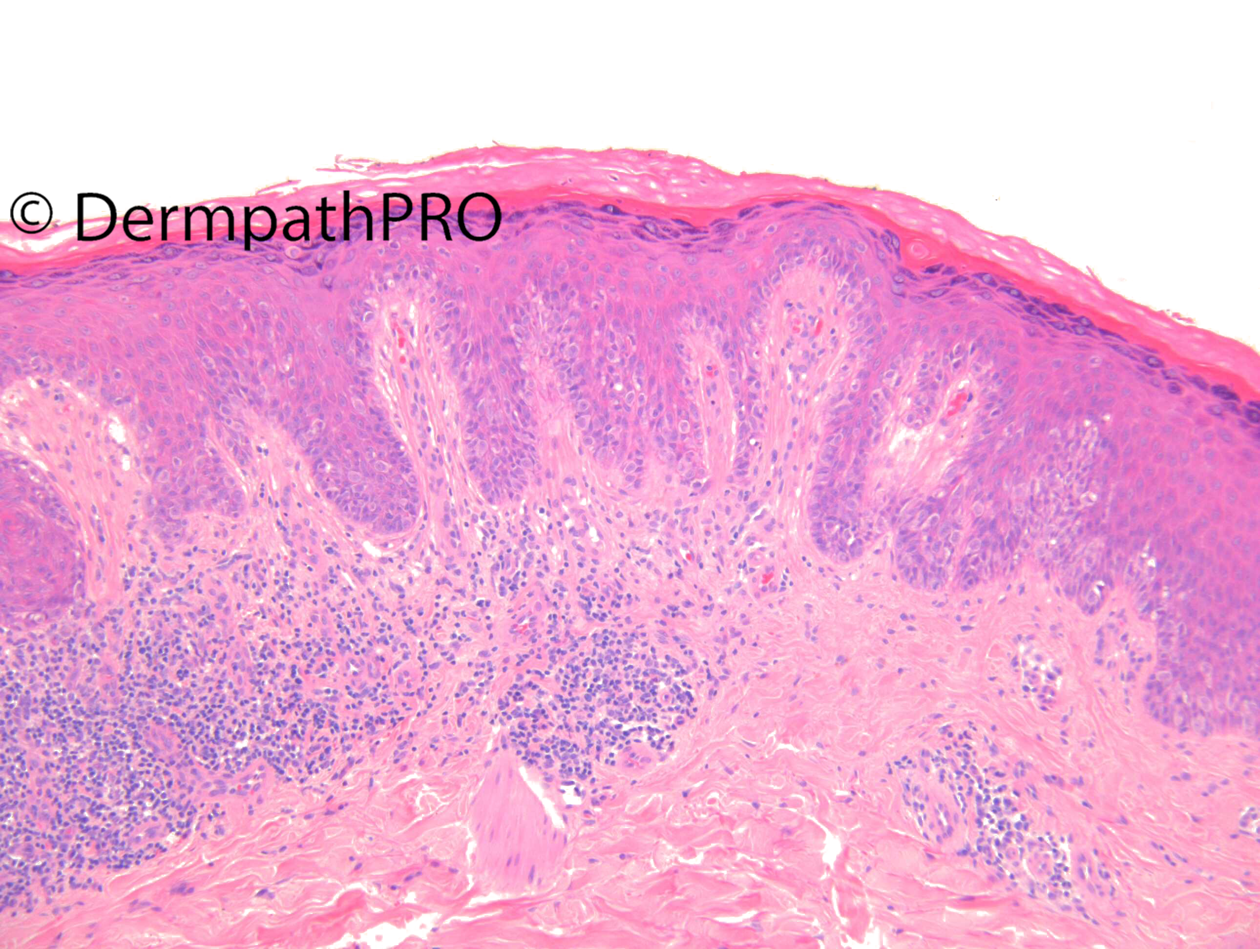
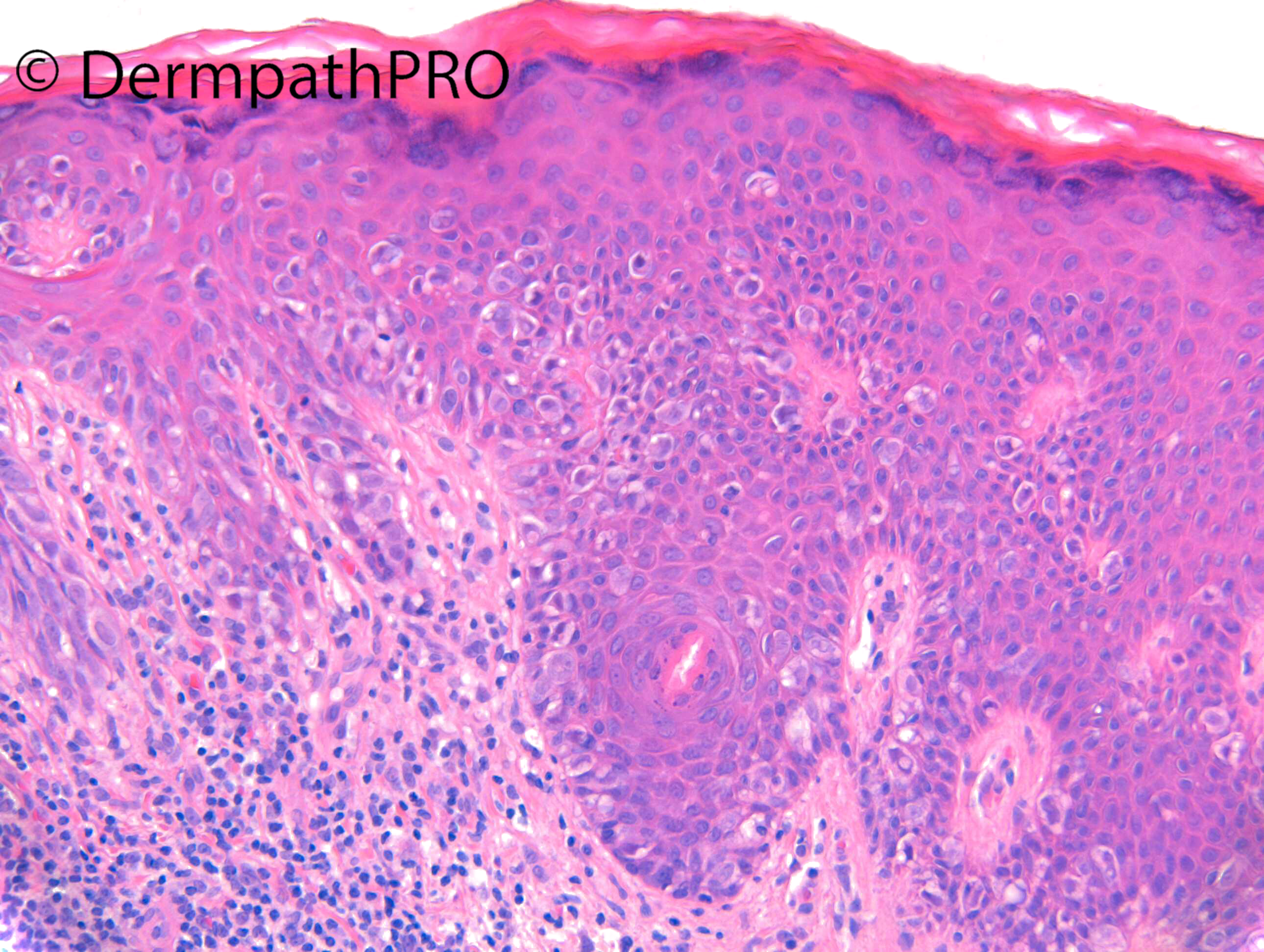
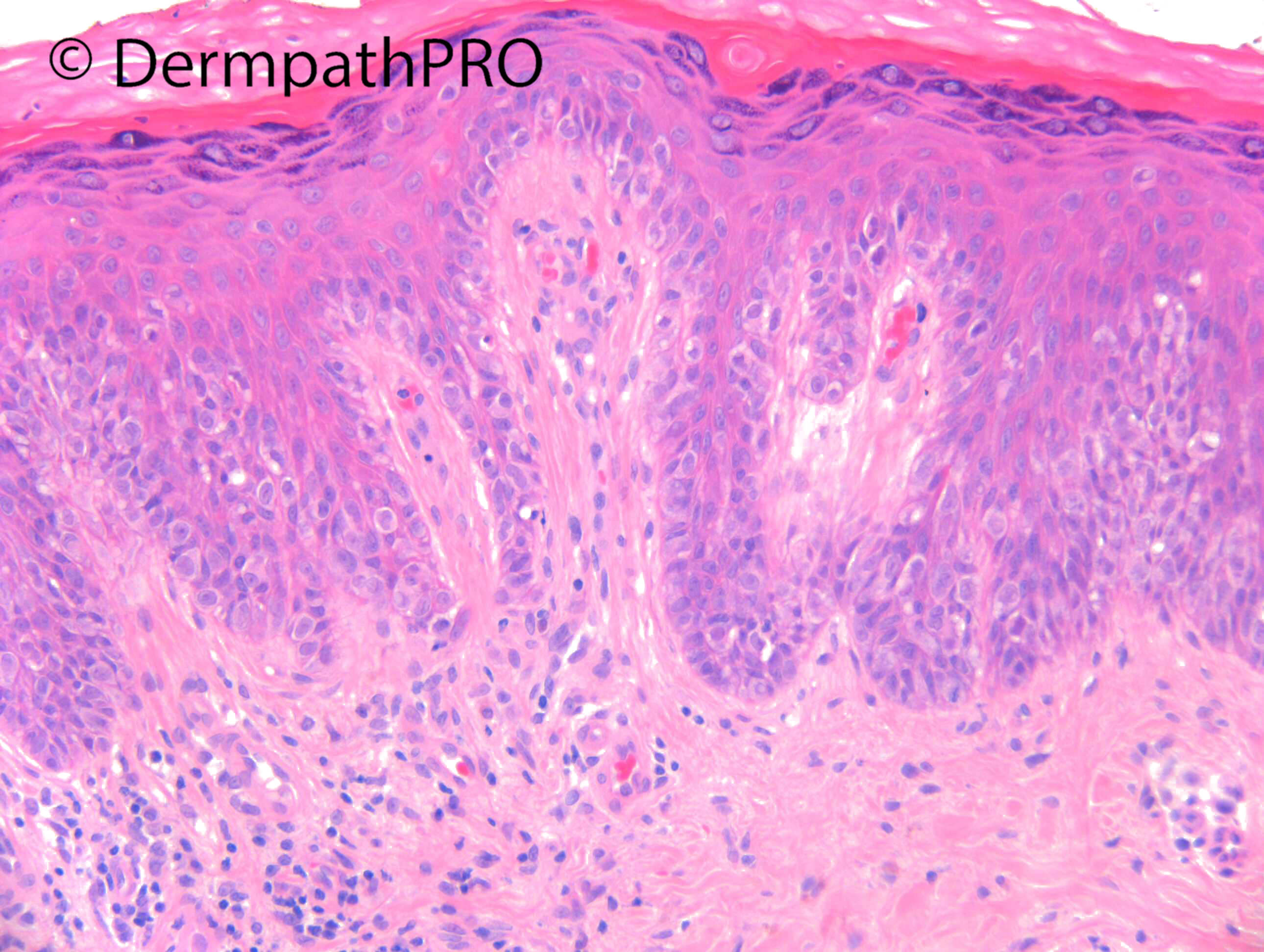
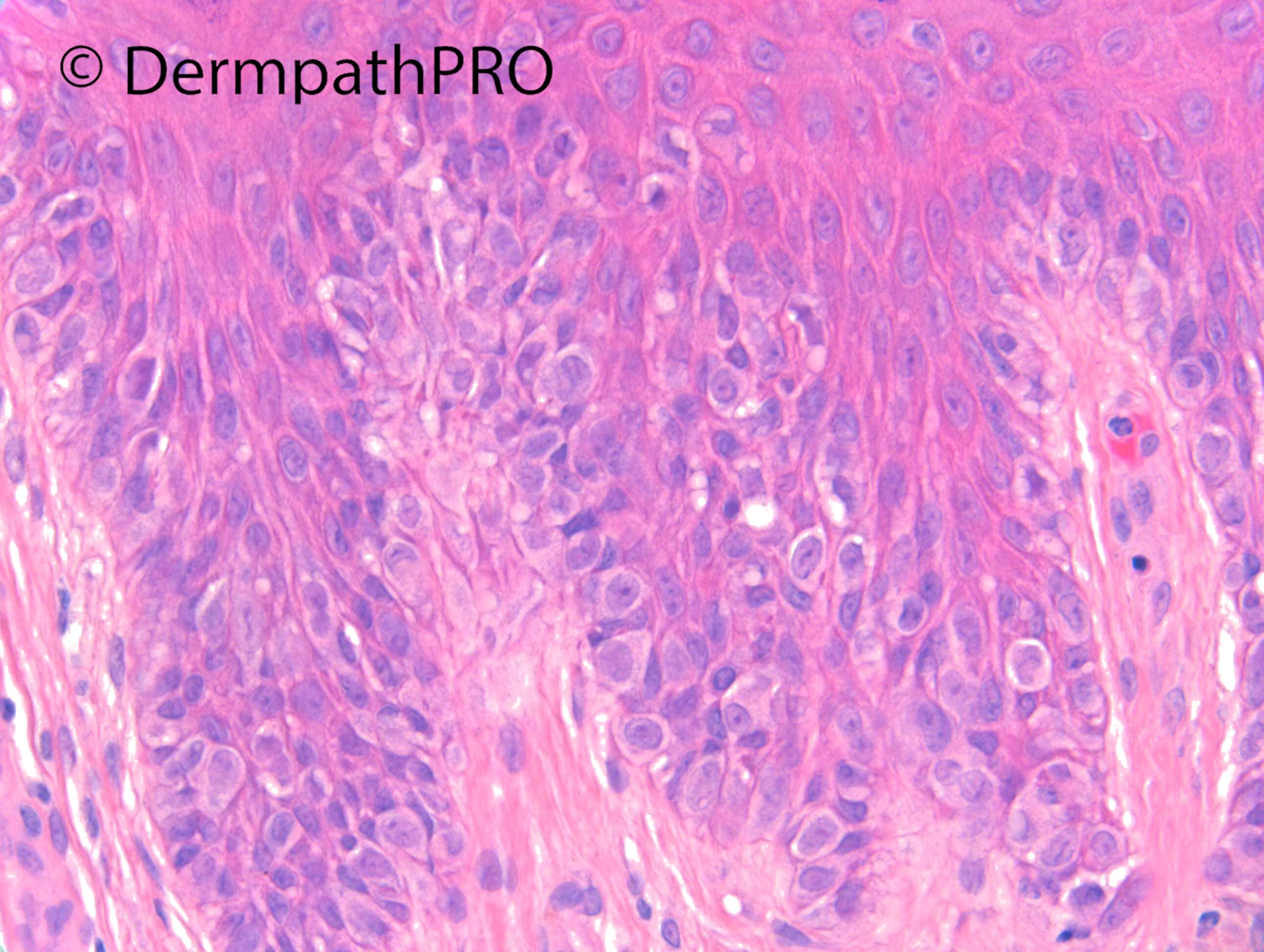
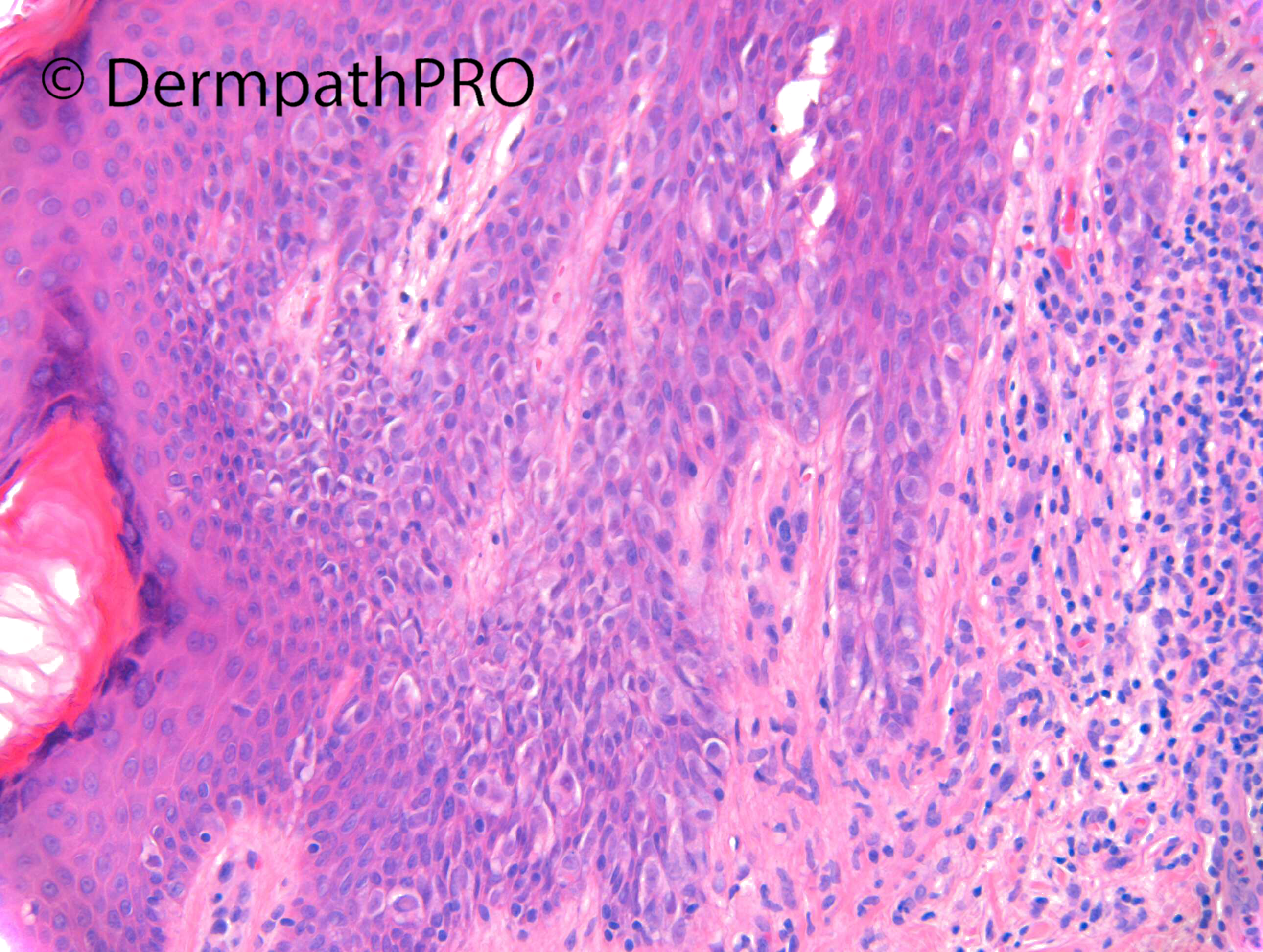
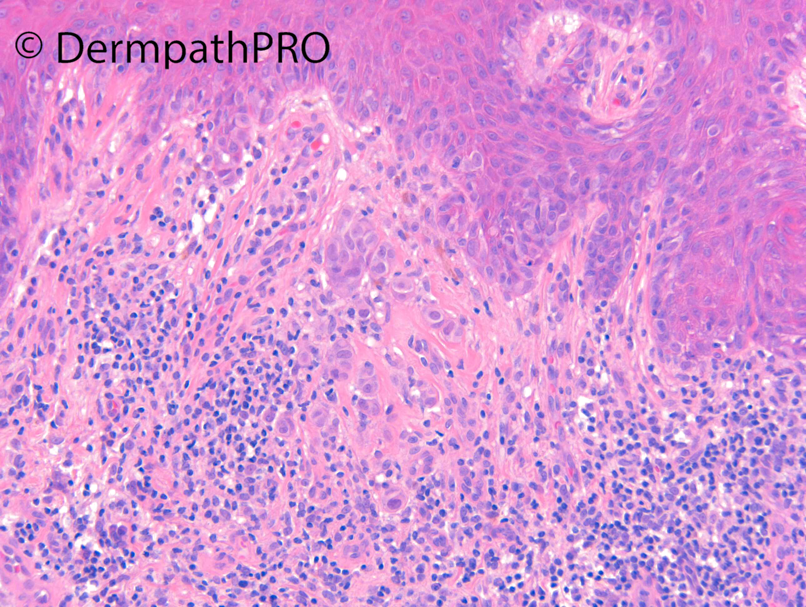
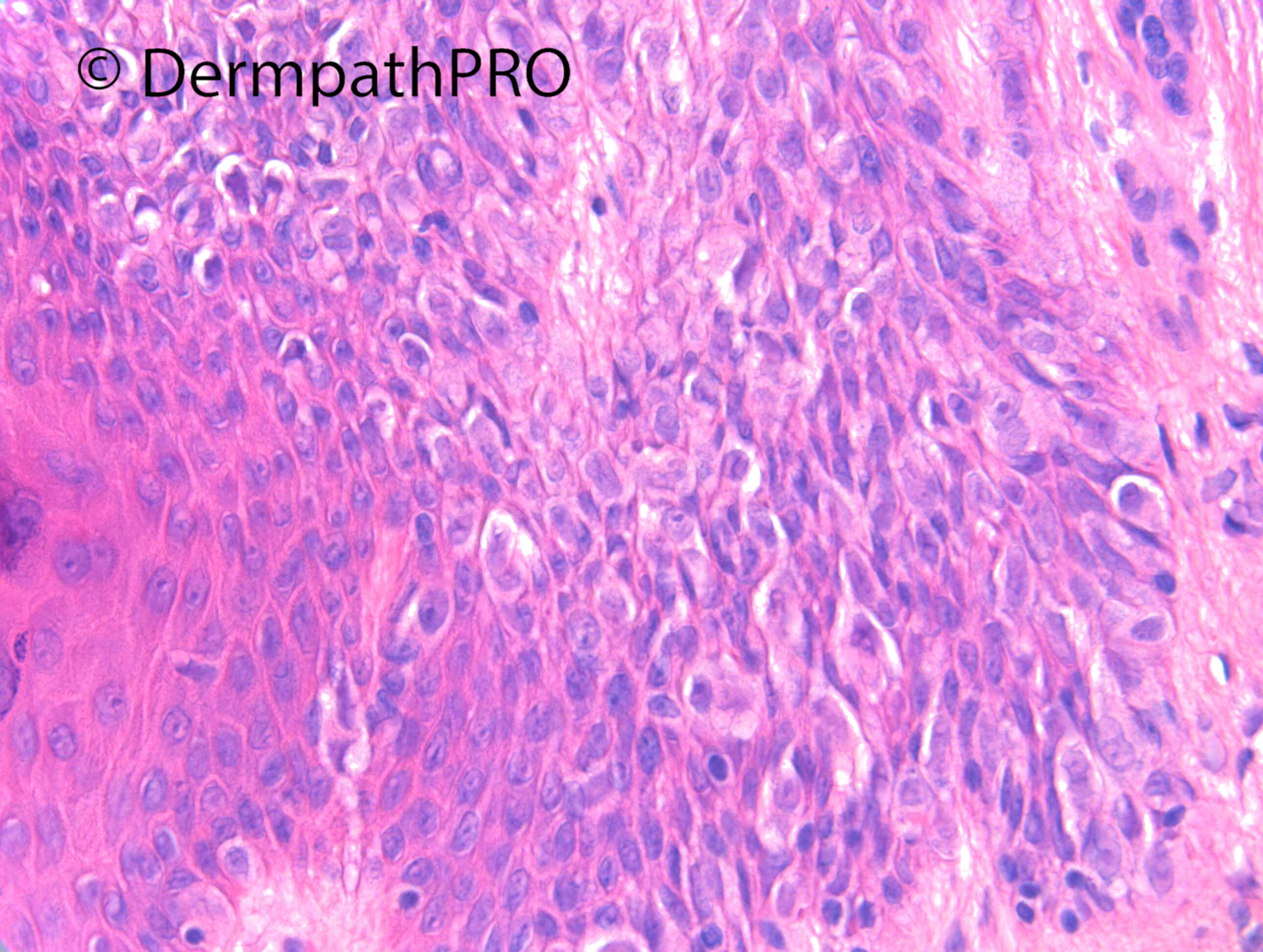
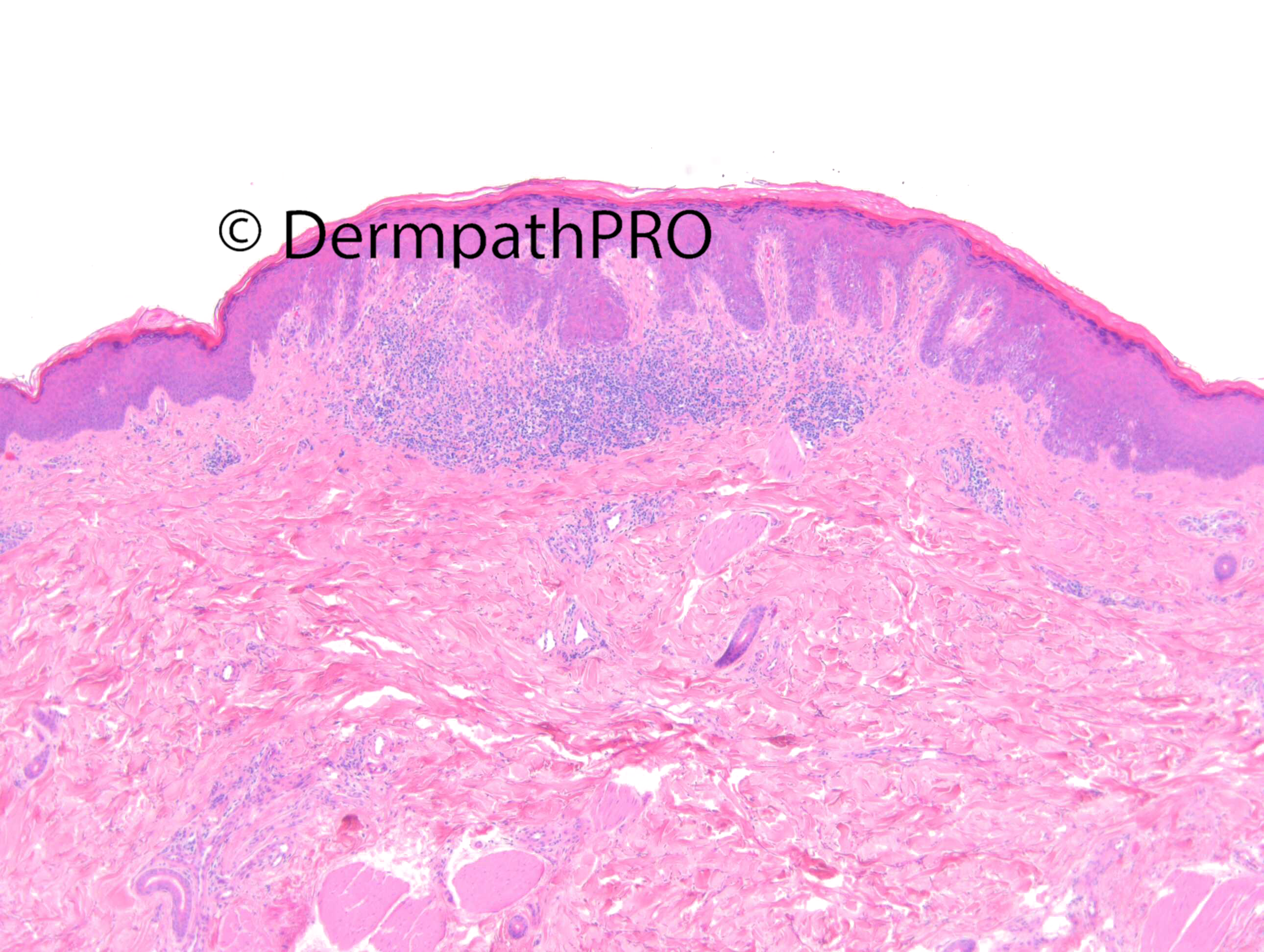
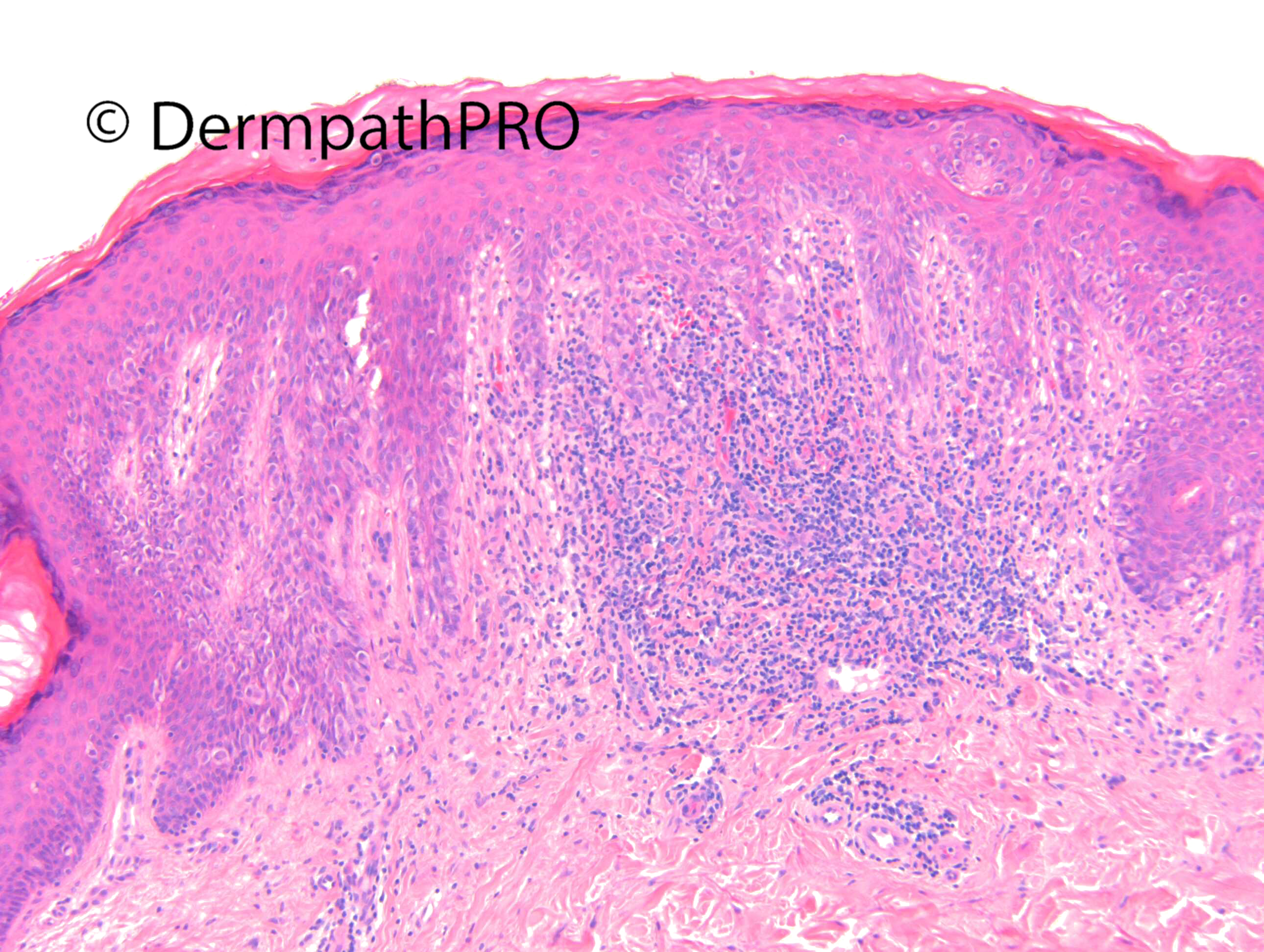
M34. Left thigh. Pink macule, changes 1/12 ago, become partially pigmented, increasing size. ?DN, ?MM, ?pigmented BCC

  Report Record

User Feedback


Monomorphic, epithelioid and atypical cells, with a lentiginous and focally pagetoid intraepidermal growth. Spitz Nevus could be a good spot, but I would know BRAF-(V600E) status. If negative ok Spitz.  

Share this comment


Link to comment
share_externally

Meenakshi Batrani

Posted (edited)

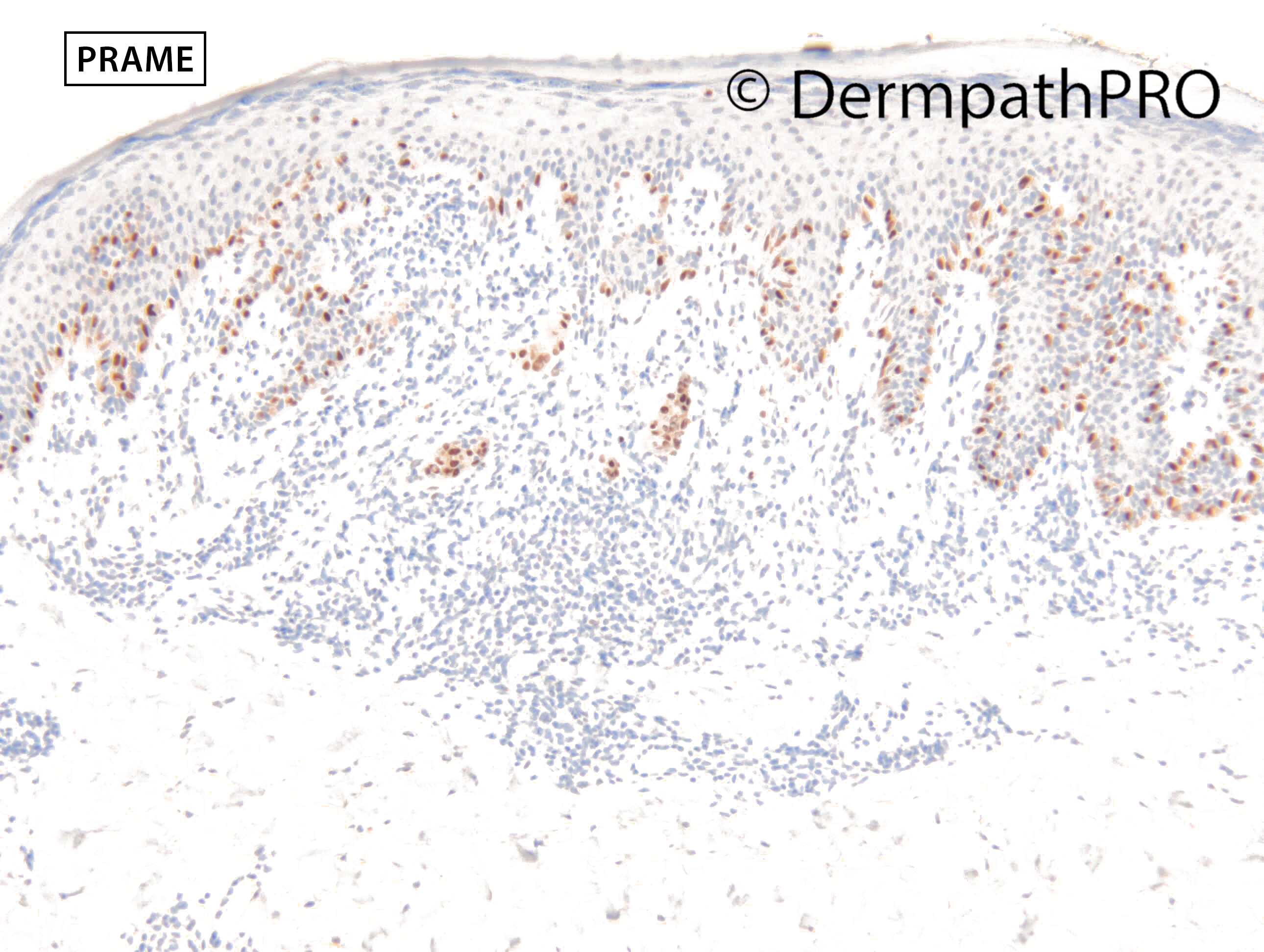
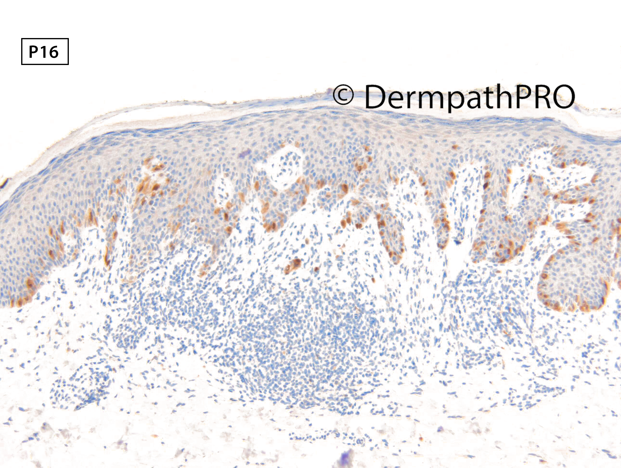
Thinking of pagetoid spitz nevus. But not sure about PRAME expression and p16, markers in Spitz nevi, does the IHC profile goes against benign nevus and is indicative of melanoma.

Edited by Meenakshi Batrani

Share this comment


Link to comment
share_externally

Dr. Richard Carr

Posted

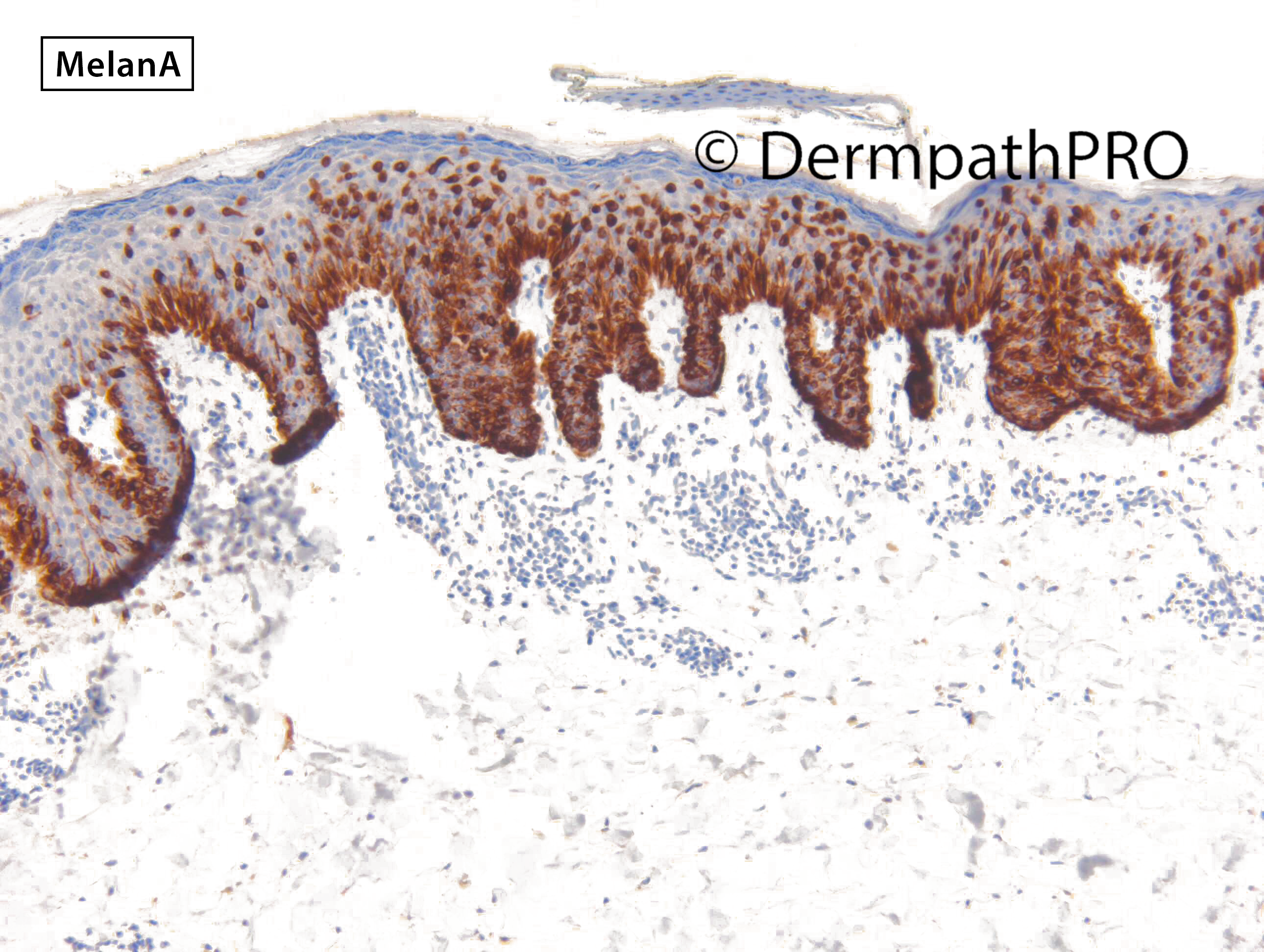
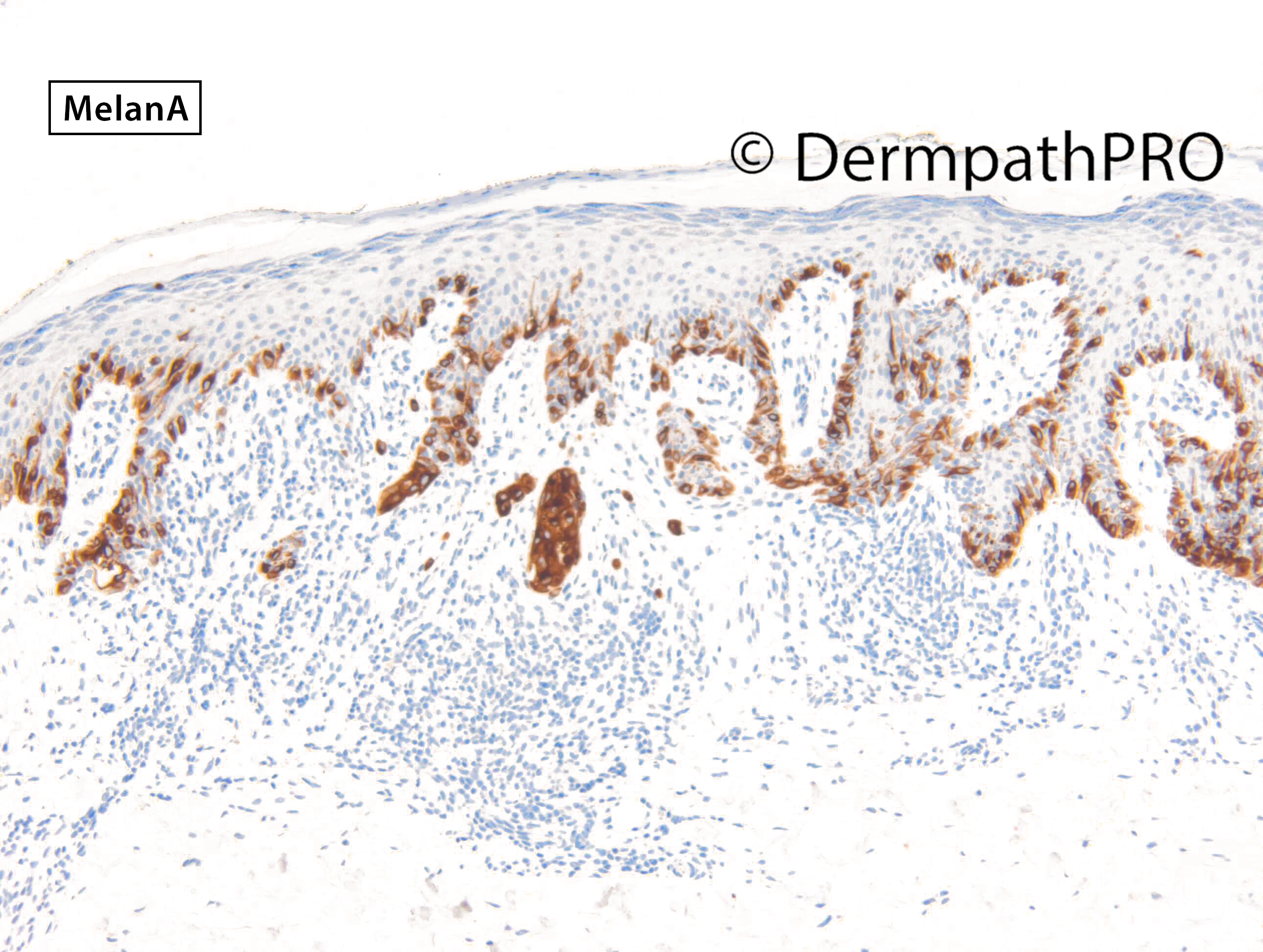
Thanks for the comments. Smallish inflamed lesions are a challenging to say the least and the honest answer in most cases should probably be I don't know. Pagetoid spread is obviously a clue to melanoma but as pointed out in the responses can be seen in Spitz lesions. I was concerned by the rather confluent atypical lentiginous component. On H&E I was in the superficial atypical melanocytic proliferation of uncertain significance (SAMPUS) realm of don't know rather than MELTUMP that I tend to use for the thicker lesions with vertical growth / tumourigenic dermal component. Such a lesion will have negligible metastatic potential and I see little to advocate calling all such lesions thin melanoma rather than admit uncertainty openly. However complete and clear excision is warranted and 3 to 5mm should suffice in most cases and this can be aided by IHC for melanA or other markers such as Sox10 and PRAME. In this case p16 is preserved which I think would lend support to a low risk biology (although more work will be required and larger studies). PRAME is clearly moderately diffusely positive and that certainly moves the goal posts towards a likely evolving or early melanoma (overwhelmingly in situ). My experience with PRAME is in the 10's rather than 100's but after around 40+ cases done in the last few months it seems to support my favoured H&E impression in around 82% and is counter to it in 18%. In 23% of cases I was in the middle of the fence so I've excluded those from these numbers. One has to be careful about over interpreting margins in sun-damaged skin where you may get occasional positive small basal melanocytes in the absence of significant proliferation or atypia but it can also be helpful in assessing margins if positive in a lesion and thus guiding the discussion of re-excision. Anecdotally I'm noticing weaker dermal staining (compared with obvious melanoma in situ) in the largely radial growth phase small cell and neavoid variants of SSMM in which I've never seen a metastasis up till now. In my opinion re-excision should be personalised to the pathology, careful, systematic bread-slice examination, preferably reported or reviewed by an expert, of the lesion (location, size, growth pattern, borders, etc etc) and the individual preferences of the patient rather than following, in my opinion, crude guidelines based on the depth of the lesion which makes no logical or scientific sense and has be elaborated by Prof. Weyers.

https://pubmed.ncbi.nlm.nih.gov/31490196/


Share this comment


Link to comment
share_externally

Meenakshi Batrani

Posted

On 12/10/2021 at 14:16, Dr. Richard Carr said:

Thanks for the comments. Smallish inflamed lesions are a challenging to say the least and the honest answer in most cases should probably be I don't know. Pagetoid spread is obviously a clue to melanoma but as pointed out in the responses can be seen in Spitz lesions. I was concerned by the rather confluent atypical lentiginous component. On H&E I was in the superficial atypical melanocytic proliferation of uncertain significance (SAMPUS) realm of don't know rather than MELTUMP that I tend to use for the thicker lesions with vertical growth / tumourigenic dermal component. Such a lesion will have negligible metastatic potential and I see little to advocate calling all such lesions thin melanoma rather than admit uncertainty openly. However complete and clear excision is warranted and 3 to 5mm should suffice in most cases and this can be aided by IHC for melanA or other markers such as Sox10 and PRAME. In this case p16 is preserved which I think would lend support to a low risk biology (although more work will be required and larger studies). PRAME is clearly moderately diffusely positive and that certainly moves the goal posts towards a likely evolving or early melanoma (overwhelmingly in situ). My experience with PRAME is in the 10's rather than 100's but after around 40+ cases done in the last few months it seems to support my favoured H&E impression in around 82% and is counter to it in 18%. In 23% of cases I was in the middle of the fence so I've excluded those from these numbers. One has to be careful about over interpreting margins in sun-damaged skin where you may get occasional positive small basal melanocytes in the absence of significant proliferation or atypia but it can also be helpful in assessing margins if positive in a lesion and thus guiding the discussion of re-excision. Anecdotally I'm noticing weaker dermal staining (compared with obvious melanoma in situ) in the largely radial growth phase small cell and neavoid variants of SSMM in which I've never seen a metastasis up till now. In my opinion re-excision should be personalised to the pathology, careful, systematic bread-slice examination, preferably reported or reviewed by an expert, of the lesion (location, size, growth pattern, borders, etc etc) and the individual preferences of the patient rather than following, in my opinion, crude guidelines based on the depth of the lesion which makes no logical or scientific sense and has be elaborated by Prof. Weyers.

https://pubmed.ncbi.nlm.nih.gov/31490196/


Thank you for valuable insights from your vast experience regarding such challenging melanocytic and squamous lesions you have been regularly posting.  

Share this comment


Link to comment
share_externally


Join the conversation

You can post now and register later. If you have an account, sign in now to post with your account.

Guest
Add a comment...

×   Pasted as rich text.   Paste as plain text instead

  Only 75 emoji are allowed.

×   Your link has been automatically embedded.   Display as a link instead

×   Your previous content has been restored.   Clear editor

×   You cannot paste images directly. Upload or insert images from URL.

Loading...

×
×
  • Create New...