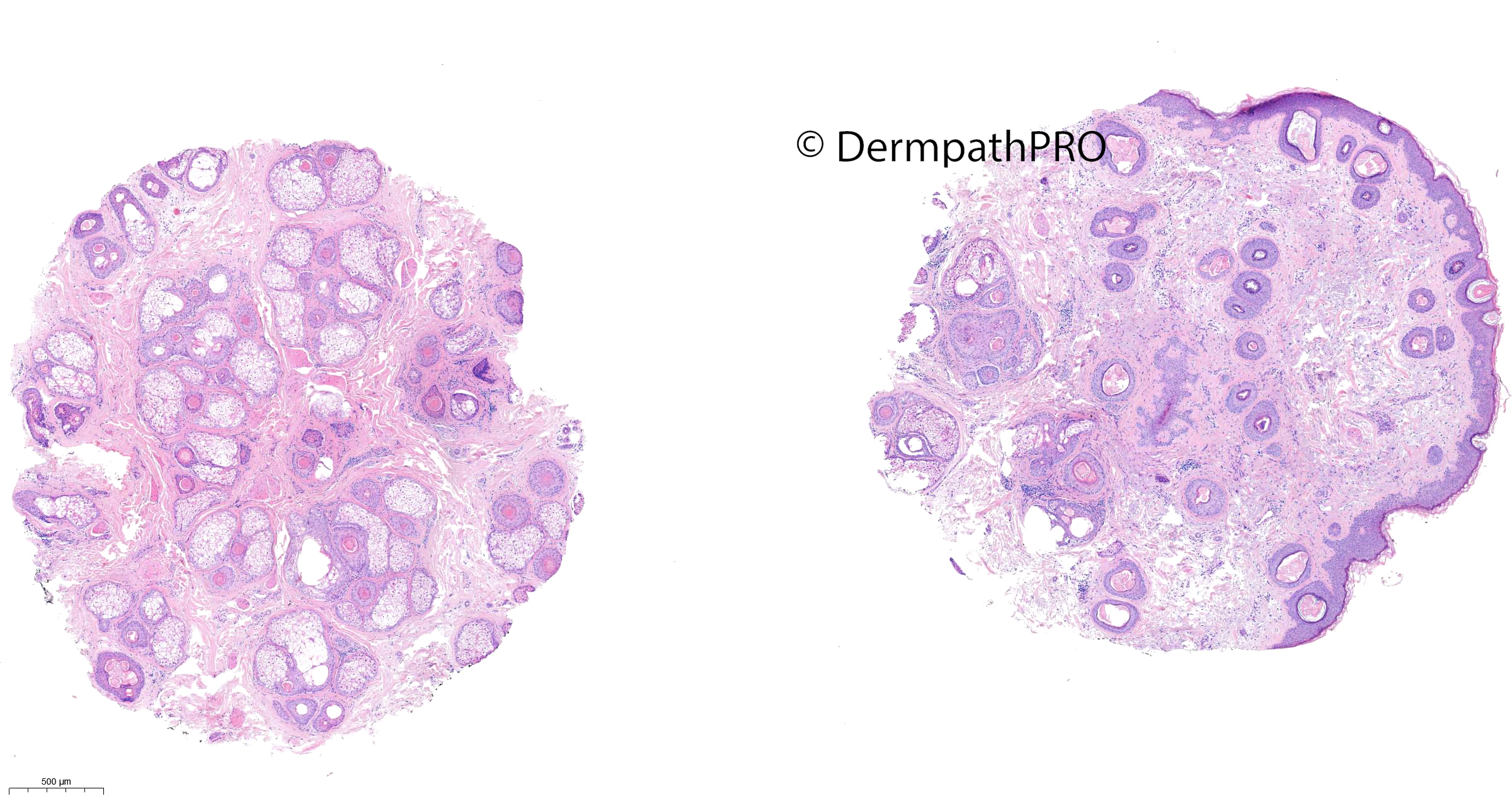
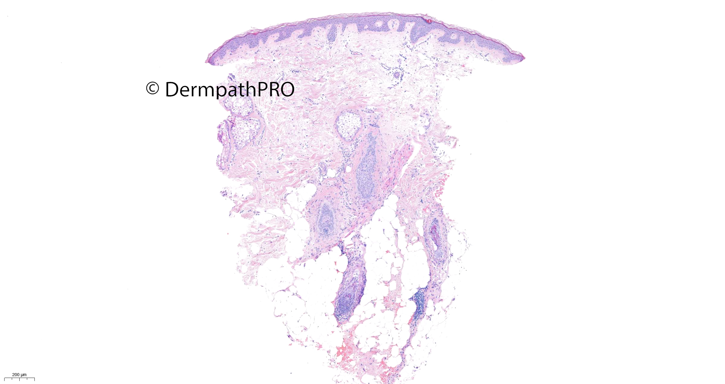
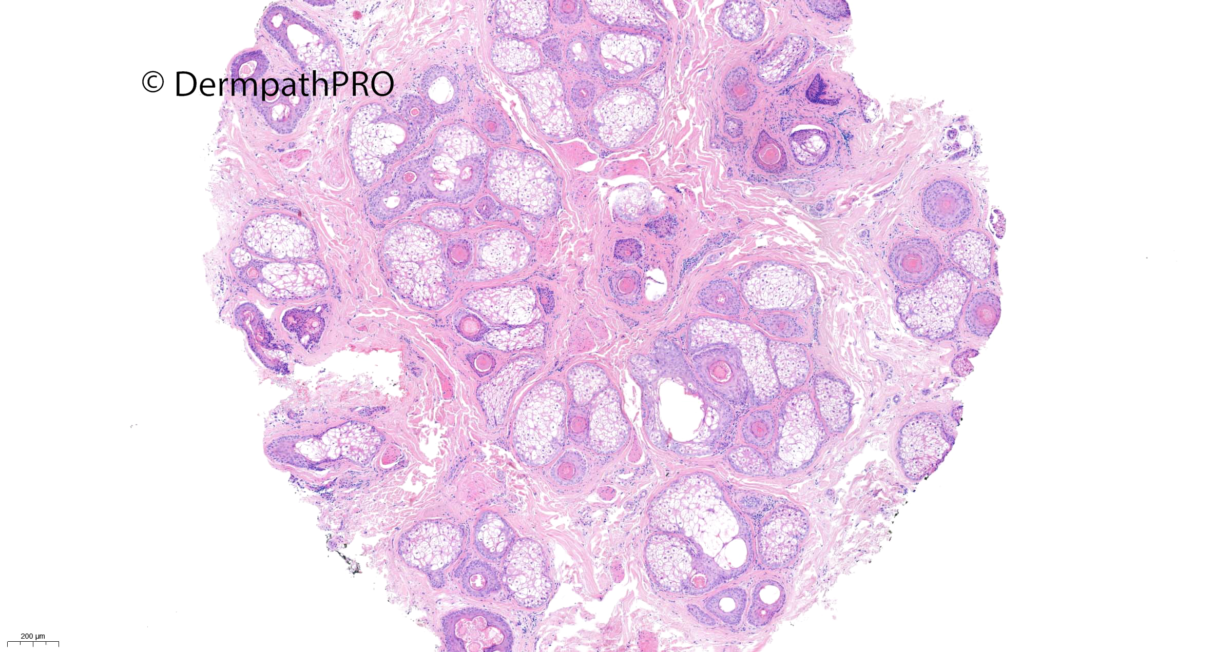
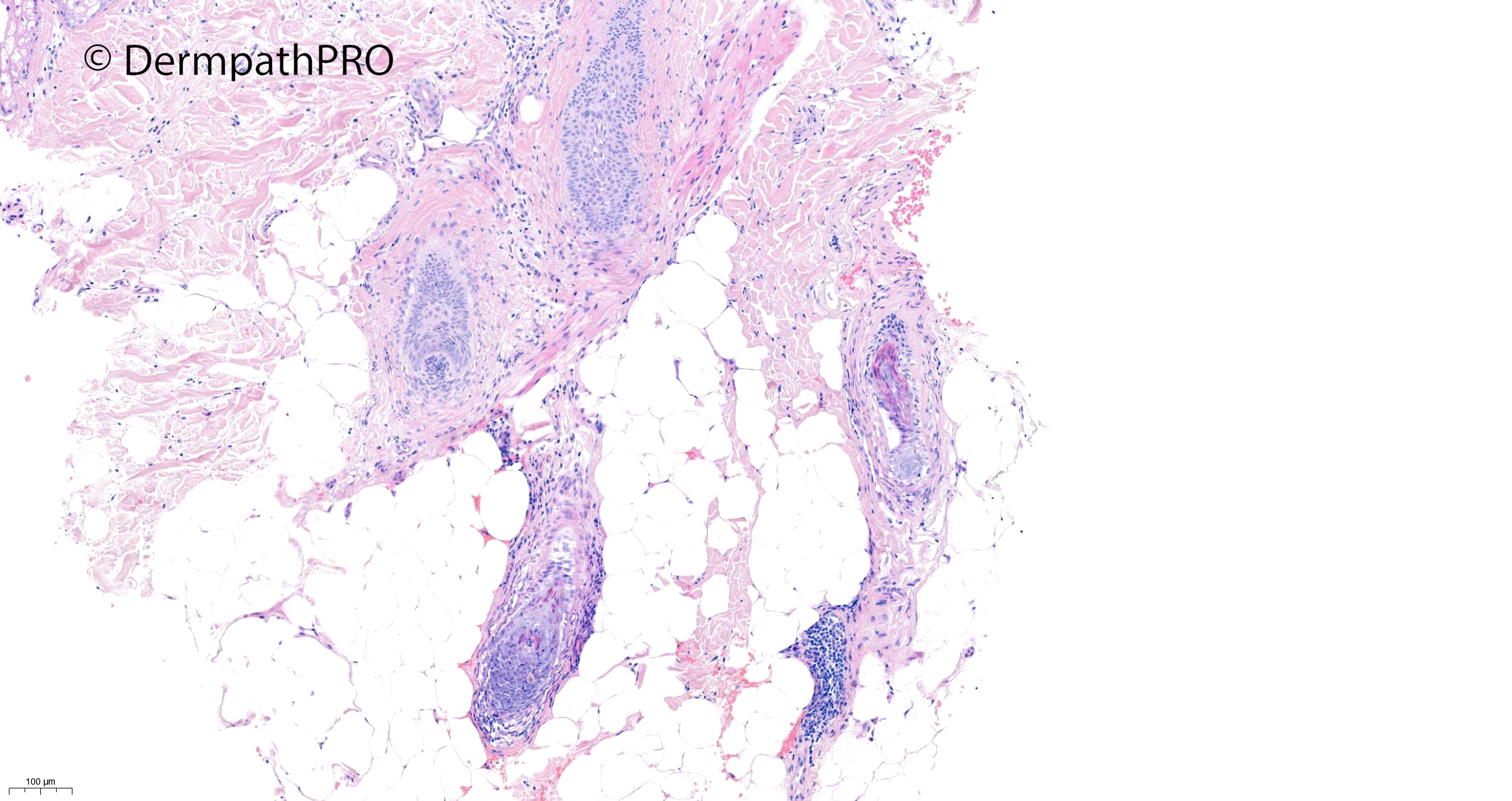
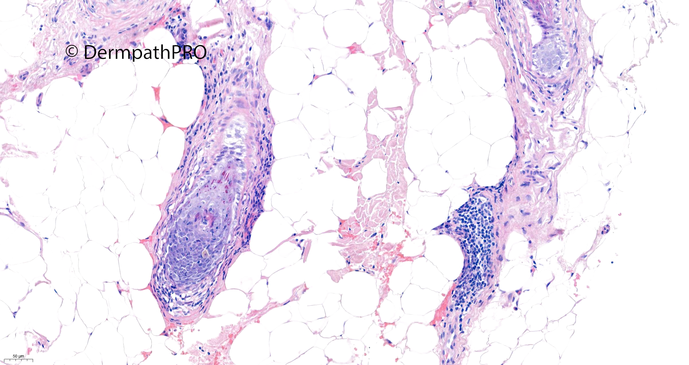
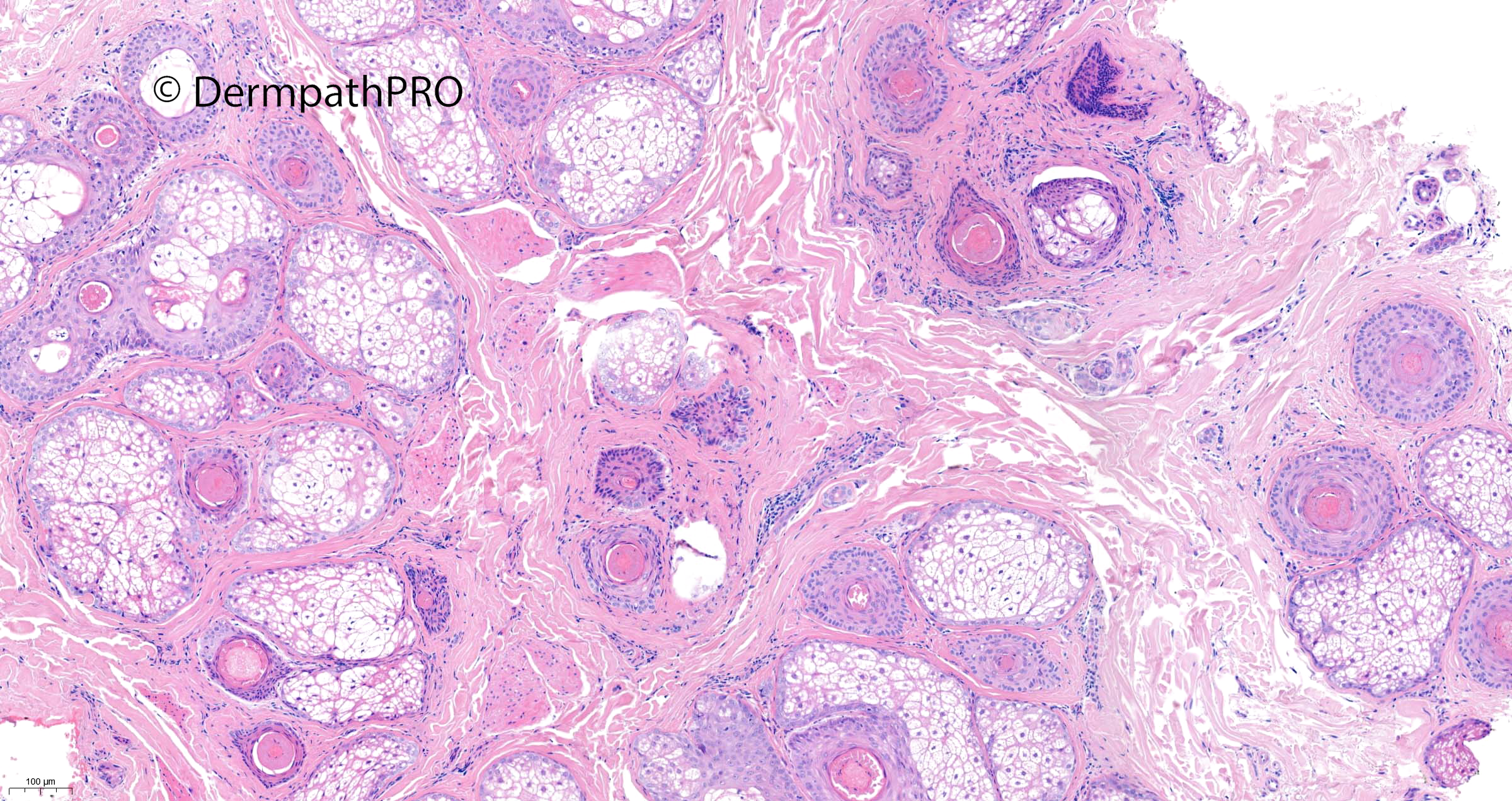
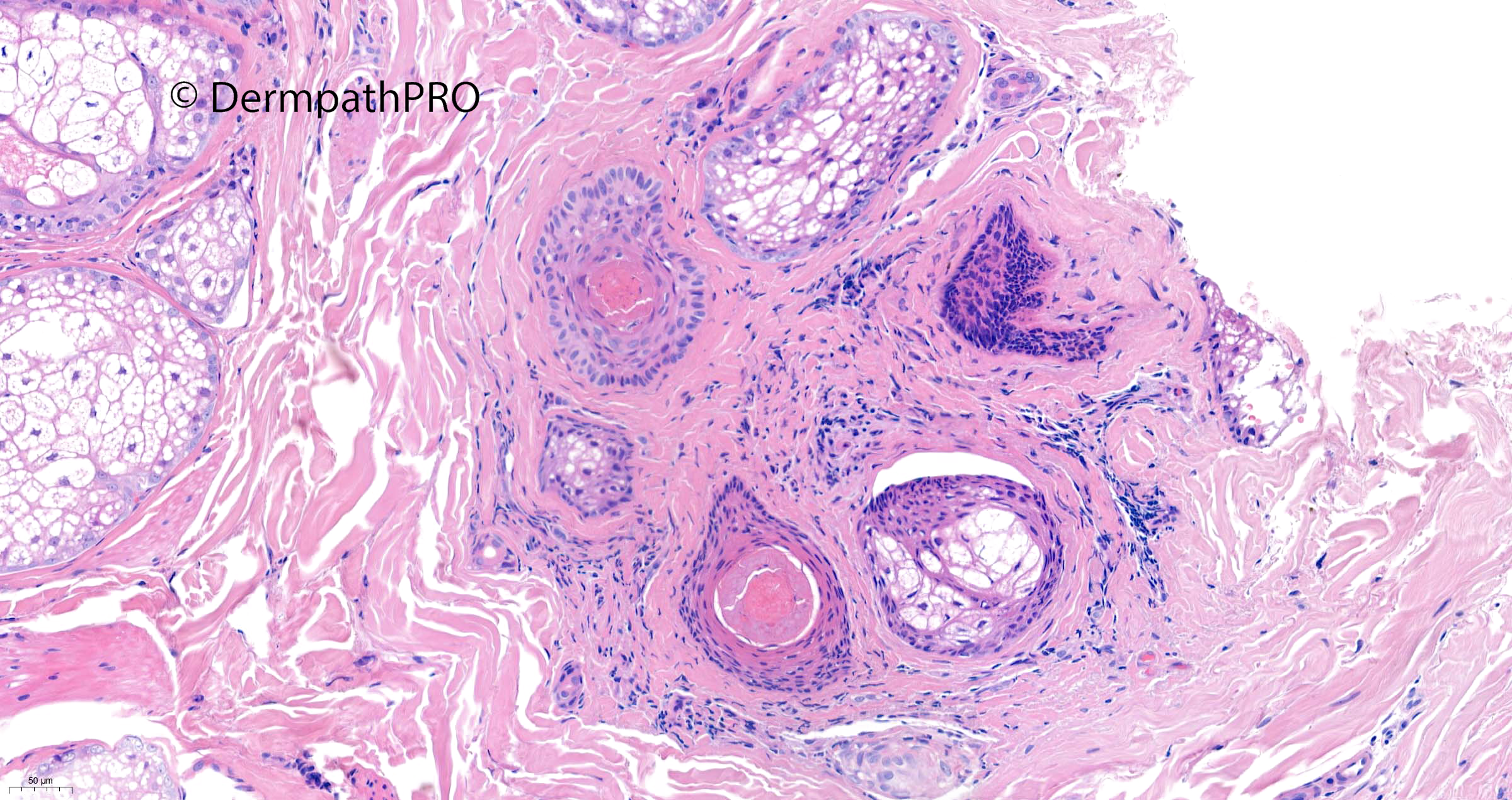
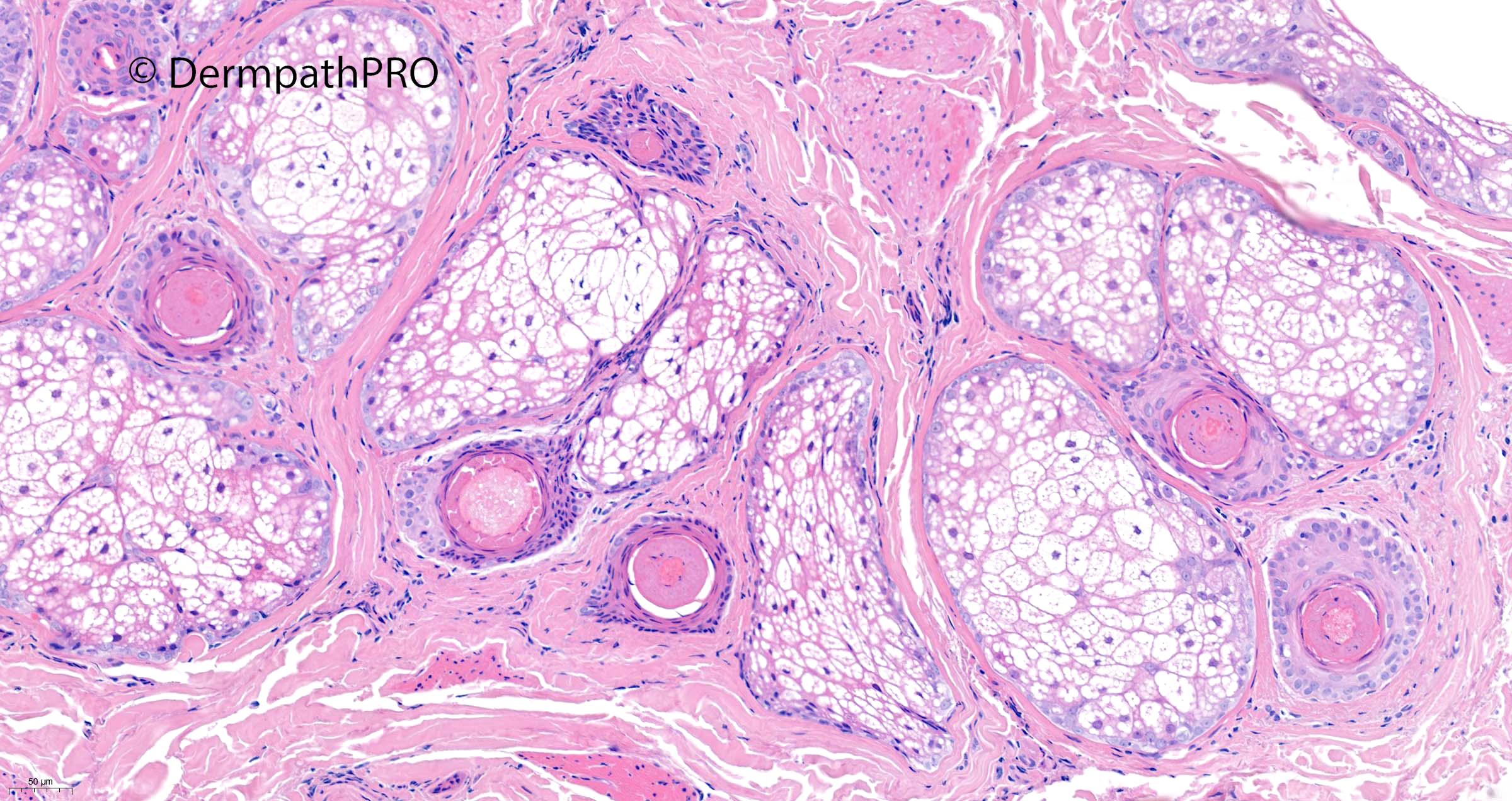
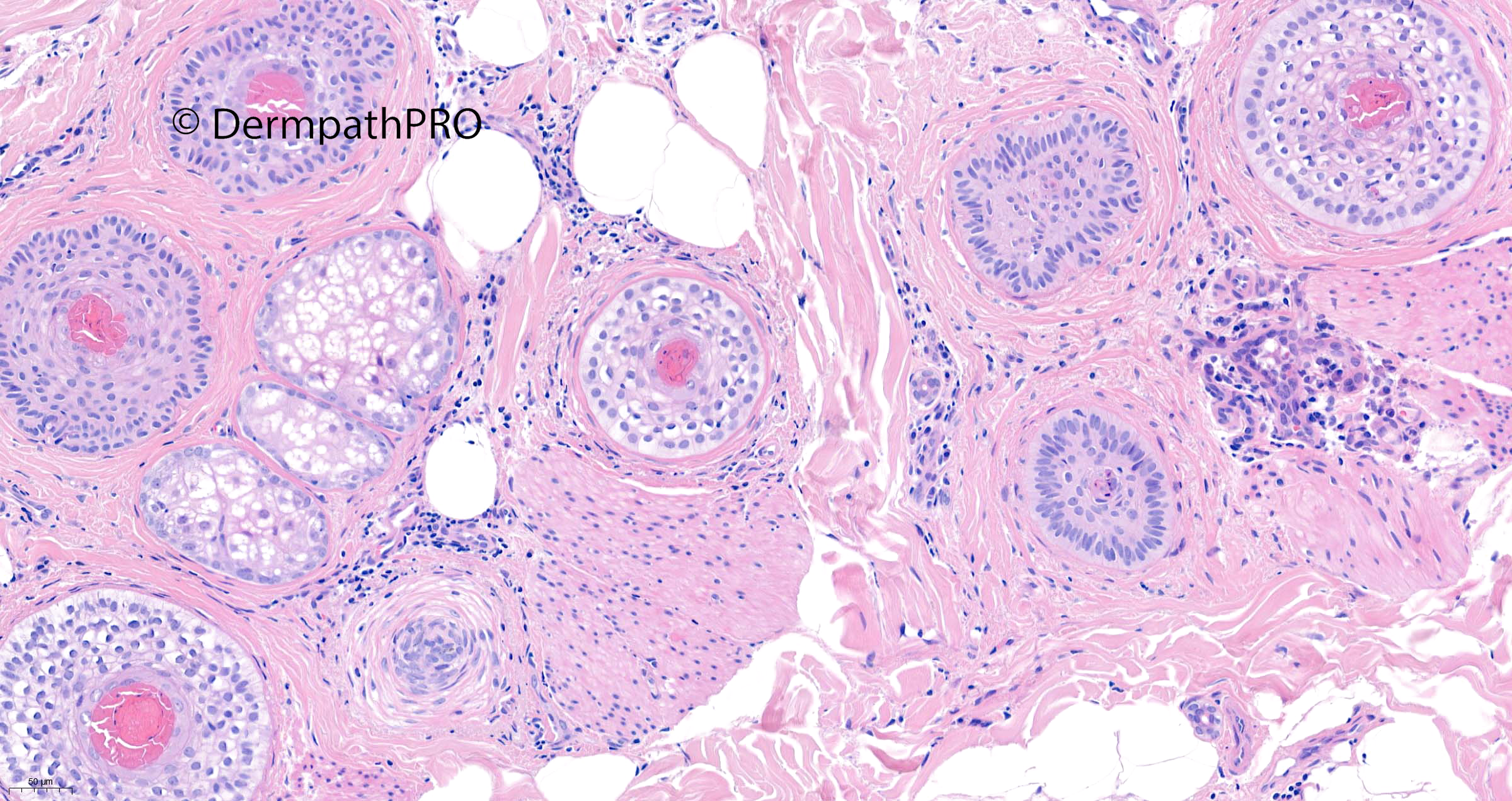
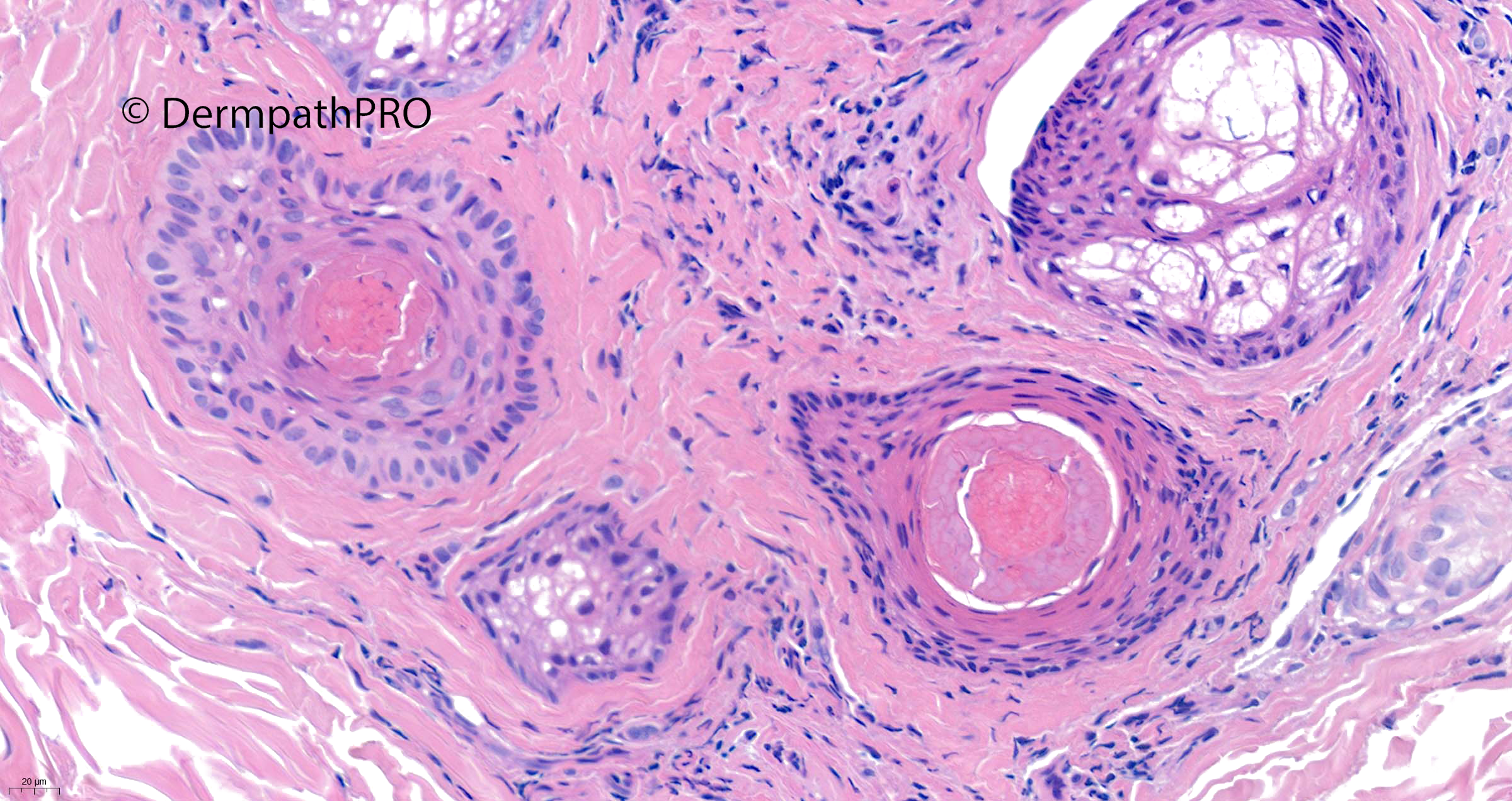
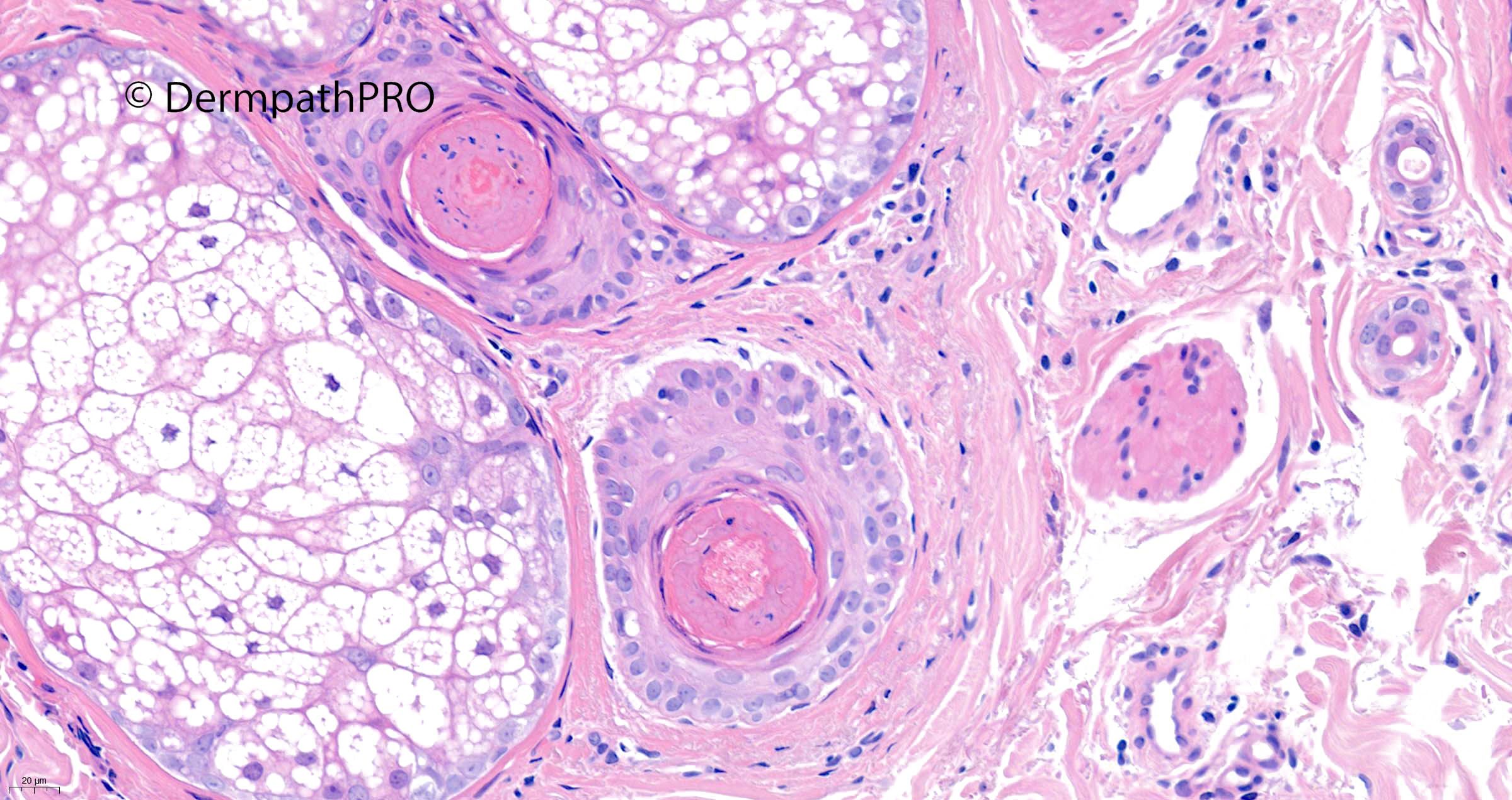
Jump to content
In this section we have spot diagnoses posted on a daily basis since June 2010, now over 4000! You can review the archived cases and read the suggested diagnoses by users and the final comment by the contributors.
Case are uploaded each week day by 10 am UK time with the correct diagnosis will generally be posted at 8 pm UK time. Why not view the most recent spot diagnosis and proffer a diagnosis?

Case Number : Case 2946 - 21 October 2021 Posted By: Saleem Taibjee

Please read the clinical history and view the images by clicking on them before you proffer your diagnosis.
Submitted Date :
   (0 reviews)

68F hairloss over months ?scarring alopecia


  • Like 1
  Report Record

User Feedback


Richard Logan

Posted

It's almost a year since the last posting of a case of alopecia, also by Saleem  (case 2697, 5th November 2020). I'm something of a novice in assessing alopecia biopsies, but have been guided by Saleem's last case and also an excellent discussion on Arti Bakshi's case 1644, 13th October 2016.

This is obviously not scarring alopecia, as there isn't any scarring and the sebaceous glands are preserved.  There is considerable miniaturisation of the follicles with an increase of telogen and catagen follicles.  Slides 4 and 5 perhaps show some evidence of peri-bulbar lymphocytic inflammation. I don't see any pigment casts.

I therefore agree with Meenakshi Batrani that this is alopecia areata, probably more in its chronic, rather than acute phase.

Share this comment


Link to comment
share_externally

4 hours ago, Richard Logan said:

It's almost a year since the last posting of a case of alopecia, also by Saleem  (case 2697, 5th November 2020). I'm something of a novice in assessing alopecia biopsies, but have been guided by Saleem's last case and also an excellent discussion on Arti Bakshi's case 1644, 13th October 2016.

This is obviously not scarring alopecia, as there isn't any scarring and the sebaceous glands are preserved.  There is considerable miniaturisation of the follicles with an increase of telogen and catagen follicles.  Slides 4 and 5 perhaps show some evidence of peri-bulbar lymphocytic inflammation. I don't see any pigment casts.

I therefore agree with Meenakshi Batrani that this is alopecia areata, probably more in its chronic, rather than acute phase.

Yes, agree, and would add a “swarm of bees” referred to lymphocytic infiltrate. 

Share this comment


Link to comment
share_externally

Saleem Taibjee

Posted

Yes, I thought this was a very nice example of alopecia areata.

You have already pointed out the many of the features.

The one other important clue, and actually the main reason for including this particular case, is that a number of follicles show a pecular pink cornified centre.

This is well-described in alopecia areata. They can be referred to (as in Whiting DA. Histopathologic features of alopecia areata. Arch Dermatol. 2003;139:1555-1559) as "nanogen hairs, found in the middle or upper dermis, are minute and cycle rapidly, showing mixed features of anagen, catagen, and telogen. They may contain central pinkish remnants of inner root sheath, but they lack hair shafts, perhaps accounting for the empty infundibula often seen in alopecia areata." They are also described in Sperling's excellent "An Atlas of Hair Pathology With Clinical Correlations". 

I have tried to highlight this phenomonen in several of the images shown above.

It probably corresponds to the yellow dots appearance which dermoscopy enthusiasts describe in alopecia areata i.e. 'empty follicles'.

BW, Saleem

Share this comment


Link to comment
share_externally


Join the conversation

You can post now and register later. If you have an account, sign in now to post with your account.

Guest
Add a comment...

×   Pasted as rich text.   Paste as plain text instead

  Only 75 emoji are allowed.

×   Your link has been automatically embedded.   Display as a link instead

×   Your previous content has been restored.   Clear editor

×   You cannot paste images directly. Upload or insert images from URL.

Loading...

×
×
  • Create New...