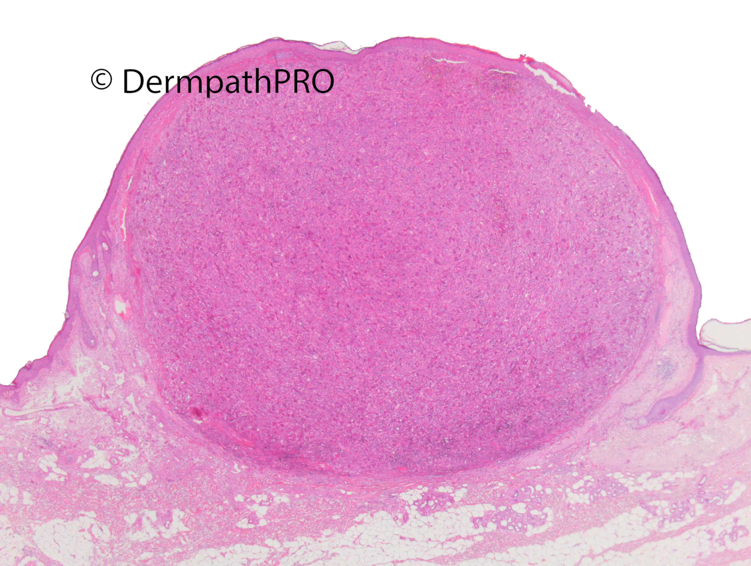
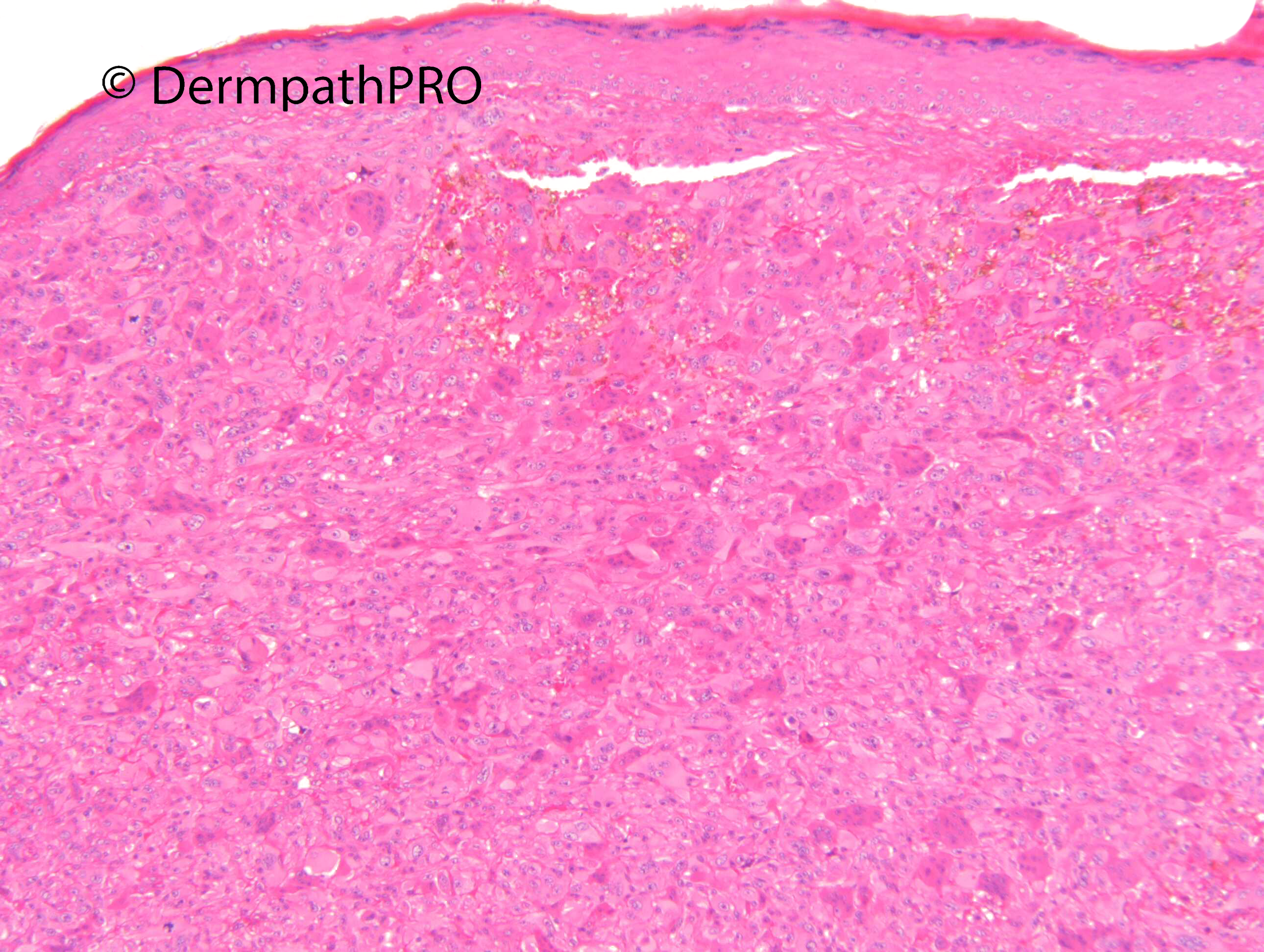
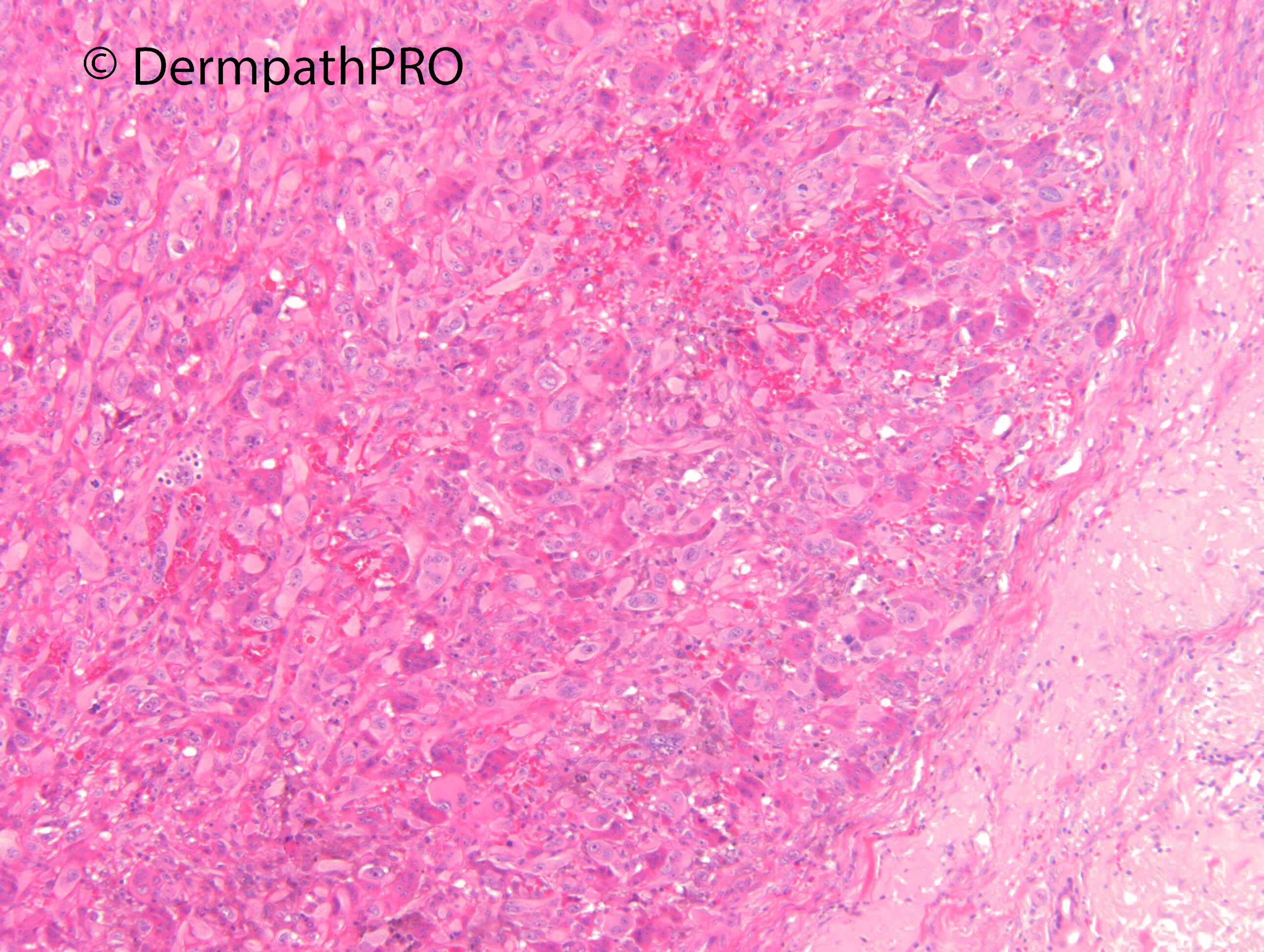
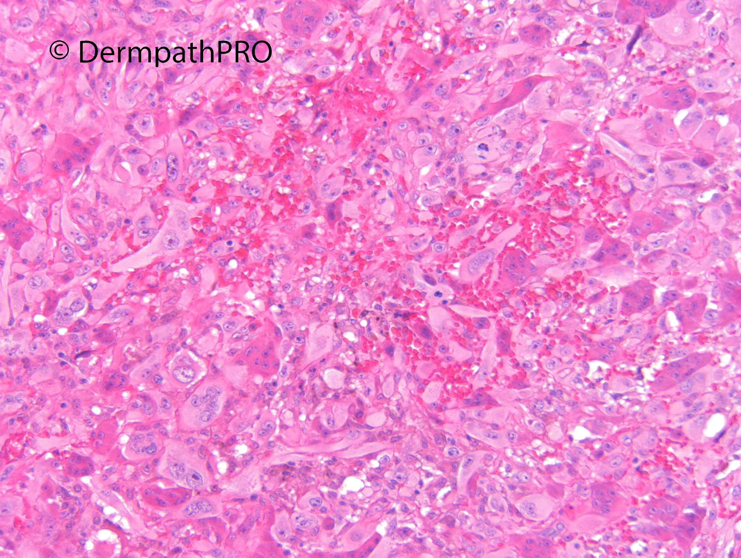
Jump to content
In this section we have spot diagnoses posted on a daily basis since June 2010, now over 4000! You can review the archived cases and read the suggested diagnoses by users and the final comment by the contributors.
Case are uploaded each week day by 10 am UK time with the correct diagnosis will generally be posted at 8 pm UK time. Why not view the most recent spot diagnosis and proffer a diagnosis?

Case Number : Case 2947 - 22 October 2021 Posted By: Dr. Richard Carr

Please read the clinical history and view the images by clicking on them before you proffer your diagnosis.
Submitted Date :
   (0 reviews)

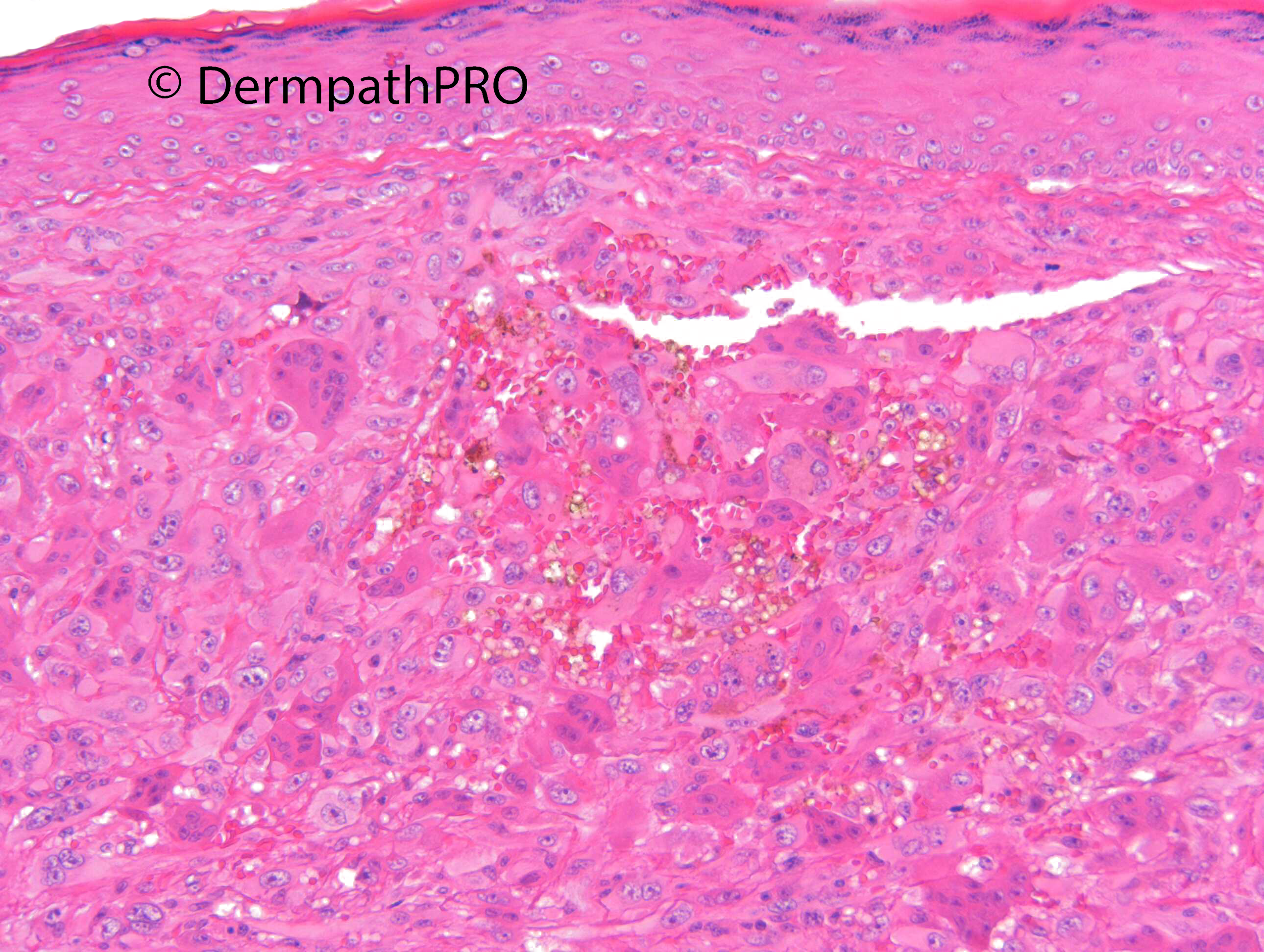
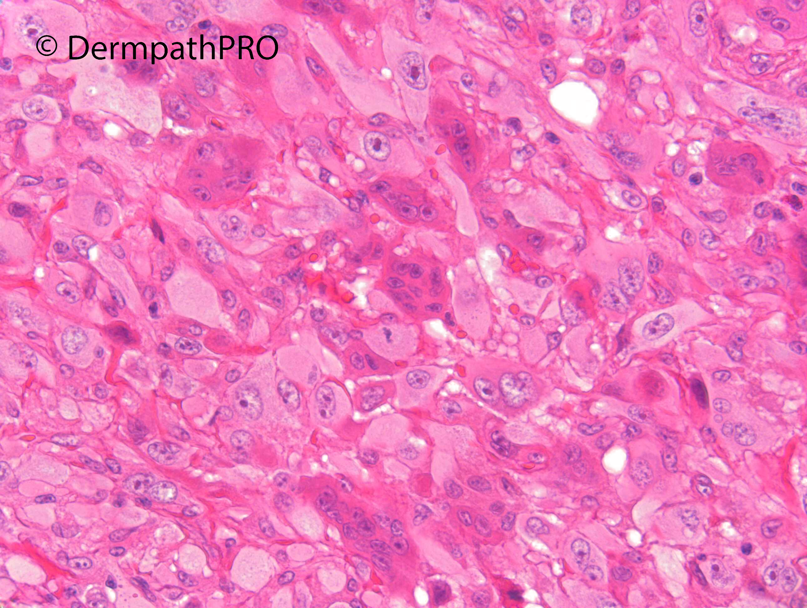
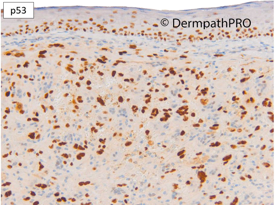
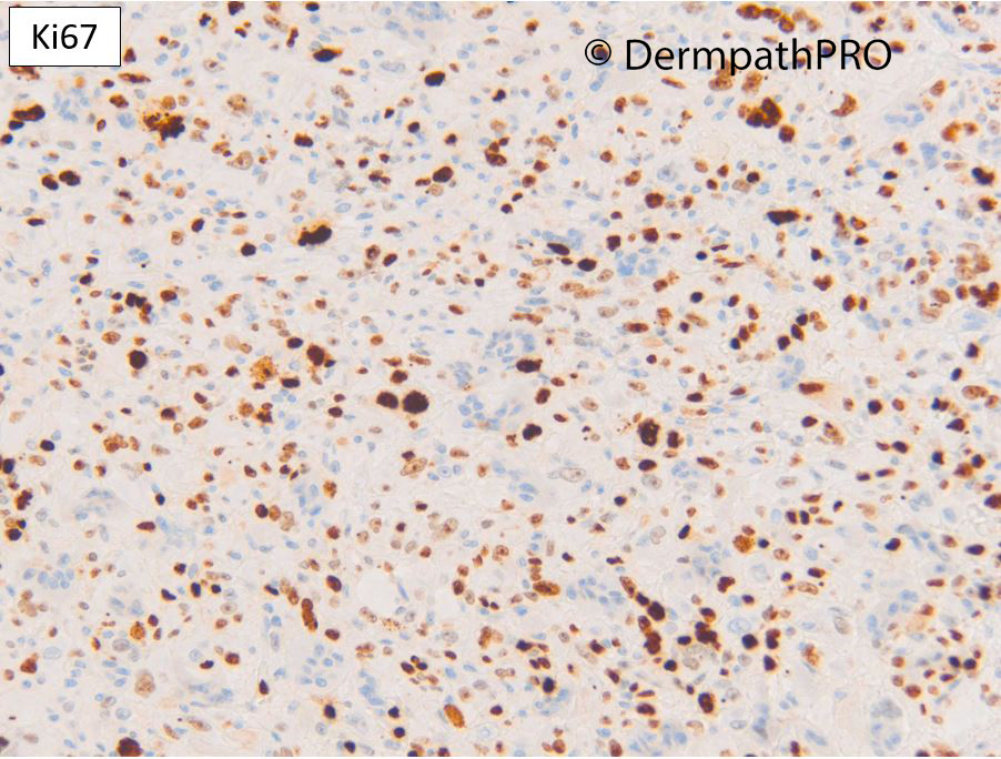
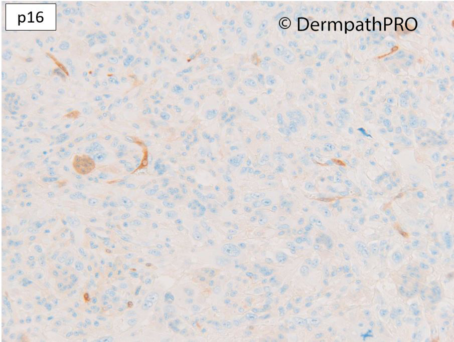
Site not stated. ?BCC ?Melanoma. Case c/o Dr Laszlo Igali


  • Like 1
  Report Record

User Feedback


Richard Logan

Posted (edited)

Agree.


See other examples on this website:

Spot Diagnosis Case 541  5th July 2012

More/Resources/On-line course/Case 45 18th February 2012

More/Resources/On-line course/Case157 24th February 2012


Edited by Richard Logan
Additional informaiton

Share this comment


Link to comment
share_externally

Dr. Richard Carr

Posted

This is case 541 5th July 2012 - thanks Richard L. Can you spot the difference?

image.thumb.png.53d76dbdca867ae73f2689402560fdcf.png

Share this comment


Link to comment
share_externally

Richard Logan

Posted

Richard, your case seems to have two different populations of cells.  The more eosinophilic cells contain more, smaller nuclei than the larger cells with glassy cytoplasm.  The cytology  in case 541 is rather more uniform.

Share this comment


Link to comment
share_externally

Dr. Richard Carr

Posted

That's entirely correct. I've asked dermpathpro team to replace the low power as I fear the over indulgent whiting to my original image has obscured a vital clue (present in 2nd image down on the left). Although the age and anatomic location is not stated I'd expect you to guess.  I also apologise for omitting the sex = female and the age = 97 years (but you could have guessed "old"). 

Share this comment


Link to comment
share_externally

Richard Logan

Posted

Seems like I'm the only one playing this game!

There are a lot of extravasated red cells, so I'm wondering about an aneurysmal fibrous histiocytoma, but that doesn't really explain the two different types of giant cells.  As far as anatomical location, perhaps trunk?

Share this comment


Link to comment
share_externally

But...why not a nodular melanoma? SOX10? If sox10 neg AFX could be a good spot. 

Share this comment


Link to comment
share_externally

Cem Leblebici

Posted

Melanoma, SCC, smooth muscle tumors and AFX are my differentials.  Need to perform more IHC, such as p40, PanCK, SMA, SOX10. I think osteoclastic type giant cells are associated with this epitheloid tumor. 


Share this comment


Link to comment
share_externally

Dr. Richard Carr

Posted

Okay we got there eventually. This is an AFX with osteoclast type giant cells. The severe solar elastosis would be a clue to sun-exposed head & neck skin. To my great surprise I had not posted an AFX on the platform.

I have tended to use only 4 markers (up till recently): CD10 (diffuse positive in nearly all AFX and uncommonly diffusely strongly positive in the main diffferentials which are SCC and Melanoma). p16 and a pan-keratin for carcinoma and S100 for melanoma. I reserved other markers like CD30 & vascular markers for cases with other clues. Leiomyosarcomas on the severely sun-damaged skin of the H&N are very uncommon and management is same as AFX or AFX-type cutaneous sarcoma (at term which I strongly prefer to "pleomorphic dermal sarcoma") so it's not really an important differential so I don't do muscle markers but be aware AFX / AFX-type cutaneous sarcomas can be SMA positive.  

For the more monomorphic spindle cell AFX it can be difficult to distinguish from cellular scar or a benign fibrohistiocytic lesion and I've been routinely running p16, p53 & Ki67 for a while. Most AFX bar 1 have been highly aberrant null or weak cytoplasmic only for p16 (similar to SCC on severely sun-damaged skin) only 1 of 20 cases showed a nice mosaic pattern. p53 often shows widespread positive staining or a null pattern that is not compatible with a benign lesion in my experience. Ki67 is often higher in AFX than one would usually expect in a benign soft tissue tumour. In this case p16 is null (weak in osteoclast type giant cells), p53 moderately over-expressed and Ki67 moderately high all in keeping with a malignant lesion. The tumour was diffusely positive for CD10 and negative for other more specific histogenetic markers and is therefore an atypical fibroxanthoma with reactive osteoclastic giant cells.

You may see reactive osteoclastic giant cells in any tumour that is accompanied by interstitial haemorrhage (benign or malignant). I personally don't like the term pleomorphic dermal sarcoma for lesions that are AFX-like but that have ill-defined or infiltrative borders and / or frankly invade the subcutis. I prefer the term AFX-like cutaneous sarcoma. I have yet to see a case metastasise (and I've reported >40 cases locally over the years, and many more AFX) and the largest published series of so-called pleomorphic dermal sarcoma (AFX-type cutaneous sarcoma) had a metastatic rate of 10% in around 30 cases but I'm sure there will be referral bias as this was from a tertiary referral centre. The more monotonous, low-grade, types of melanoma (desmoplastic / neurotropic) and spindle cell carcinoma rarely metastasise in practice so the management of all should simply be complete excision with clear margins as for an SCC although sometimes extensive surgery is required to achieve complete excision due to indolent and extensive growth of some of these types of lesions that may also recur after several years. 

Share this comment


Link to comment
share_externally


Join the conversation

You can post now and register later. If you have an account, sign in now to post with your account.

Guest
Add a comment...

×   Pasted as rich text.   Paste as plain text instead

  Only 75 emoji are allowed.

×   Your link has been automatically embedded.   Display as a link instead

×   Your previous content has been restored.   Clear editor

×   You cannot paste images directly. Upload or insert images from URL.

Loading...

×
×
  • Create New...