Jump to content
In this section we have spot diagnoses posted on a daily basis since June 2010, now over 4000! You can review the archived cases and read the suggested diagnoses by users and the final comment by the contributors.
Case are uploaded each week day by 10 am UK time with the correct diagnosis will generally be posted at 8 pm UK time. Why not view the most recent spot diagnosis and proffer a diagnosis?

Case Number : Case 2952 - 29 October 2021 Posted By: Dr. Richard Carr

Please read the clinical history and view the images by clicking on them before you proffer your diagnosis.
Submitted Date :
   (0 reviews)

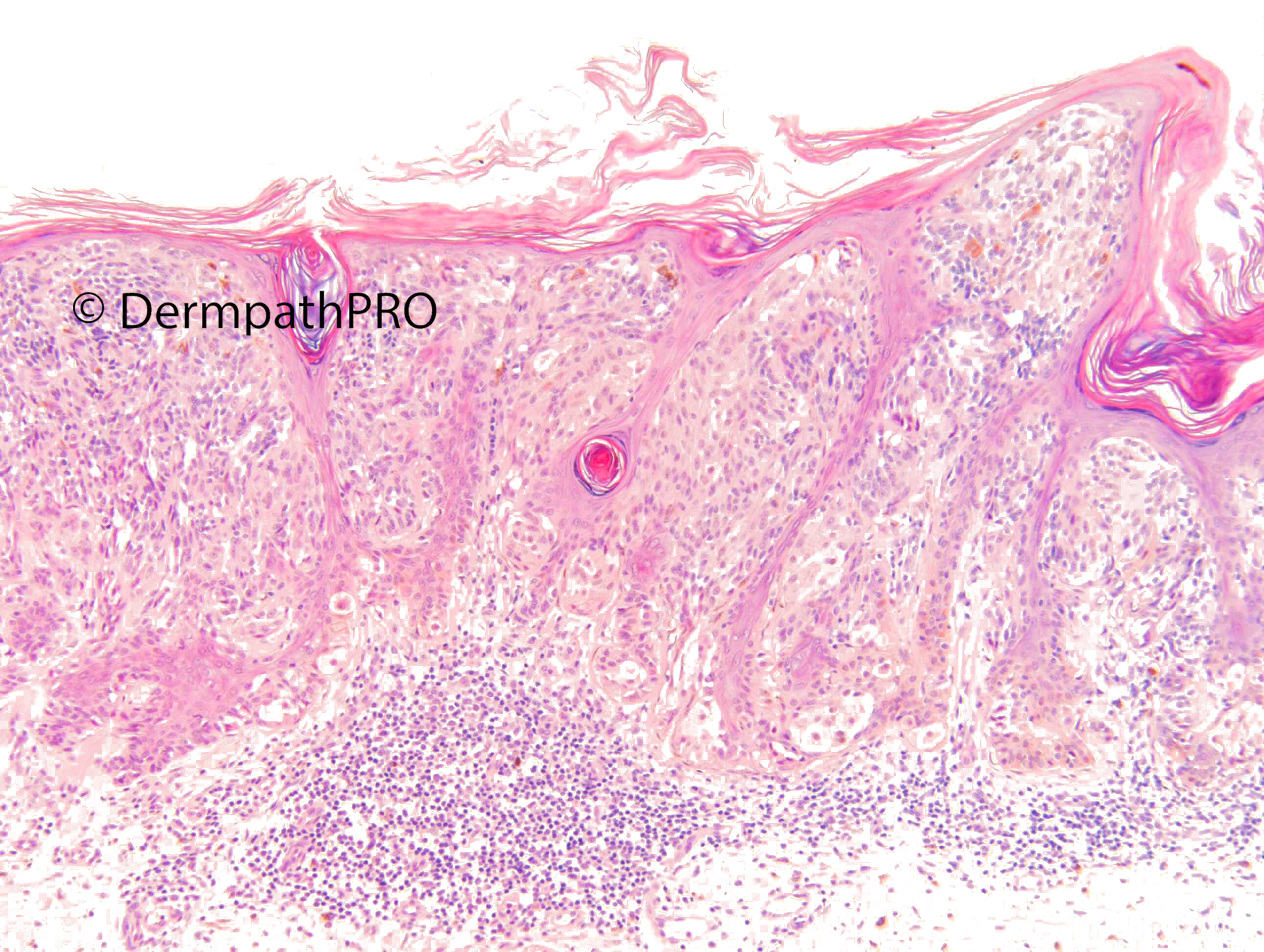
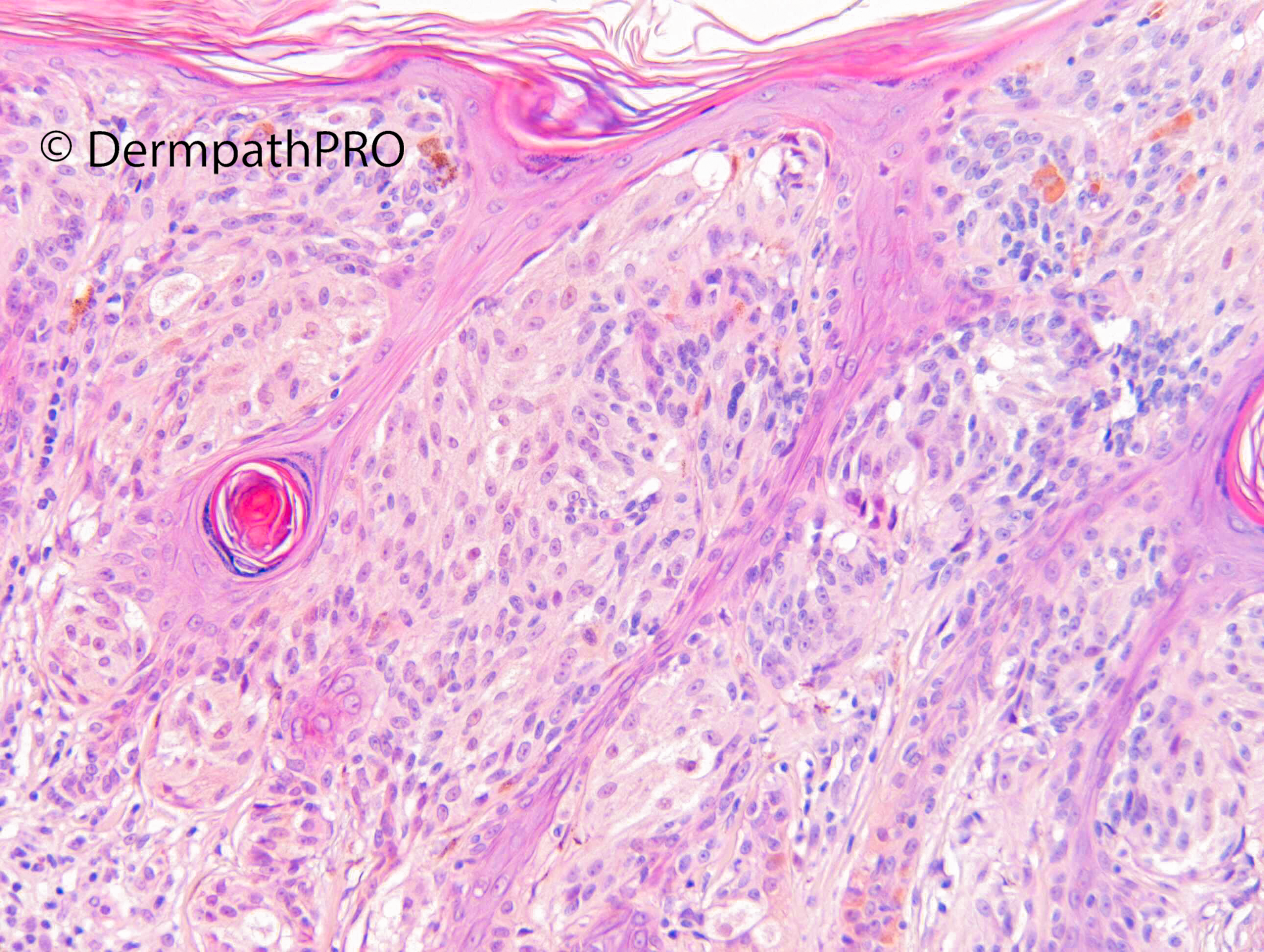
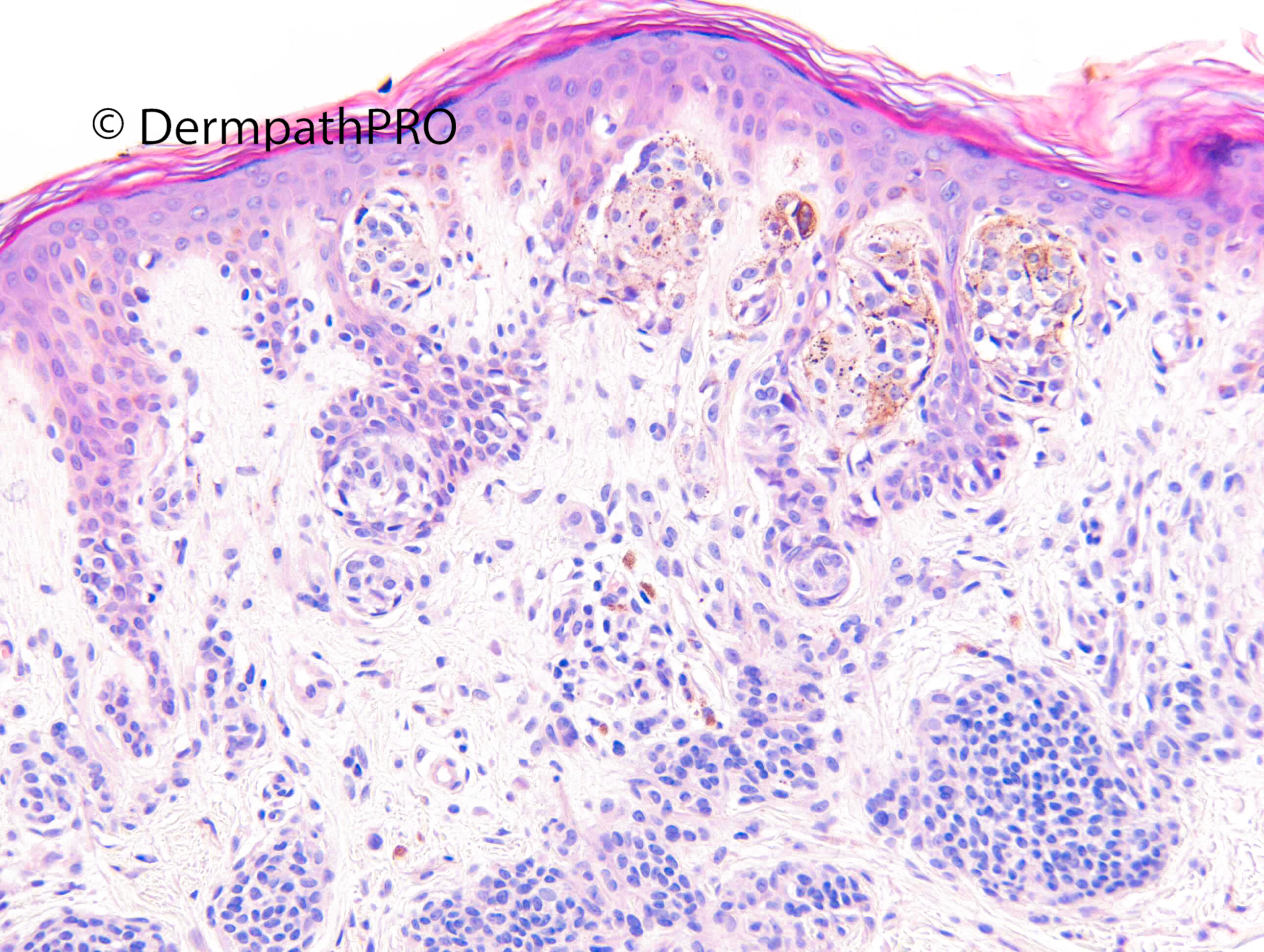
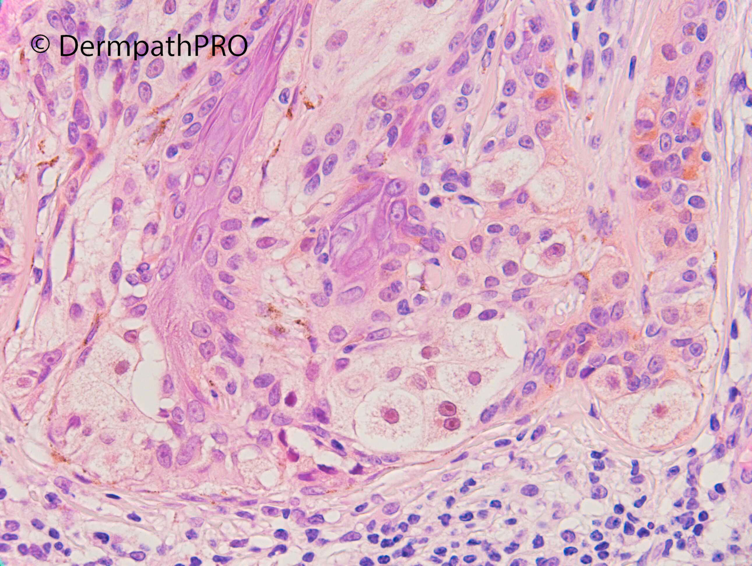
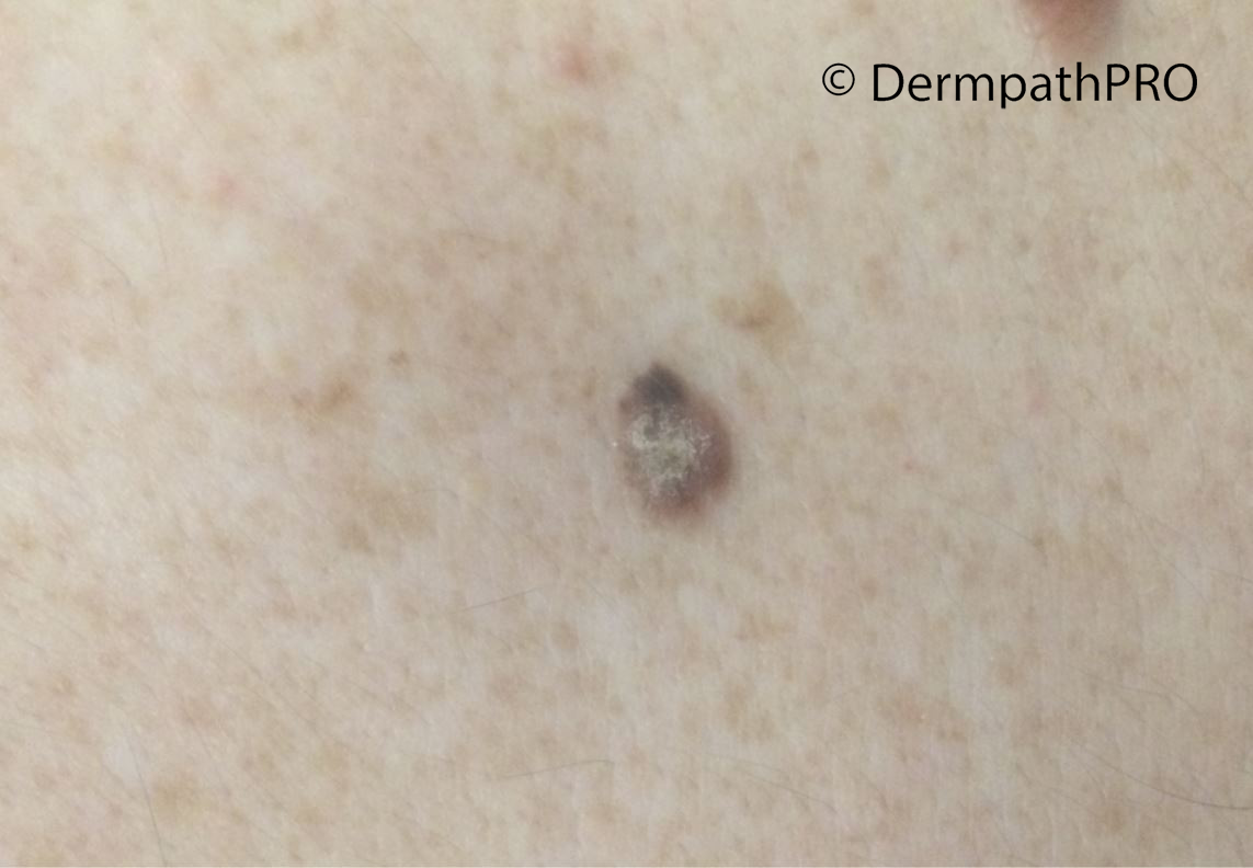
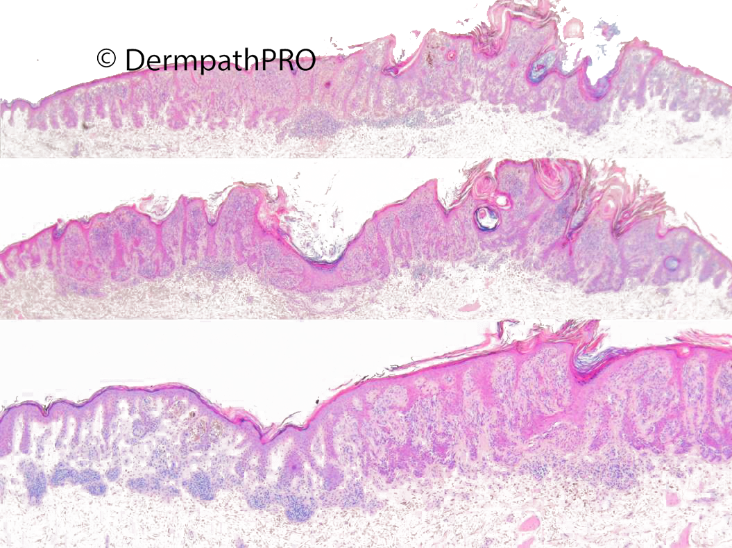
M50. Lower back. 1/52 ago noticed lesion. 10 x 7mm erythematous, hyperkeratotic, papillomatous papule, irregular pigmentation (dermoscopy) ?SEBK ?Mole

  • Like 1
  Report Record

User Feedback


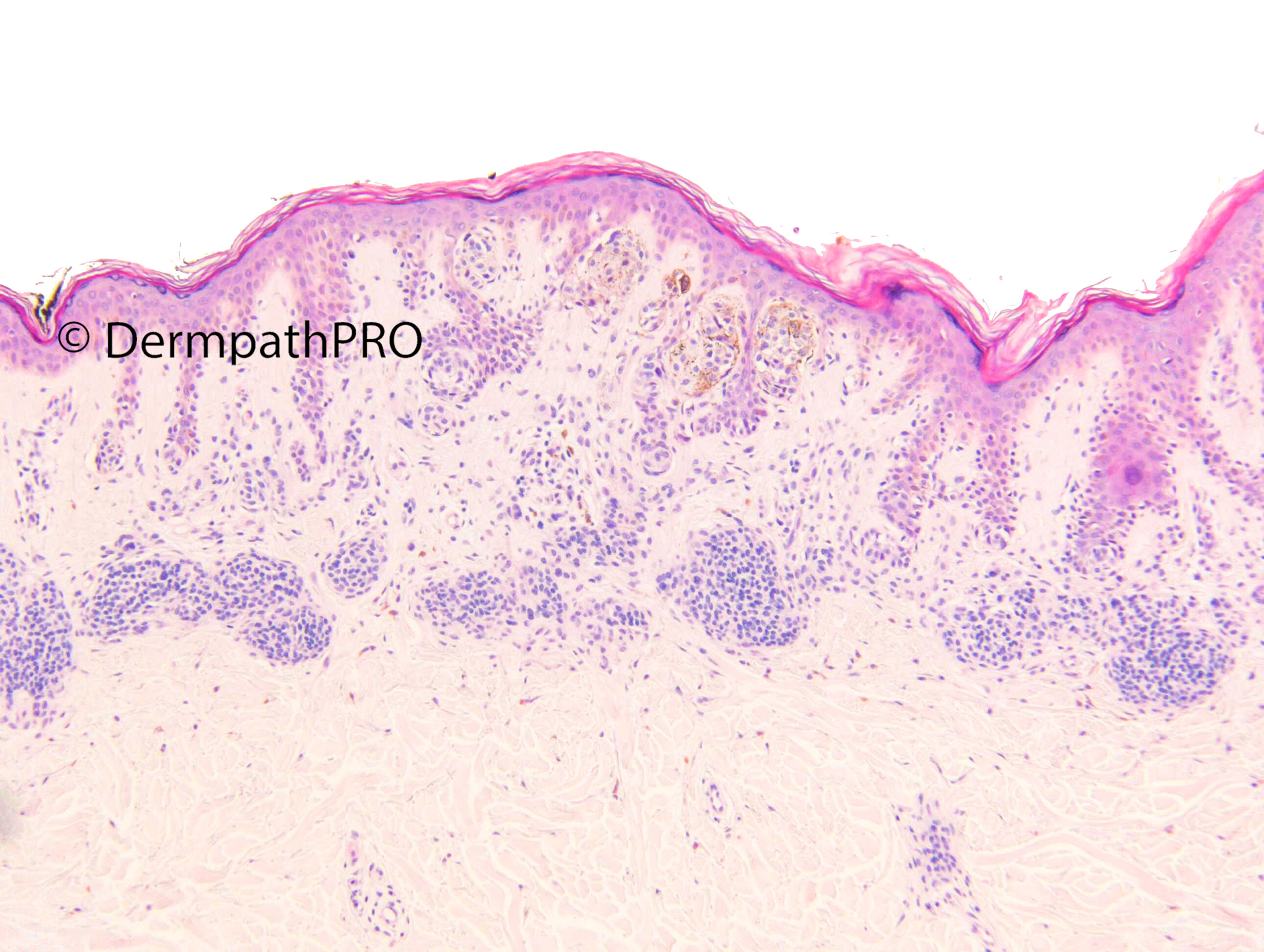
The melanocytic lesional combination of a Spitz nevus ( spindle cell type in this case ) overlying a common acquired nevus, is an unusual but well known event. I think this could be the case. 

Share this comment


Link to comment
share_externally

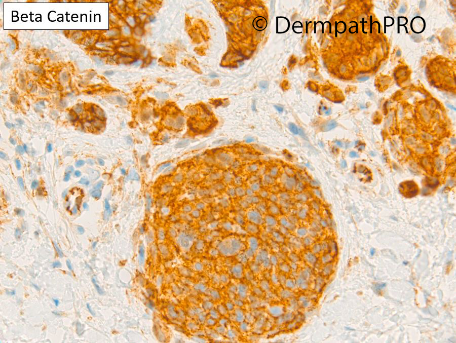
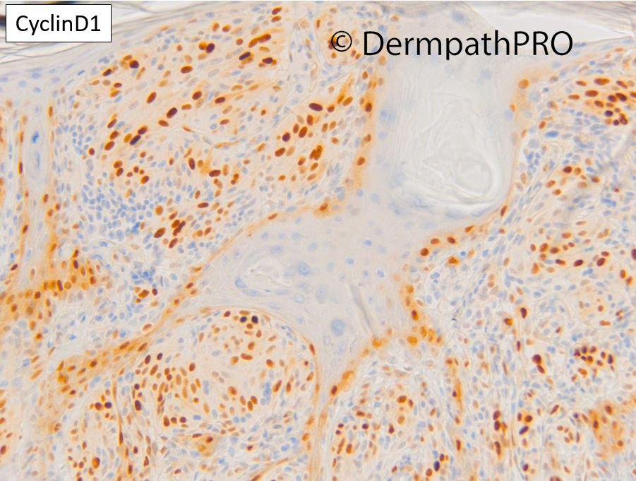
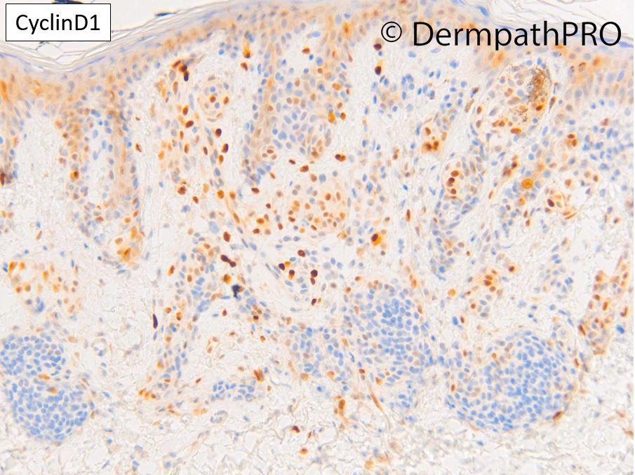
Beta Catenin is negative. CyclinD1 mosaic pattern. I was a bit suggested by a weird junctional variant of DPN, but with this IHC…—>combined nevus. 

Share this comment


Link to comment
share_externally

Meenakshi Batrani

Posted

Difficult to classify. Melanocytic nevus with atypical epithelioid component which some believe is a superficial variant of DPN or it could be a combined nevus  IDMN+Spitiz or BAP -1 inactivated tumor (but Beta catenin is not overexpressed in sptiz), MELTUMP.

Share this comment


Link to comment
share_externally

Dr. Richard Carr

Posted

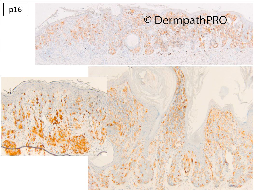
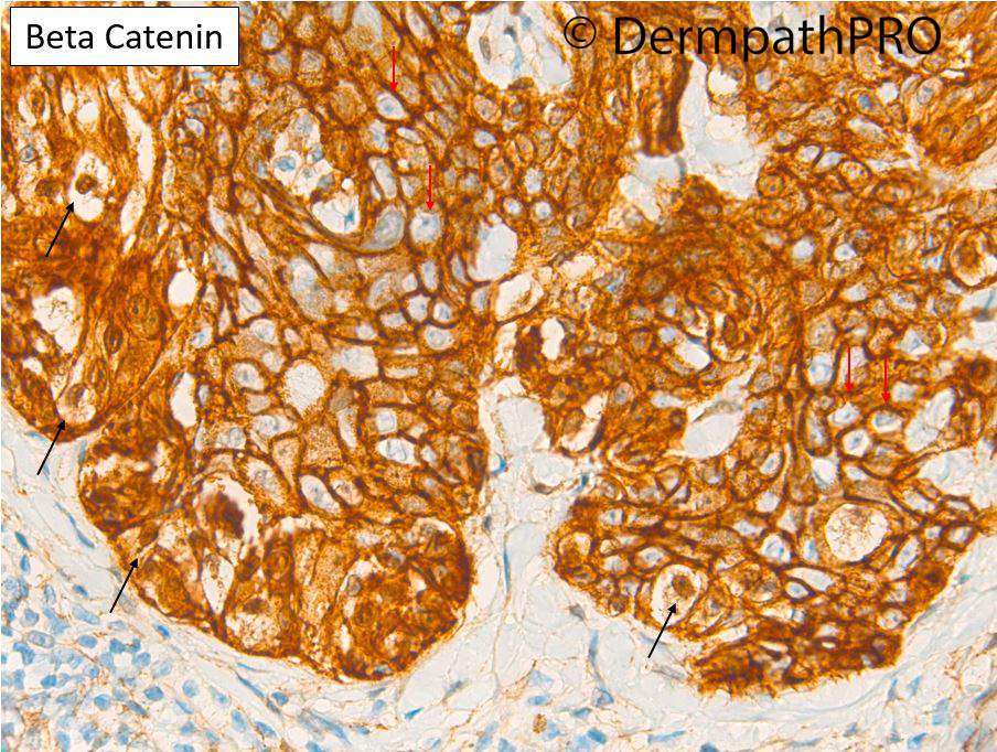
Some lesions are hard to classify. I thought there was a small ordinary naevocellular naevus (nuclear beta-catenin negative, cyclinD1 low) and a rather "Spitzoid" predominantly junctional clone with some DPN-like features and indeed there is some nuclear Beta-catenin in the clone (small black arrows) to compare with the membranous only pattern in the epidermal keratinocytes (red arrows). The increased cyclinD1 fits with DPN. p16 is retained mosaic but stronger in the naevus (being a marker of cellular senescence we can see reduced expression in juncationally active lesions that are benign). The cytology is pretty bland and the PRAME negative so I thought the lesion was essentially benign and did not want raise the suspicions to a MELTUMP so my final classificatioin favours a combined naevocellular & superficial / junctional DPN clonal type naevus! 

Share this comment


Link to comment
share_externally


Join the conversation

You can post now and register later. If you have an account, sign in now to post with your account.

Guest
Add a comment...

×   Pasted as rich text.   Paste as plain text instead

  Only 75 emoji are allowed.

×   Your link has been automatically embedded.   Display as a link instead

×   Your previous content has been restored.   Clear editor

×   You cannot paste images directly. Upload or insert images from URL.

Loading...

×
×
  • Create New...