Jump to content
In this section we have spot diagnoses posted on a daily basis since June 2010, now over 4000! You can review the archived cases and read the suggested diagnoses by users and the final comment by the contributors.
Case are uploaded each week day by 10 am UK time with the correct diagnosis will generally be posted at 8 pm UK time. Why not view the most recent spot diagnosis and proffer a diagnosis?

Case Number : Case 2916 - 09 September 2021 Posted By: Saleem Taibjee

Please read the clinical history and view the images by clicking on them before you proffer your diagnosis.
Submitted Date :
   (0 reviews)

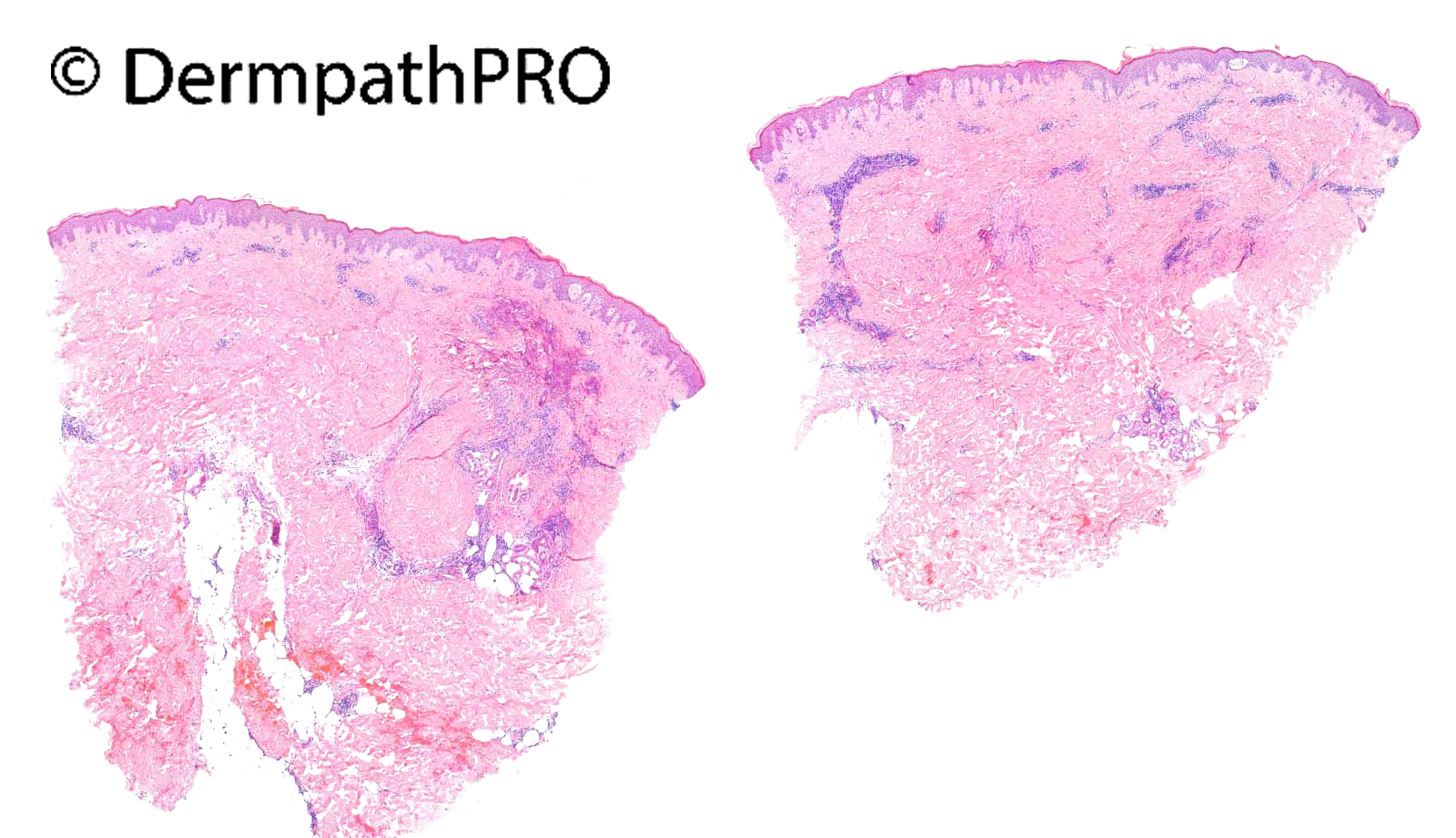
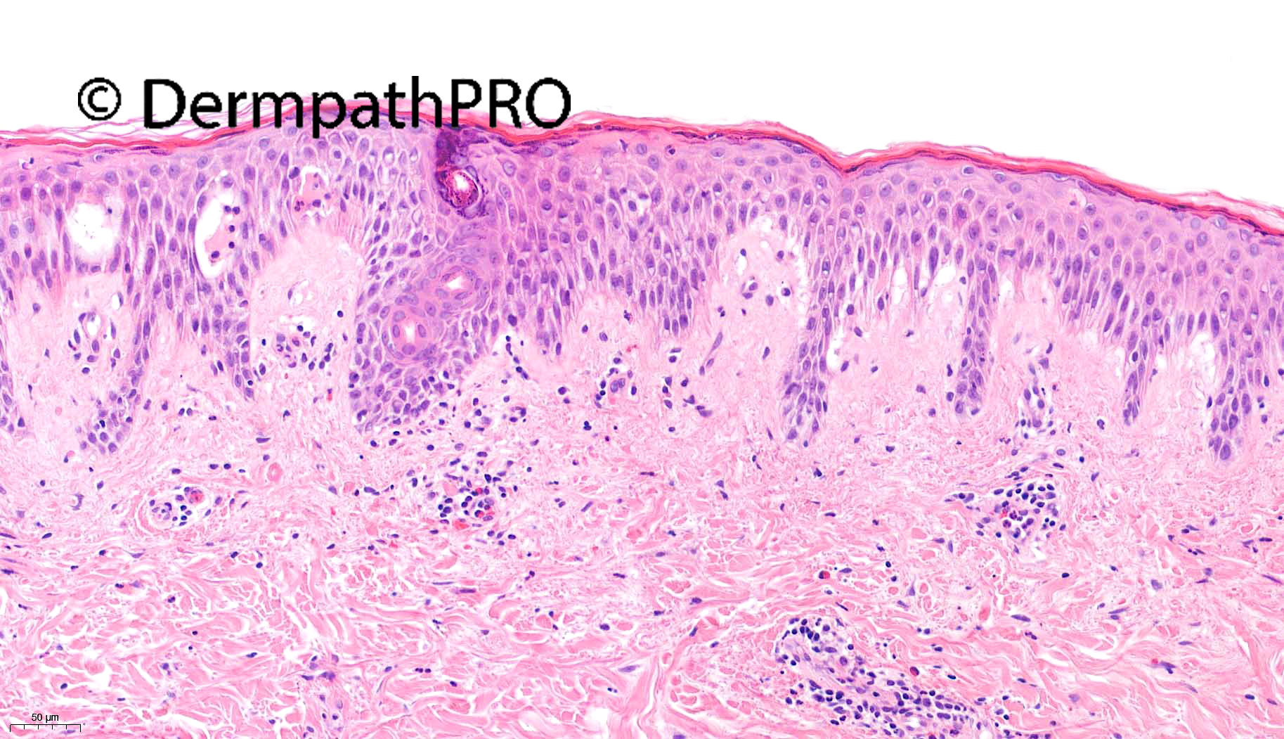
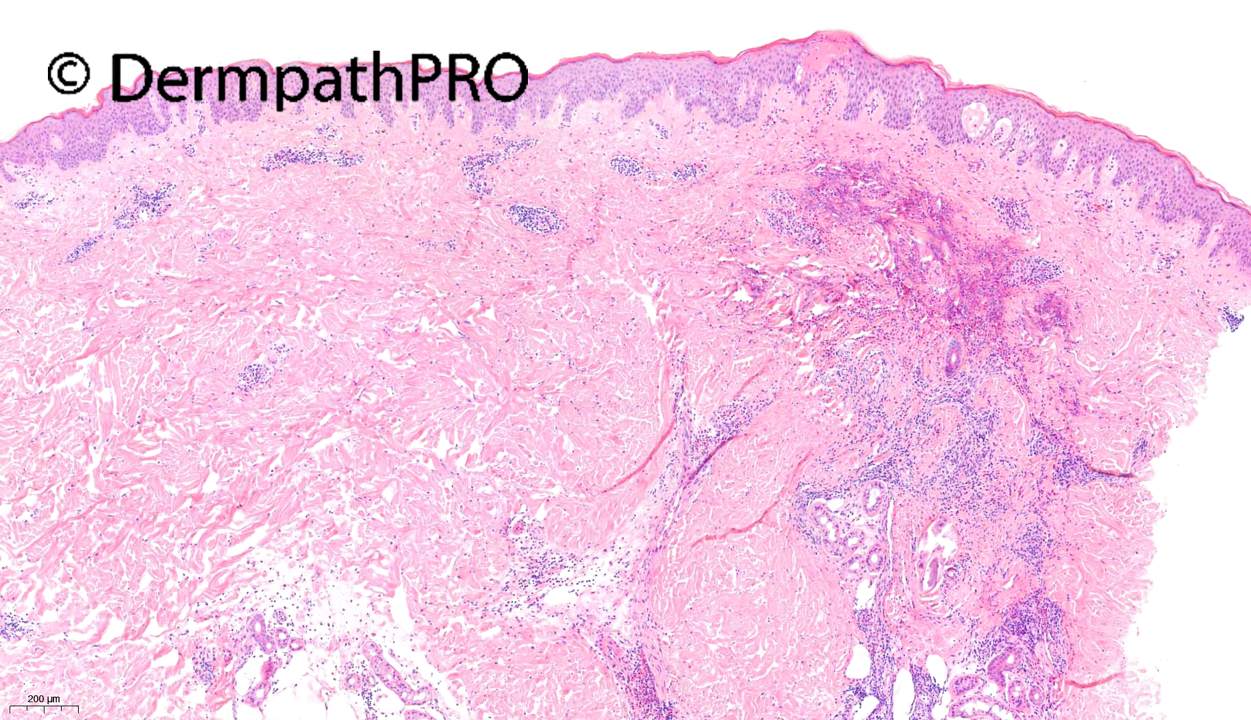
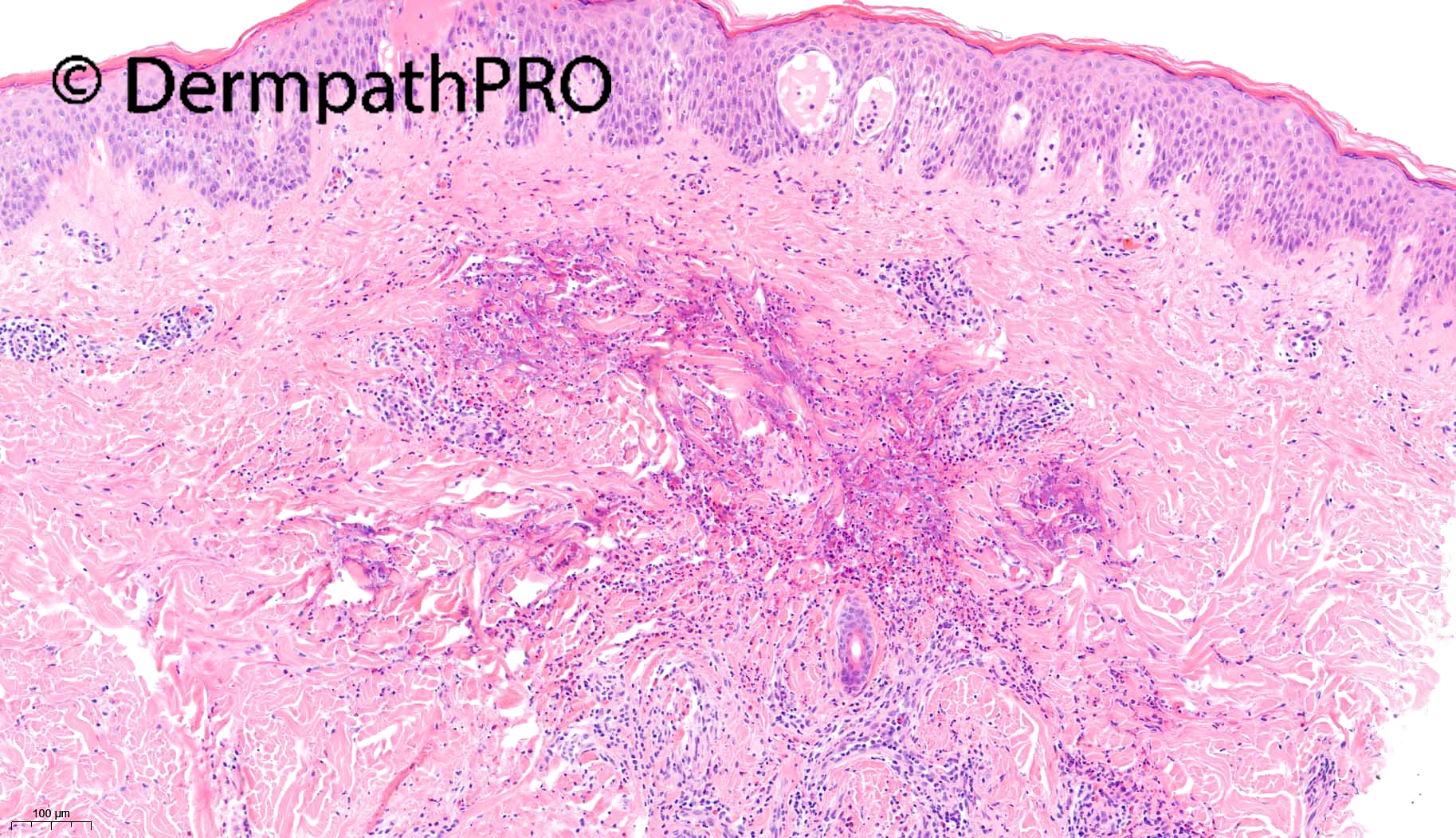
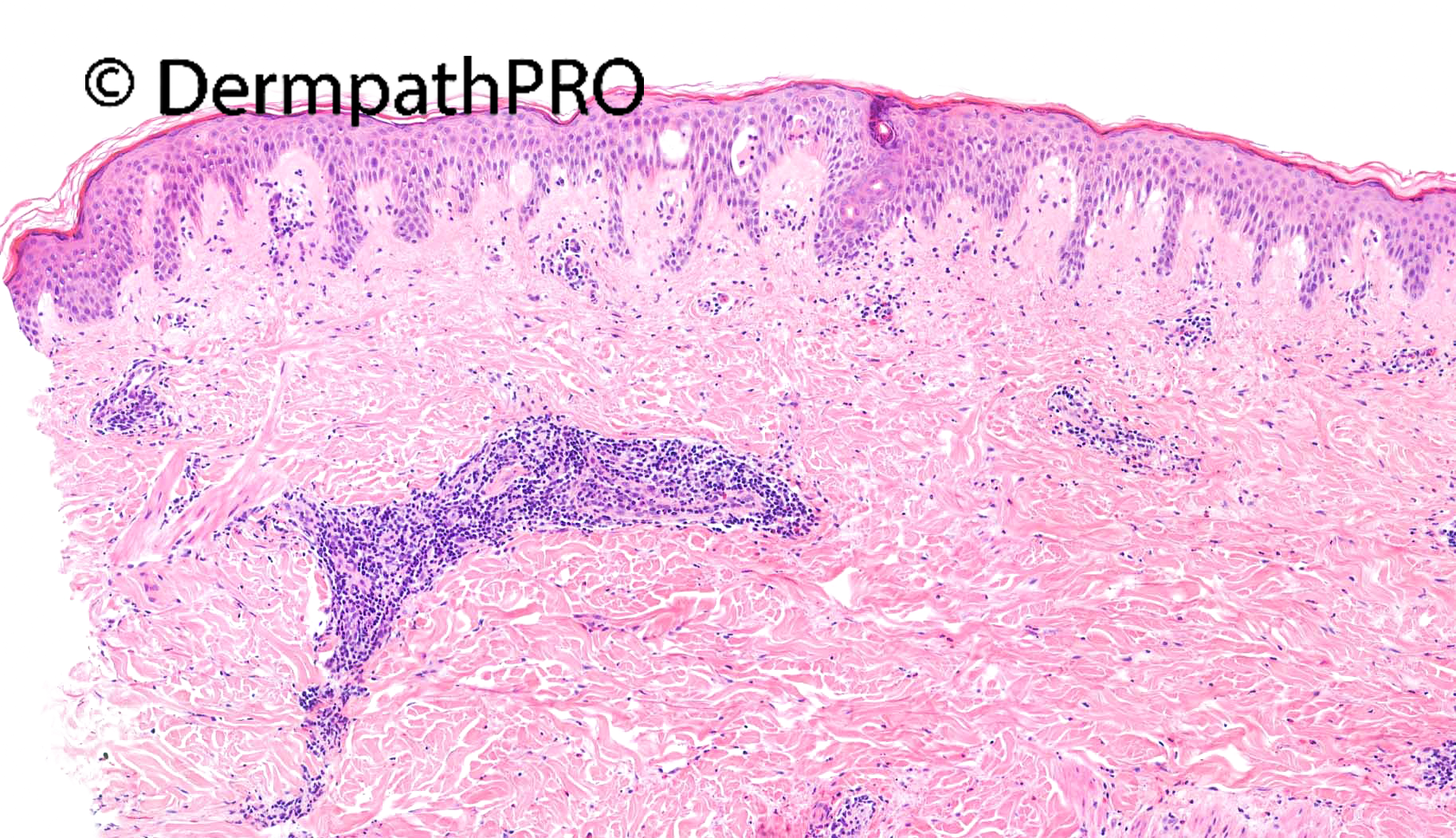
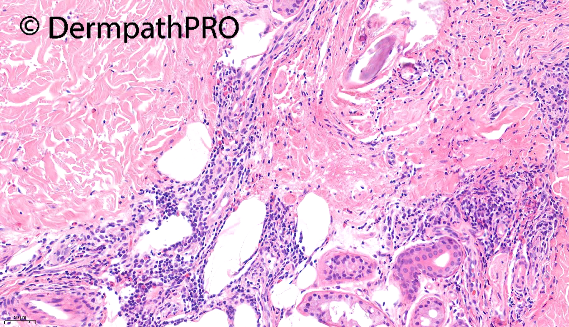
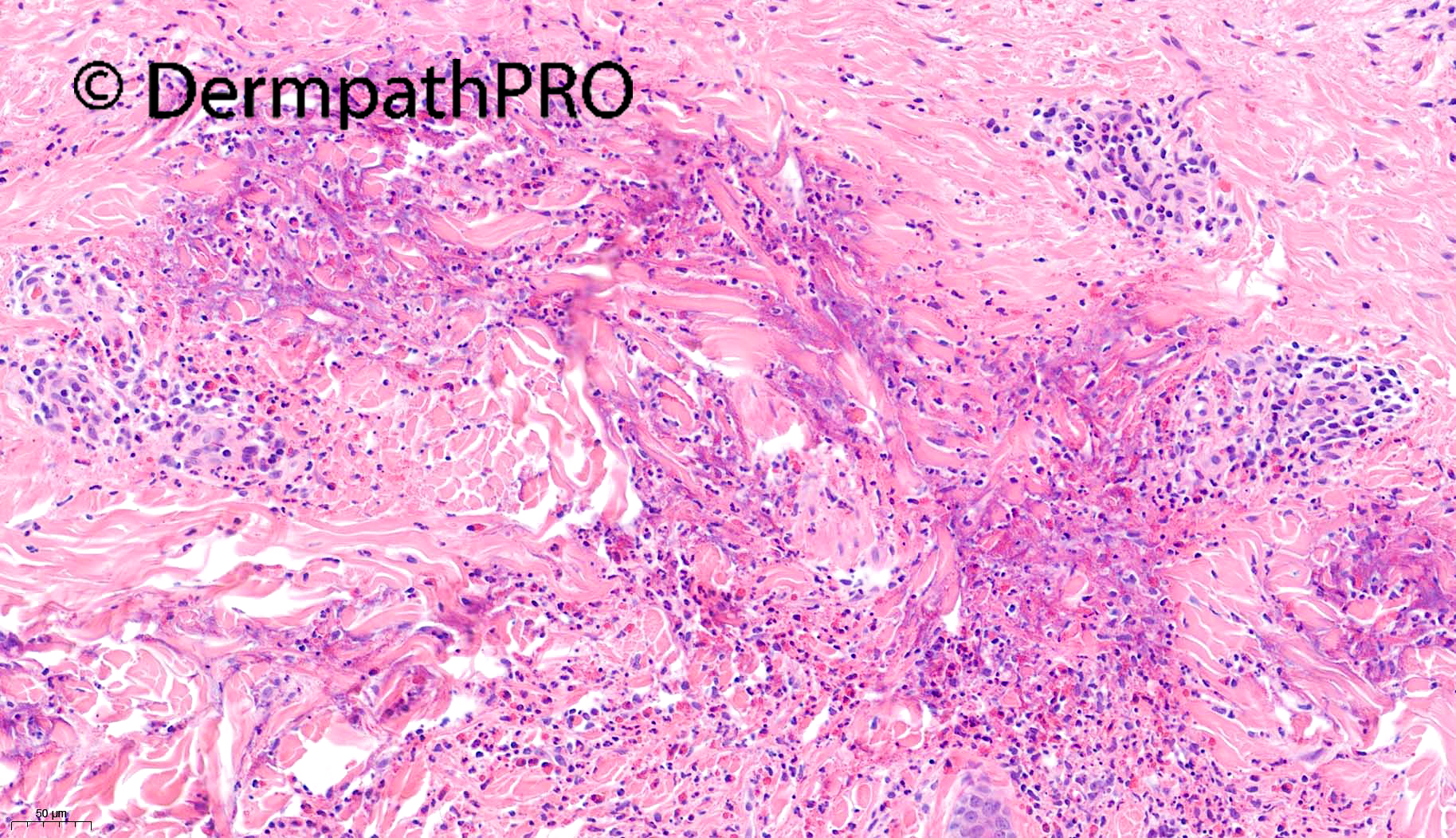
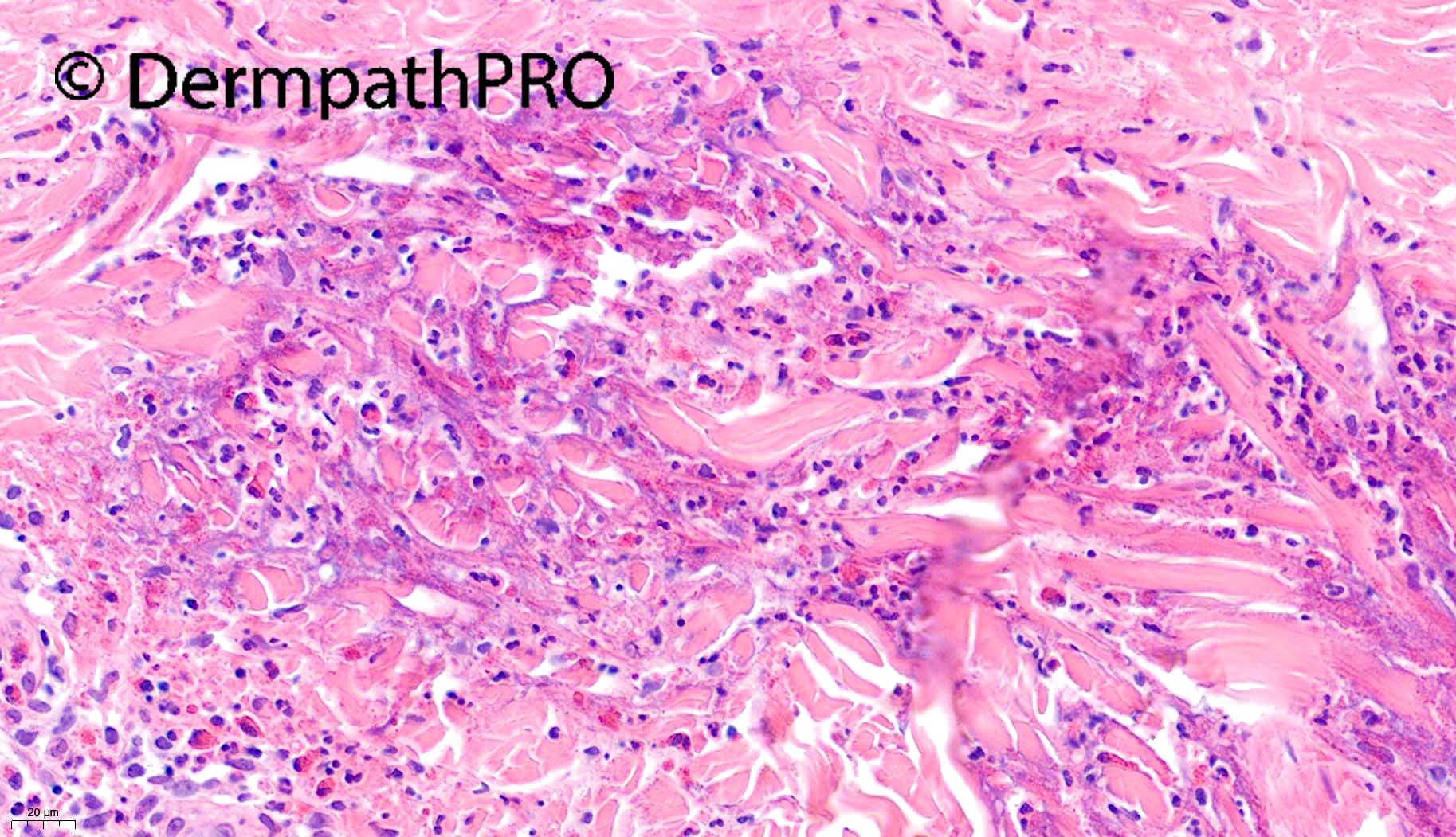
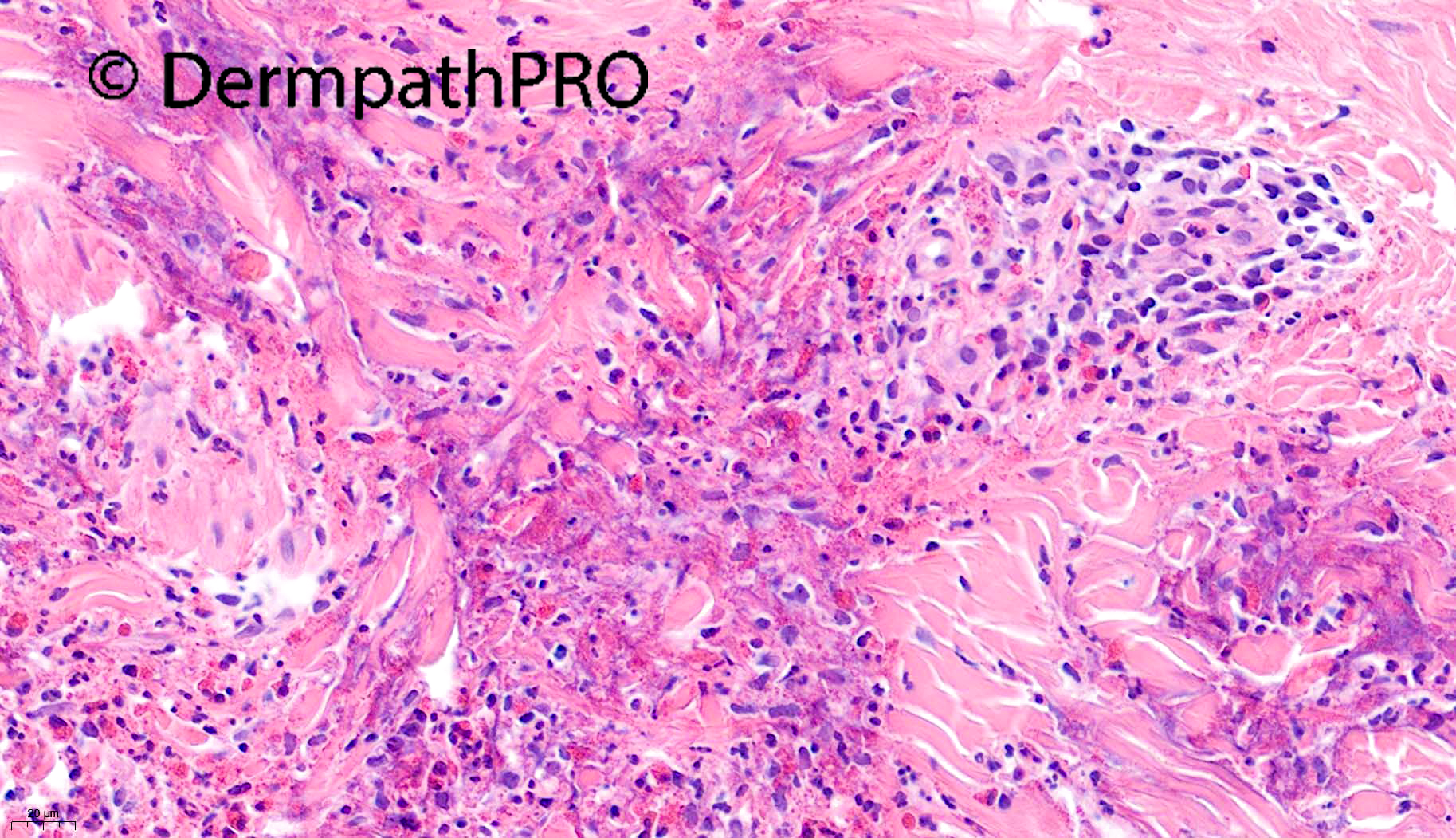
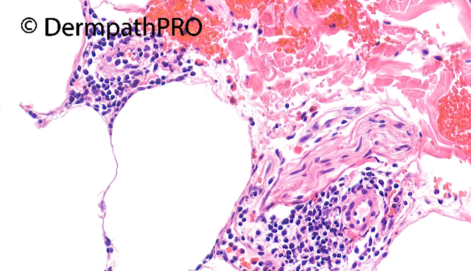
46M Polymorphic rash affecting elbows, lower back and legs since Sept 2020 ?dermatitis herpetiformis ?bullous pemphigoid ?autoimmune bullous dermatosis. History of renal cancer.


  • Like 1
  Report Record

User Feedback


Richard Logan

Posted

This is a fascinating mixture of pathological changes. 

There is a predominantly perivascular, lymphocytic infiltrate with prominent eosinophils, and an area of flame figure formation in the upper dermis.  The dermal papillae show vacuolar change and oedema, with some small neutrophilic microabscesses.

I was caught out by a previous case of Saleem's (2781, 4th March) where the diagnosis was dermatitis herpetiformis with prominent eosinophils, so I wonder if this might be another similar case.  The anatomical distribution would do for it.  Of course bullous pemphigoid and a drug reaction would need to be considered (especially with the history of renal cancer), and scabies must be excluded.   Eosinophilic cellulitis is unlikely given the widespread anatomical involvement.

I would suggest deeper sections and immunofluorescence.

Share this comment


Link to comment
share_externally

Found this: Rajpara A, Liolios A, Fraga G, Blackmon J. Recurrent paraneoplastic wells syndrome in a patient with metastatic renal cell cancer. Dermatol Online J. 2014 Jun 15;20(6):13030/qt35w8r1g3. PMID: 24945651.

Share this comment


Link to comment
share_externally

...but agree with Richard: clinical feature doesn’t fit well EC. On low magnification flame figure sounds me like lined up with the vesicular epidermal lesion. So Scabies would be a good diff. 

Share this comment


Link to comment
share_externally

Saman Fatah

Posted

This pattern has a number of differential as suggested by earlier comments. Those variably-sized merging little white spaces within oedematous papillary dermis in image 2, may well be incipient sub-epidermal separation/vesiculation rather than vaculoar interface changes.

BP worth exclusion with IMF +/-ELISA for BP180/230 though he is relatively young age but odd paraneoplastic antigen cross reactivity can occur.

Eosinophilic Dermatosis of Haematological Malignancy (EDHM) and Churg-Strauss are a couple of additional histological DDx that requires CPC. Not aware of EDHM (as the name suggests) in other neoplastic conditions and this used to be called insect-bite like reaction in CLL in earlier literature, but such nomenclature loses sensitivity and specificity with time as it is described in other diseases. 


Share this comment


Link to comment
share_externally

Saleem Taibjee

Posted

Hi. I'm afraid I don't have a final answer for this case. I have not had any update from the clinician.

My own thoughts are... yes there is focal intraepidermal and subepidermal clefting, but the most striking changes are in the reticular dermis with a dense perivascular and interstitial infiltrate comprising lymphocytes, histiocytes, neutrophils and numerous eosinophils with flame figures. I also feel that there is eosinophilic degeneration of the collagen itself, with a hint of a palisaded arrangement. There is some deeper perivascular inflammation also.

To me eyes, the additional neutrophils and, in particular, eosinophilic collagen degeneration (contrast with basophilic degeneration in Wegeners) is quite suggestive of allergic granulomatosis with polyangiitis (formerly Churg-Strauss). I also considered drug reaction, arthropod reaction (e.g. scabies), Well's and hypereosinophilic syndrome. Unfortunately we don't have immunofluorescence to further assess for immunobullous disease (e.g. DH, pemphigoid), but I felt the dermal changes made this less likely anyway.

I will let you know if I do get any further update.

BW

Saleem

Share this comment


Link to comment
share_externally


Join the conversation

You can post now and register later. If you have an account, sign in now to post with your account.

Guest
Add a comment...

×   Pasted as rich text.   Paste as plain text instead

  Only 75 emoji are allowed.

×   Your link has been automatically embedded.   Display as a link instead

×   Your previous content has been restored.   Clear editor

×   You cannot paste images directly. Upload or insert images from URL.

Loading...

×
×
  • Create New...