Jump to content
In this section we have spot diagnoses posted on a daily basis since June 2010, now over 4000! You can review the archived cases and read the suggested diagnoses by users and the final comment by the contributors.
Case are uploaded each week day by 10 am UK time with the correct diagnosis will generally be posted at 8 pm UK time. Why not view the most recent spot diagnosis and proffer a diagnosis?

Case Number : Case 2922 - 17 September 2021 Posted By: Dr. Richard Carr

Please read the clinical history and view the images by clicking on them before you proffer your diagnosis.
Submitted Date :
   (0 reviews)

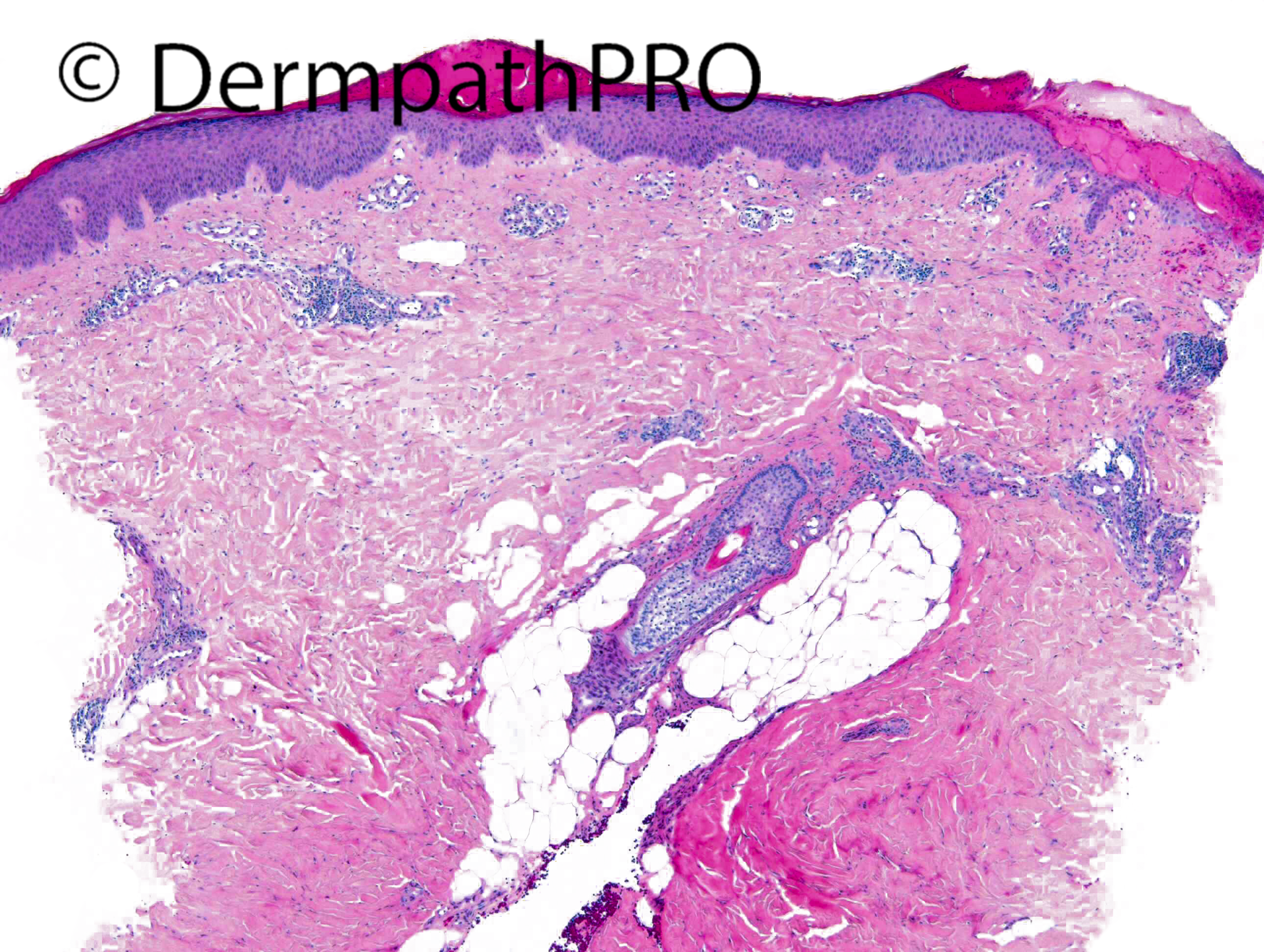
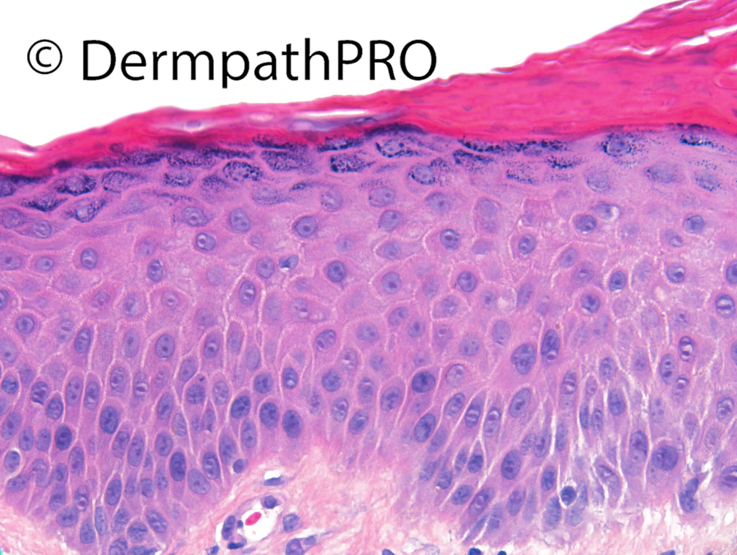
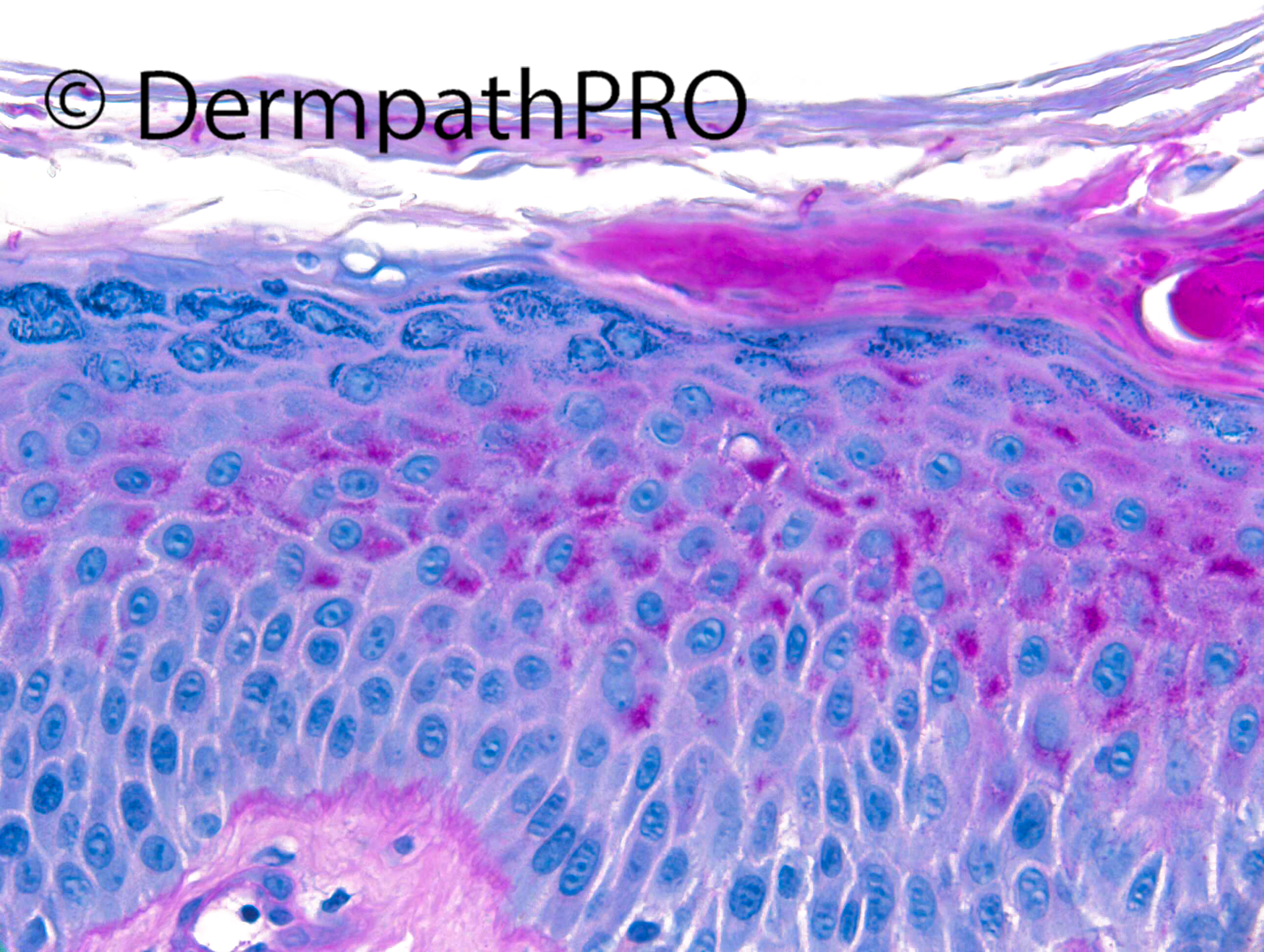
F40. Right calf. 3 yr hx itchy extending rash up right foot and leg, biopsy from active edge.


  Report Record

User Feedback


Richard Logan

Posted

A unilateral inflammatory rash should immediately raise suspicion of fungal infection.  There is sero-sanguinous exudation in the stratum corneum which would fit with that diagnosis.  However, there are quite a few eosinophils in the infiltrate which I wouldn't normally associate with fungal infection.  I suppose a parasitic condition such as larva migrans would have to be considered, but the length of the history is too long, and that diagnosis is usually obvious clinically.  PAS please.

Share this comment


Link to comment
share_externally

5 hours ago, Richard Logan said:

A unilateral inflammatory rash should immediately raise suspicion of fungal infection.  There is sero-sanguinous exudation in the stratum corneum which would fit with that diagnosis.  However, there are quite a few eosinophils in the infiltrate which I wouldn't normally associate with fungal infection.  I suppose a parasitic condition such as larva migrans would have to be considered, but the length of the history is too long, and that diagnosis is usually obvious Agree clinically.  PAS please.

First time I see a larva migrans in stratum corneum, instead of papillary dermis ( really I saw few cases until now ). Totally agree with Richard’s comment.  

Share this comment


Link to comment
share_externally

Saman Fatah

Posted

Is that a hyphae in the middle of stratum corneum in image 5? Clinically it is suggestive of a fungal infection until proven other wise. We learned from previously shared cases on Dermpathpro that the dermal changes can be very variable and sometime mimic other dermatoses. 
Looking forward to hear more and see PAS. 

Share this comment


Link to comment
share_externally

Dr. Richard Carr

Posted

Apologies for the Dermpathpro copyright covering the hyphae. Yes well done and excellent discussion. I'd read that eosinophils can be seen in tinea but I'd never seen a case in practice. I'm not sure if the eosinophils are therefore a bystander or due to some other aetiology but I suppose one should not dismiss tinea in the presence of eosinophils if there are other reasons to consider the diagnosis such as unilateral rash as discussed by Richard Logan. The hyphae are present in the H&E high power but barely discernible. I always do the PAS at step levels on the smaller biopsies (unless they are obvious on the H&E).

Share this comment


Link to comment
share_externally


Join the conversation

You can post now and register later. If you have an account, sign in now to post with your account.

Guest
Add a comment...

×   Pasted as rich text.   Paste as plain text instead

  Only 75 emoji are allowed.

×   Your link has been automatically embedded.   Display as a link instead

×   Your previous content has been restored.   Clear editor

×   You cannot paste images directly. Upload or insert images from URL.

Loading...

×
×
  • Create New...