Jump to content
In this section we have spot diagnoses posted on a daily basis since June 2010, now over 4000! You can review the archived cases and read the suggested diagnoses by users and the final comment by the contributors.
Case are uploaded each week day by 10 am UK time with the correct diagnosis will generally be posted at 8 pm UK time. Why not view the most recent spot diagnosis and proffer a diagnosis?

Case Number : Case 2925 - 22 September 2021 Posted By: Saleem Taibjee

Please read the clinical history and view the images by clicking on them before you proffer your diagnosis.
Submitted Date :
   (0 reviews)

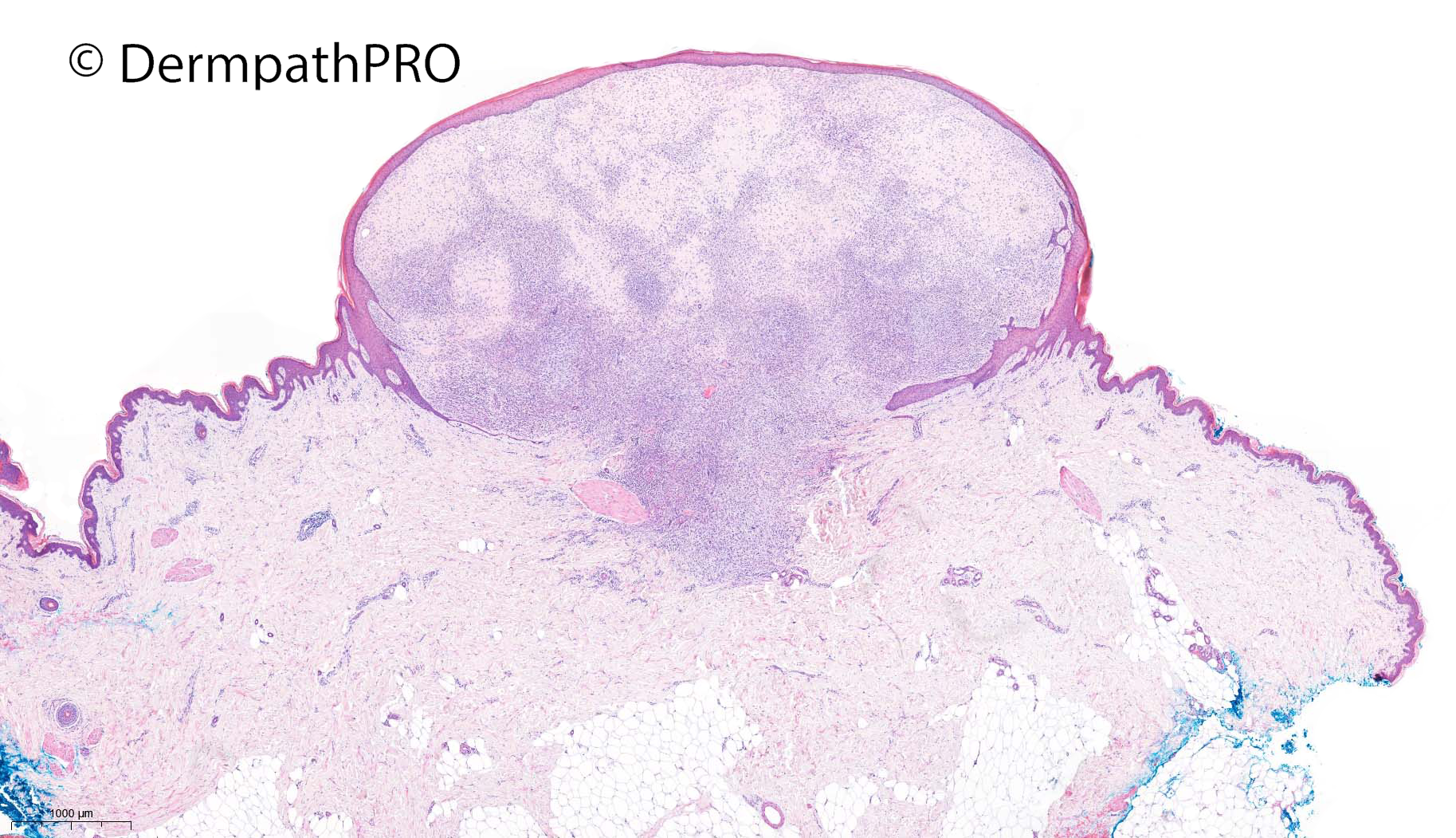
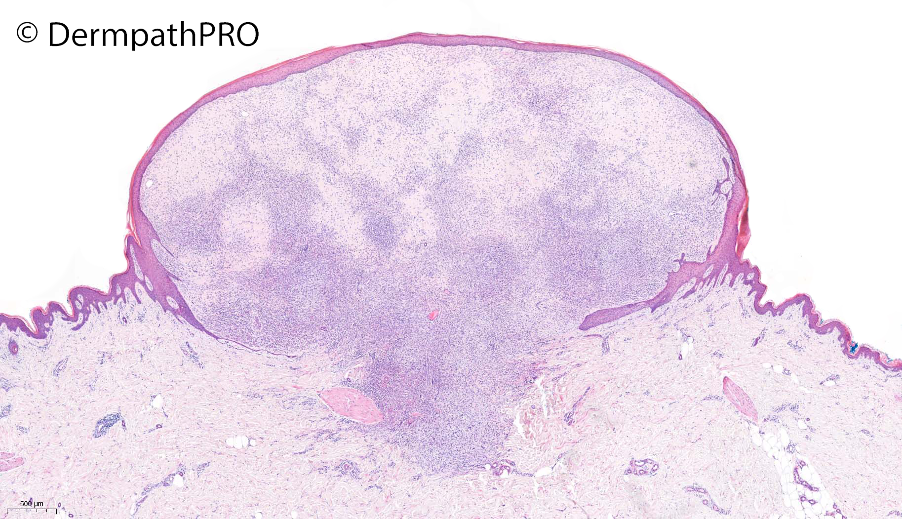
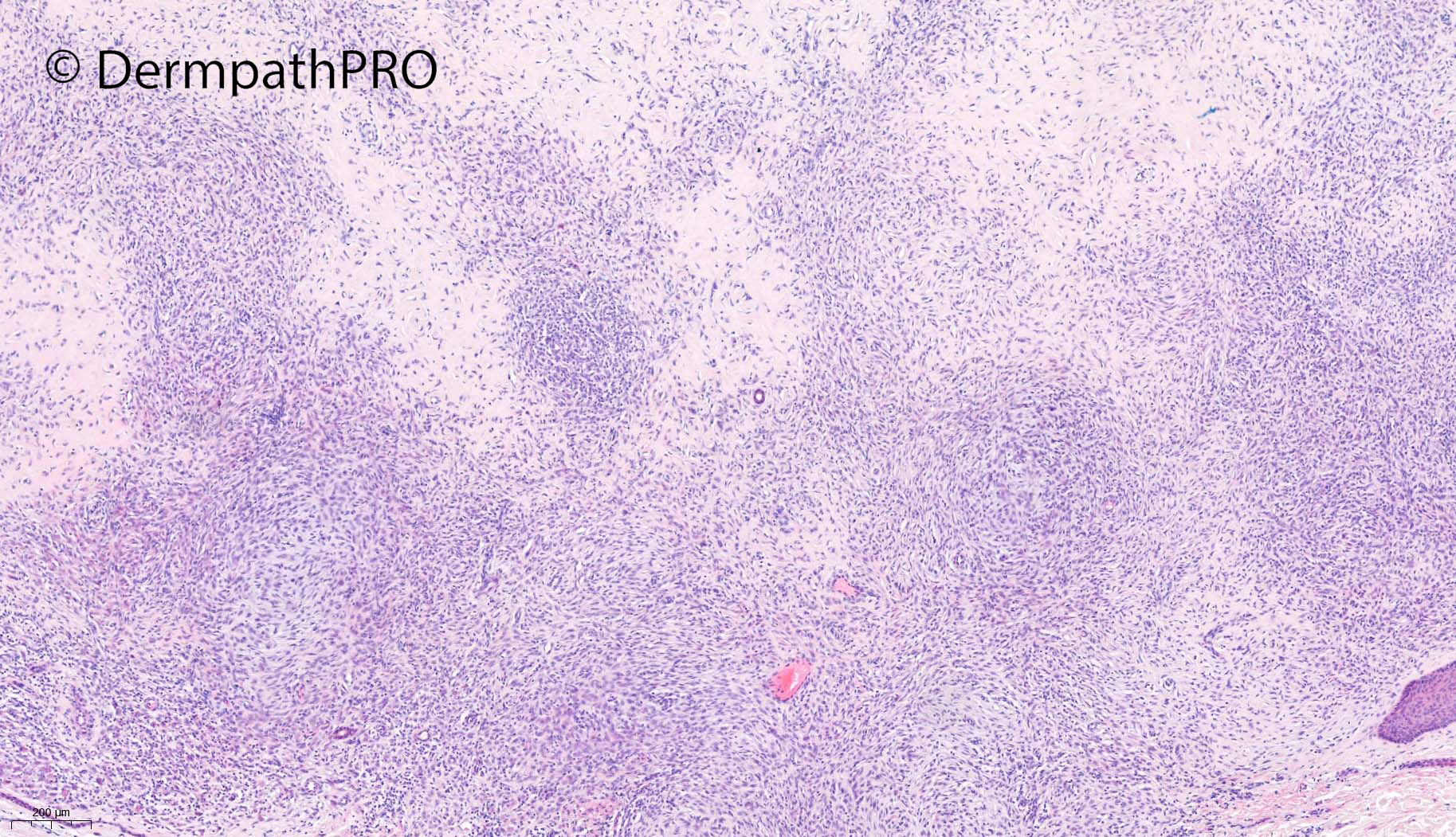
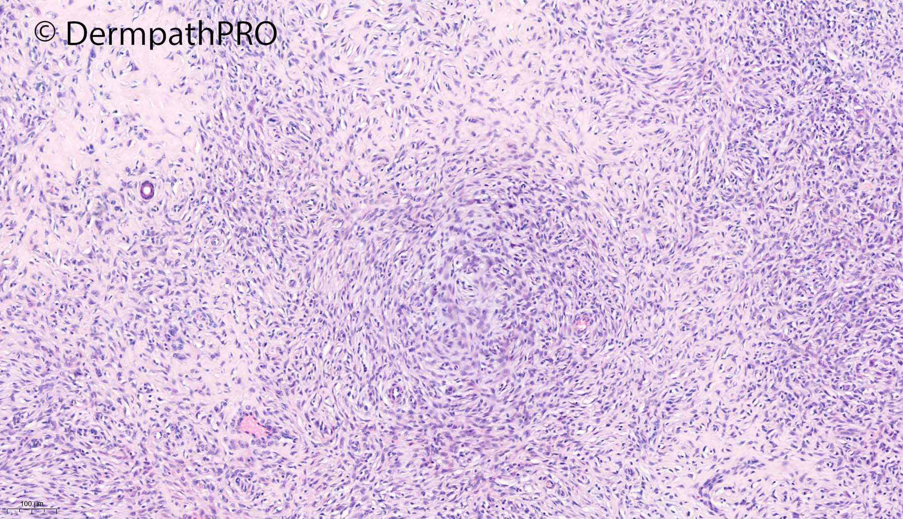
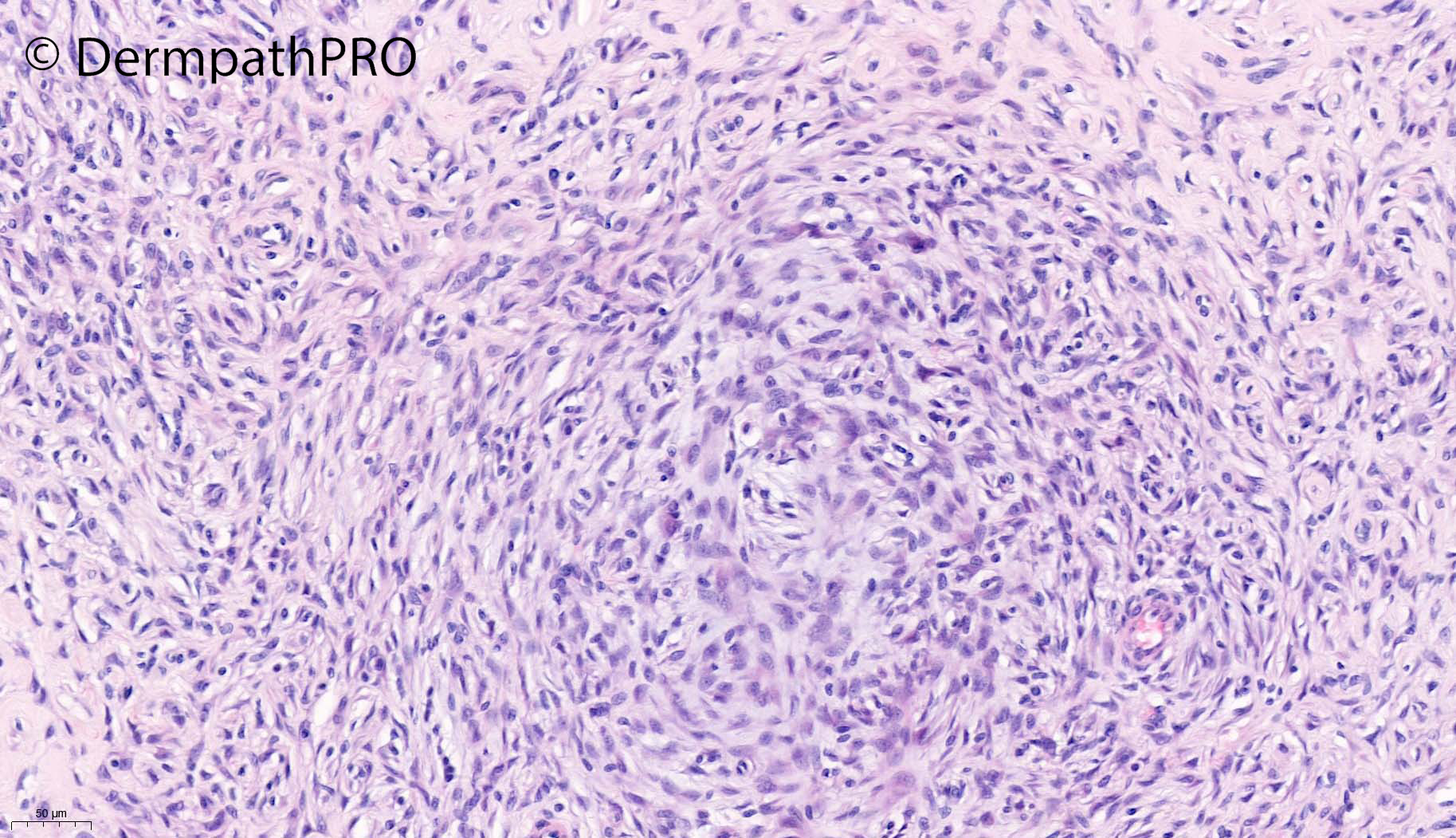
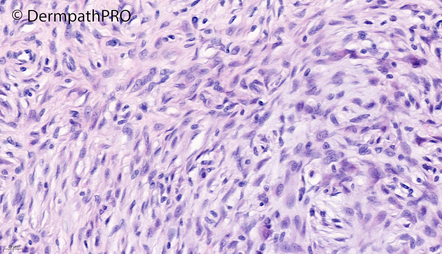
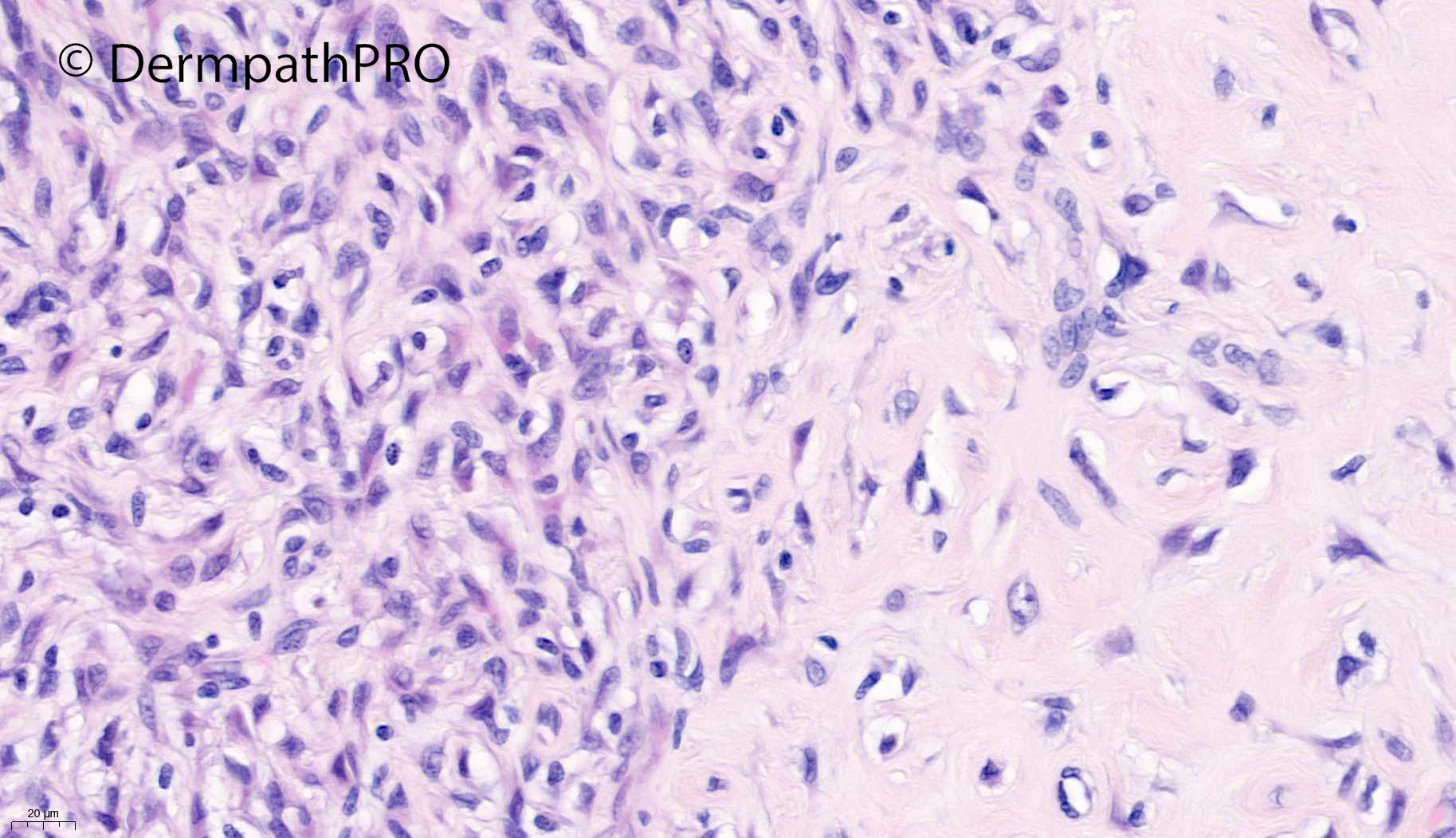
46F, excision left upper thigh ?amelanotic melanoma ?BCC ?Spitz ?BAPoma


  • Like 1
  Report Record

User Feedback


Victor Delgado

Posted

Neural vs. Myofibroblastic tumor, may be hyalinized myofibroma? Dermal Nodular Fascitis?

Share this comment


Link to comment
share_externally

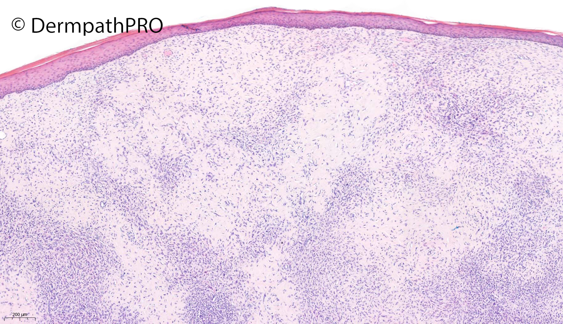
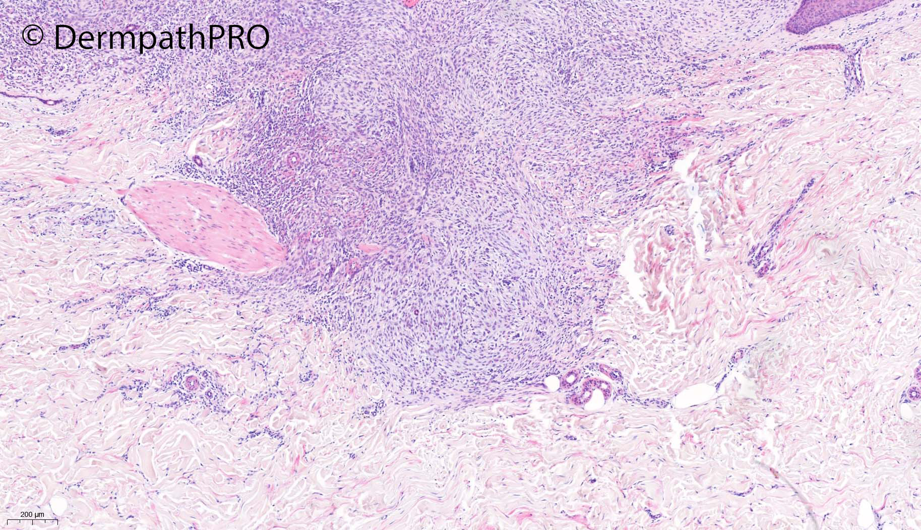
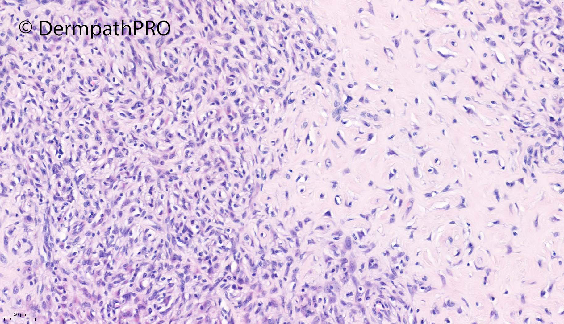
Epithelioid Fibrous Histiocytoma. ALK? Typical hyalinized stroma of older lesions and typical epidermal collarette

Share this comment


Link to comment
share_externally

There are cellular and myxoid zones with spindle cells vaguely showing nested or whorled pattern. I think that this will require a battery of IHC stains. Mixed type of neurothekeoma comes to my mind.

Share this comment


Link to comment
share_externally

Meenakshi Batrani

Posted

Could this be a newly described spindle cell variant of epithelioid cell histiocytoma (spindle cell histiocytoma). IHC is needed. 


Kazlouskaya V, Ho J, Jedrych J, Karunamurthy A. Spindle cell variant of epithelioid cell histiocytoma (spindle cell histiocytoma) with ALK gene fusions: Cases series and review of the literature. J Cutan Pathol. 2021 Jul;48(7):837-841

Share this comment


Link to comment
share_externally

Saleem Taibjee

Posted

Yes, absolutely spot on ... this is epithelioid fibrous histiocytoma, and I think could be justifiably deemed an example of the spindle cell variant. The ALK immunostain (positive) is shown below. There are frequently misdiagnosed as haemangiomas or melanocytic lesions clinically. The thigh seems to be a common anatomical site.

BW Saleem

10921_10.0x ALK.jpg10921_20.0x ALK.jpg


10921_2.5x ALK.jpg

Share this comment


Link to comment
share_externally


Join the conversation

You can post now and register later. If you have an account, sign in now to post with your account.

Guest
Add a comment...

×   Pasted as rich text.   Paste as plain text instead

  Only 75 emoji are allowed.

×   Your link has been automatically embedded.   Display as a link instead

×   Your previous content has been restored.   Clear editor

×   You cannot paste images directly. Upload or insert images from URL.

Loading...

×
×
  • Create New...