Jump to content
In this section we have spot diagnoses posted on a daily basis since June 2010, now over 4000! You can review the archived cases and read the suggested diagnoses by users and the final comment by the contributors.
Case are uploaded each week day by 10 am UK time with the correct diagnosis will generally be posted at 8 pm UK time. Why not view the most recent spot diagnosis and proffer a diagnosis?

Case Number : Case 2926 - 23 September 2021 Posted By: Iskander H. Chaudhry

Please read the clinical history and view the images by clicking on them before you proffer your diagnosis.
Submitted Date :
   (0 reviews)

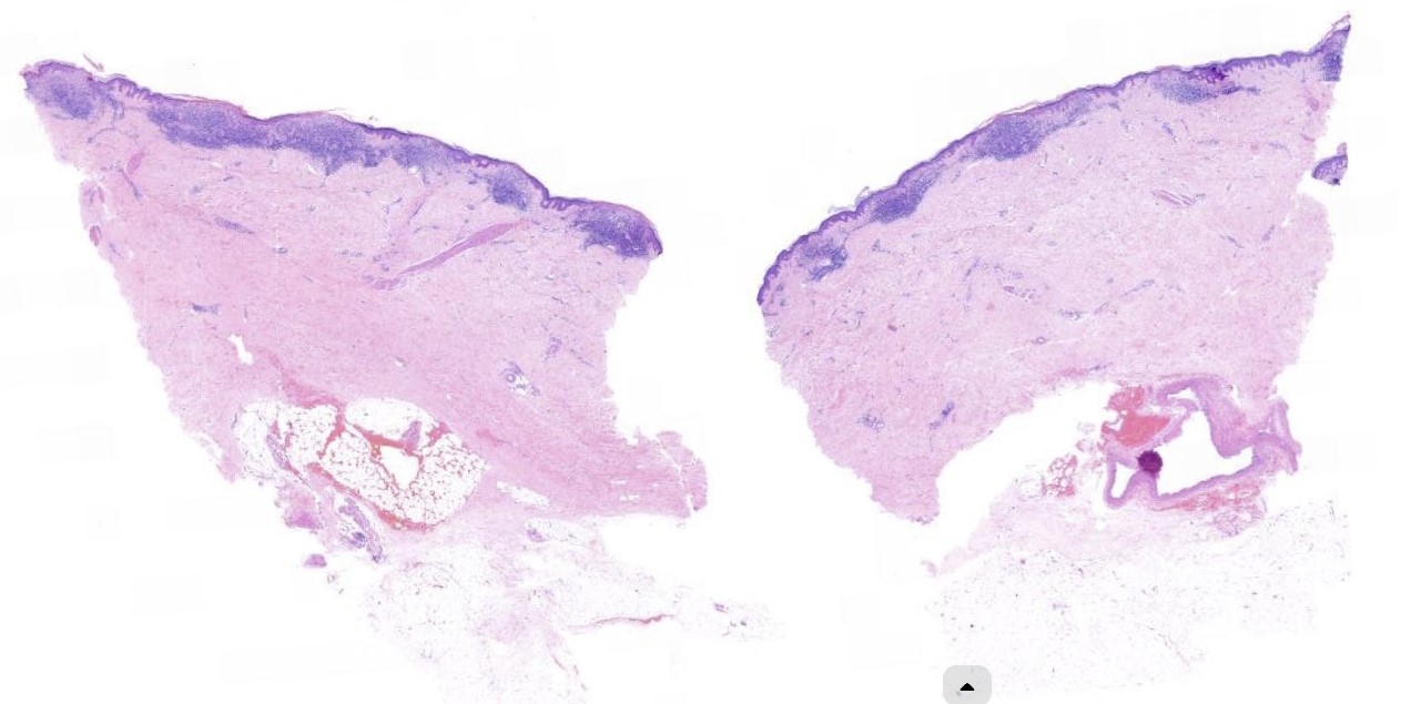
76F, Right breast, biopsy incisional - Rash right breast radiotherapy previously. ? Granulomatous

  Report Record

User Feedback


Saman Fatah

Posted

Congratulations to Dermpathpro team for this new feature of adding the digital slide itself to spot diagnosis section. 

Florid lichenoid interface dermatitis and some changes of collagen fibres in papillary dermis. I wondered about a probable early extra-genital LS confined to a vulnerable site such as radiation, CPC is essential to clarify if this fits clinically beside a simple examination of genital area for signs of LS.  

Share this comment


Link to comment
share_externally

Richard Logan

Posted

5 hours ago, Saman Fatah said:

Congratulations to Dermpathpro team for this new feature of adding the digital slide itself to spot diagnosis section. 

Florid lichenoid interface dermatitis and some changes of collagen fibres in papillary dermis. I wondered about a probable early extra-genital LS confined to a vulnerable site such as radiation, CPC is essential to clarify if this fits clinically beside a simple examination of genital area for signs of LS.  

Saman, can you remind me how to open and manipulate the slide digitally?  Thanks.

Share this comment


Link to comment
share_externally

Hi Richard, You can click over the second slide, over the empty space. 

For me it could be a Lichen Planus, atrophic variant. 

Share this comment


Link to comment
share_externally

Saman Fatah

Posted

Richard, apologies for a late response as just seen your response. It depends which device you are using to access it; PC is better as explained below.

For PC click on Dermpathpro spot diagnosis logo slide on the right side of the slide shown then that will open a separate window (digital pathology) were you will see the slide overlapped by a sign of digital slides, click on the down word triangular arrow that will get rid of the overlap to see the actual slide. Next stage up in the tool bar on the same screen next to 0 there is a double arrow click on this to orientate the slide followed by a little bit of play with the other functions at the tool bar to know what they do.

On ipad, you do the initial step but afterwards it does not provide you with the tool bar you just get rid of the overlap (if any) and magnify the slide like any other image and could not see the option of rotating the slide on there.

Hope above helps, if other has better ideas for tablet devices please share.


Share this comment


Link to comment
share_externally

Richard Logan

Posted

On 24/09/2021 at 20:32, Saman Fatah said:

Richard, apologies for a late response as just seen your response. It depends which device you are using to access it; PC is better as explained below.

For PC click on Dermpathpro spot diagnosis logo slide on the right side of the slide shown then that will open a separate window (digital pathology) were you will see the slide overlapped by a sign of digital slides, click on the down word triangular arrow that will get rid of the overlap to see the actual slide. Next stage up in the tool bar on the same screen next to 0 there is a double arrow click on this to orientate the slide followed by a little bit of play with the other functions at the tool bar to know what they do.

On ipad, you do the initial step but afterwards it does not provide you with the tool bar you just get rid of the overlap (if any) and magnify the slide like any other image and could not see the option of rotating the slide on there.

Hope above helps, if other has better ideas for tablet devices please share.


Thank you Saman.  I will ask Iskander to provide simple instruction for any further digital cases to help the technically illiterate such as me.

I think this might be CTCL (MF).  Breast is a common site of course.

Share this comment


Link to comment
share_externally

Richard Logan

Posted

Thanks to the DermpathPRO administrative staff for amending the format to include the microscope icon. No excuses now for being unable to open the digital slides!

Share this comment


Link to comment
share_externally

Admin_Dermpath

Posted

Thank you for your comments. This is skin with basket-weave keratin, epidermal atrophy, and intense lichenoid dermatitis. Moderate chronic inflammation is seen in the superficial dermis. 

The features are those of a lichenoid reaction pattern in keeping with radiation dermatitis - there was a notable past history !

Share this comment


Link to comment
share_externally


Join the conversation

You can post now and register later. If you have an account, sign in now to post with your account.

Guest
Add a comment...

×   Pasted as rich text.   Paste as plain text instead

  Only 75 emoji are allowed.

×   Your link has been automatically embedded.   Display as a link instead

×   Your previous content has been restored.   Clear editor

×   You cannot paste images directly. Upload or insert images from URL.

Loading...

×
×
  • Create New...