Jump to content
In this section we have spot diagnoses posted on a daily basis since June 2010, now over 4000! You can review the archived cases and read the suggested diagnoses by users and the final comment by the contributors.
Case are uploaded each week day by 10 am UK time with the correct diagnosis will generally be posted at 8 pm UK time. Why not view the most recent spot diagnosis and proffer a diagnosis?

Case Number : Case 2927 - 24 September 2021 Posted By: Dr. Richard Carr

Please read the clinical history and view the images by clicking on them before you proffer your diagnosis.
Submitted Date :
   (0 reviews)

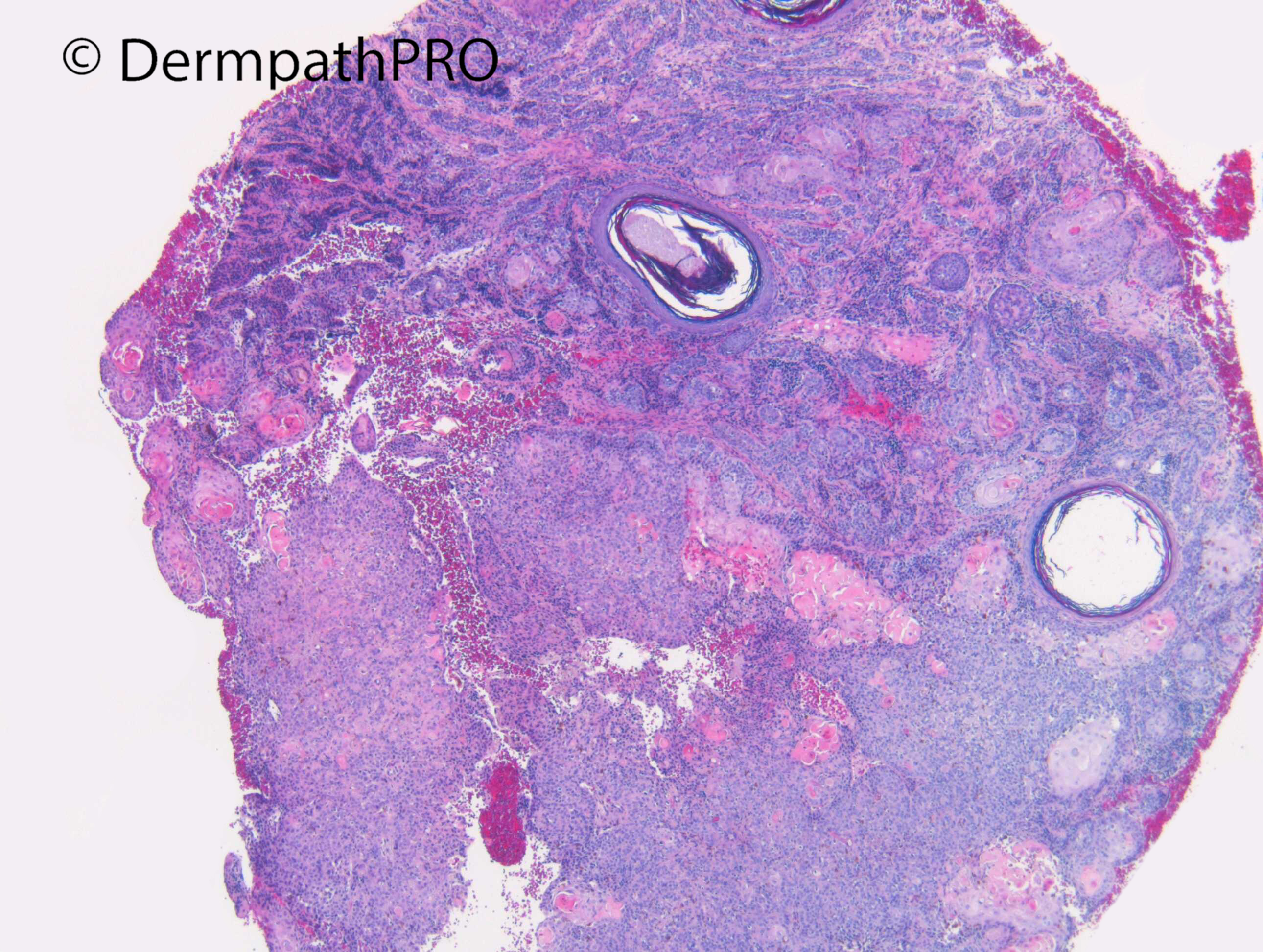
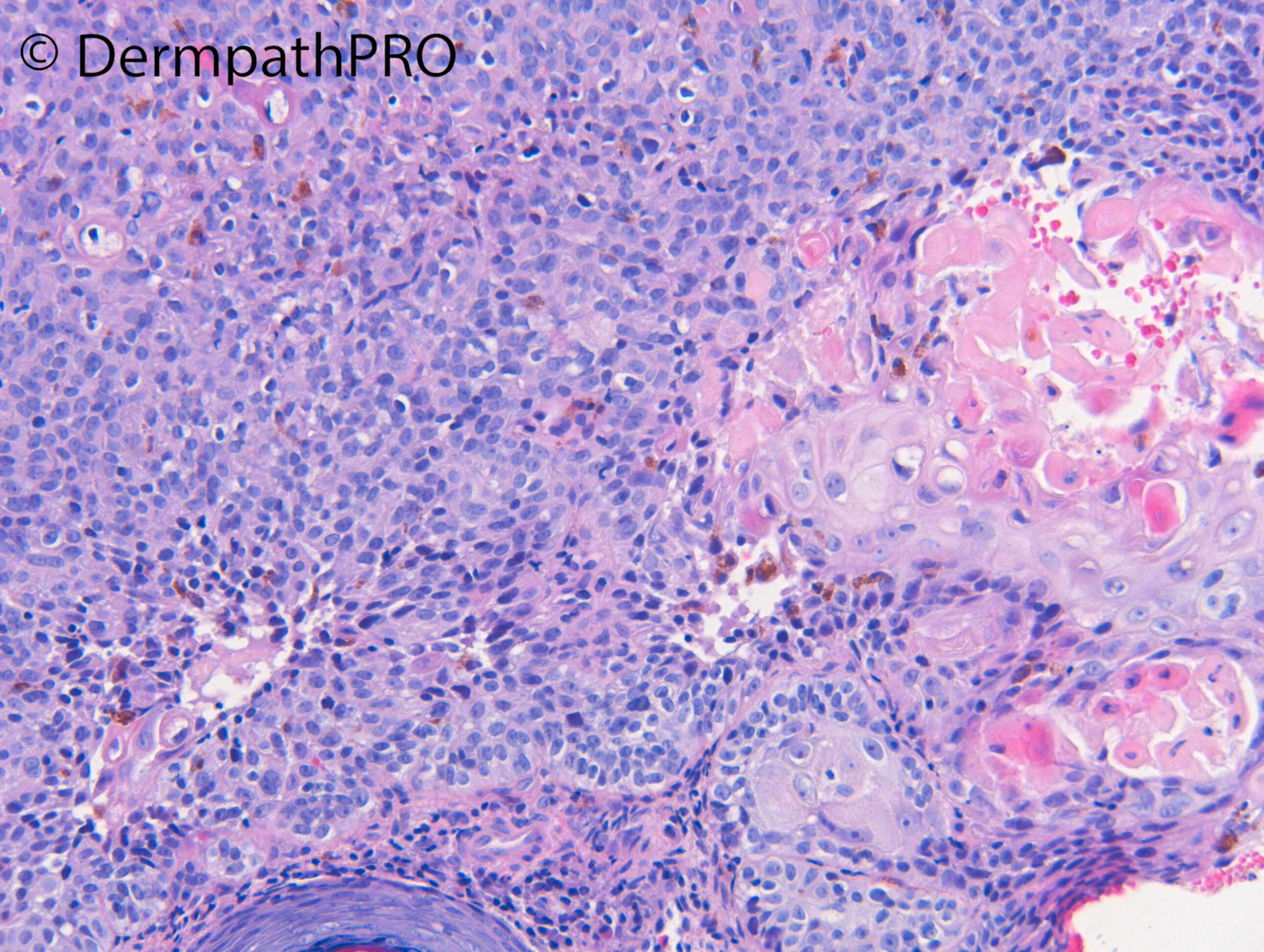
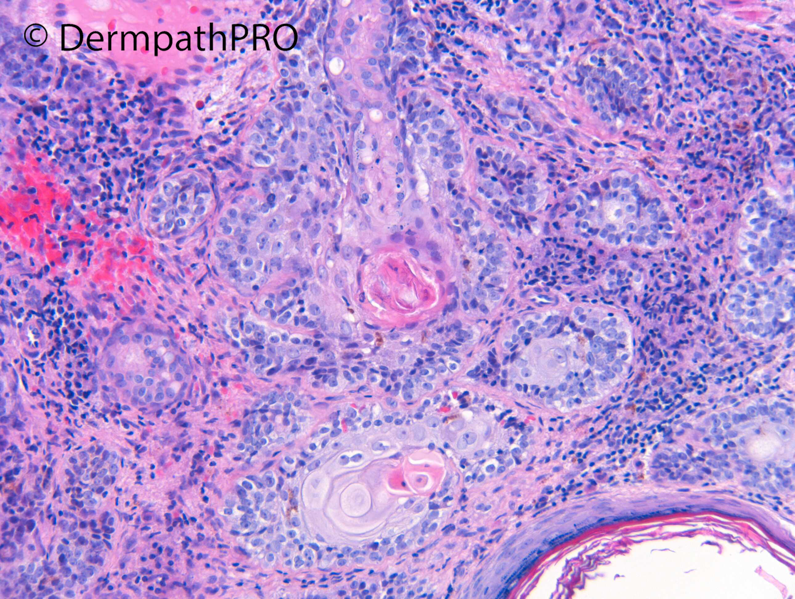
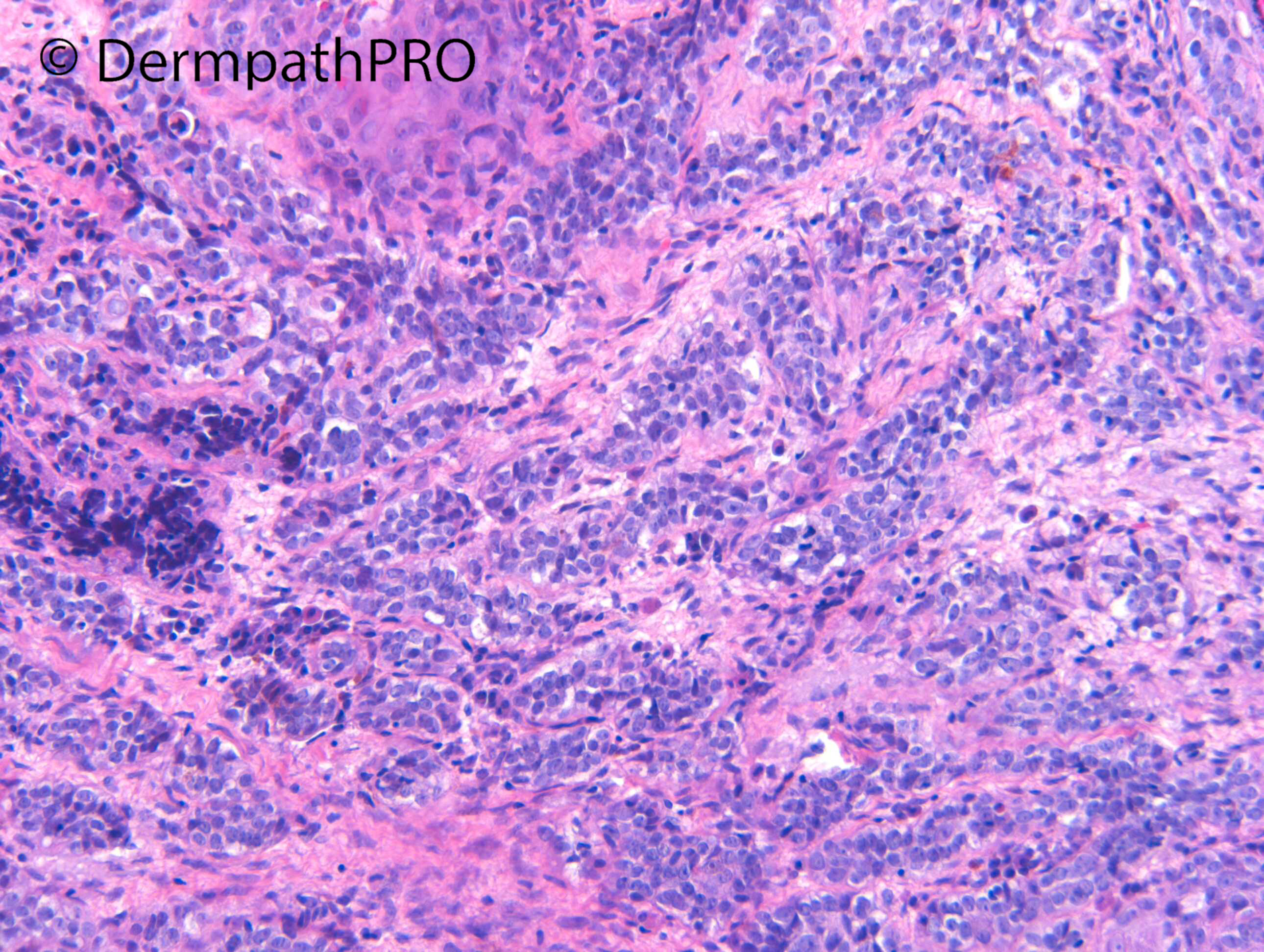
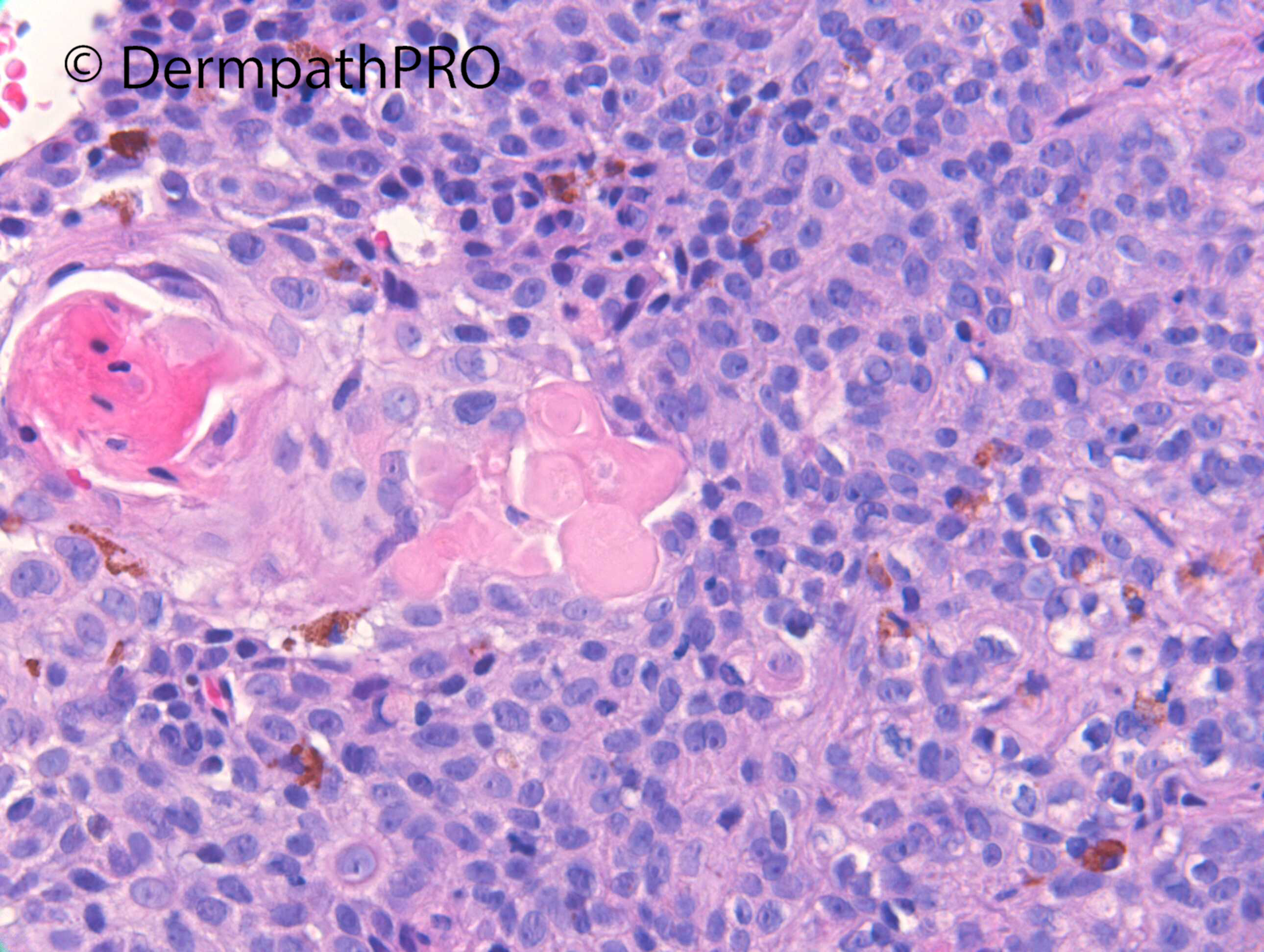
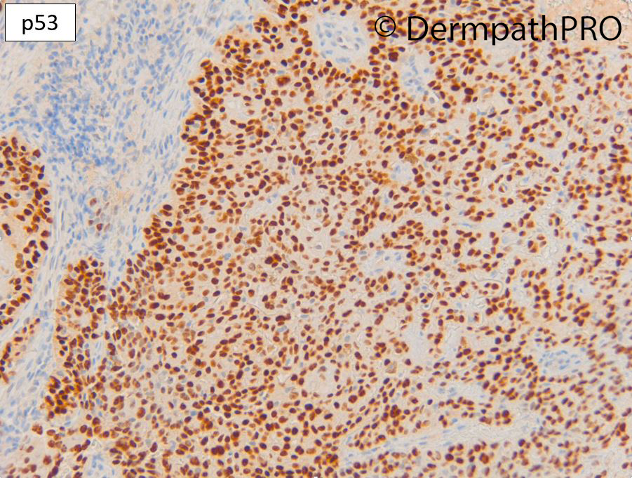
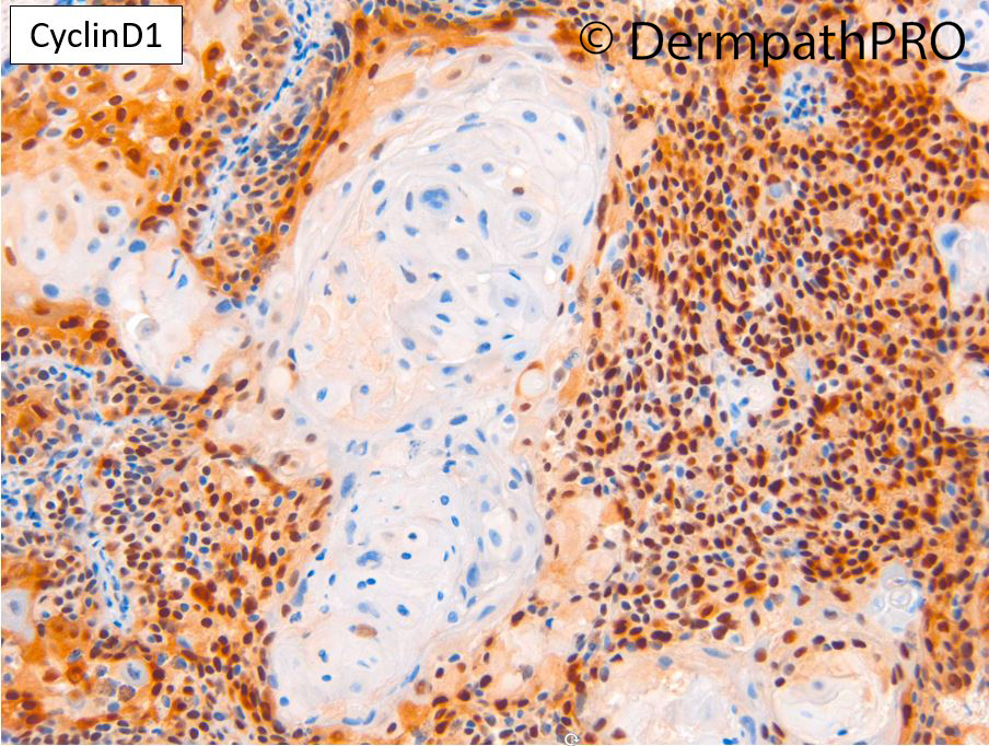
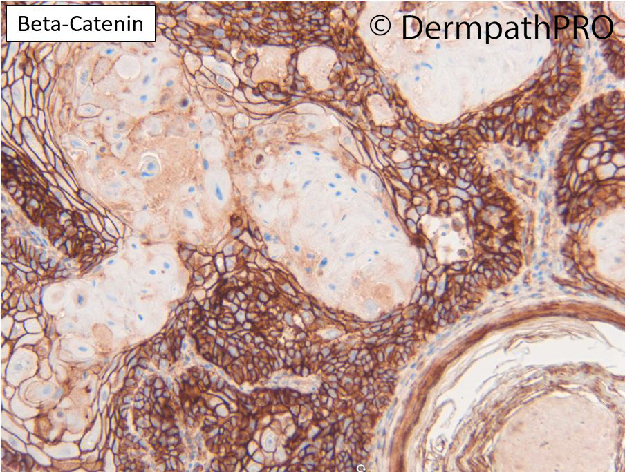
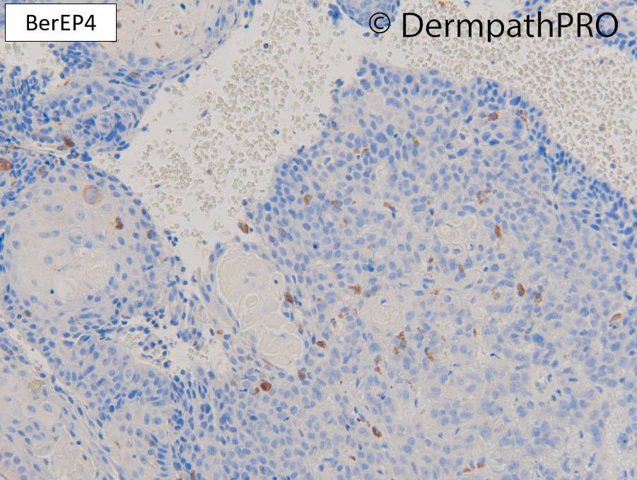
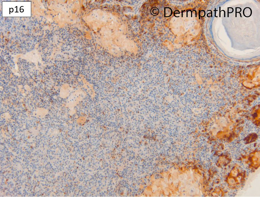
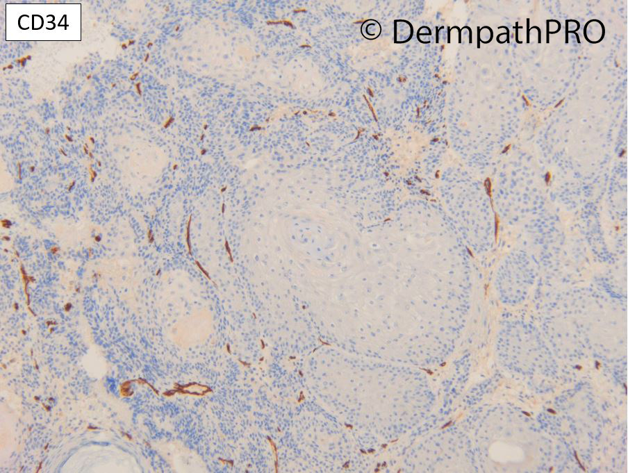
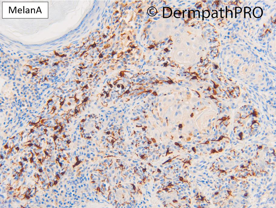
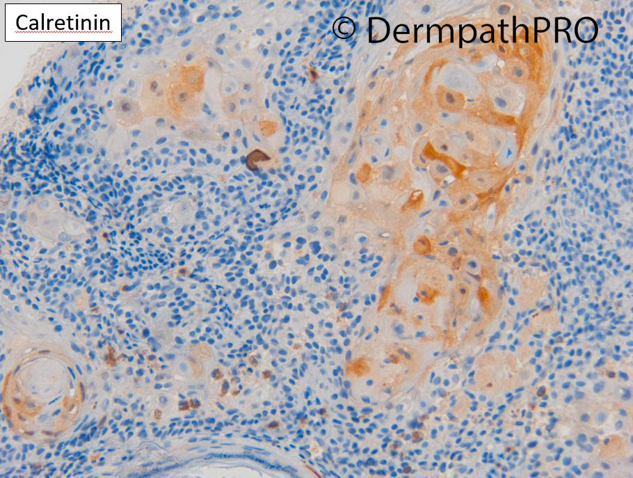
M70 Vertex scalp. 8mm nodule grew in 2/52. ?AFX ?SCC


  • Like 1
  Report Record

User Feedback


Squamomelanocytic tumor or pigmented scc? This is a chapter I’m still figuring out. 

Share this comment


Link to comment
share_externally

Dr. Richard Carr

Posted

40+ views and 1 commnent so far. This is a difficult case so thank you to Vincenzo. Other thoughts are very welcome and we all learn from the comments. IHC is now posted. 

Share this comment


Link to comment
share_externally

Looks as if benign melanocytes have colonised a follicular neoplasm. Can you please explain us βcatenin?

Share this comment


Link to comment
share_externally

Pigmented pilomatricoma. Aberrant membranous expression of beta catenin is reported in tumors with shadow cells.

Share this comment


Link to comment
share_externally

Dr. Richard Carr

Posted

Okay this is not a chapter in any book and seems quite a muddled area. Both basomelanocytic and squamomelanocytic tumour get scant mention in Weedon and Mckee.

I think we can have any combination of basaloid tumour benign or malignant (e.g. trichoblastoma or BCC) with benign or malignant melanocytes and any squamomelanocytic similarly. There are very rare reports of a few tumours that appeared to show both melanocytic and epithelial marker expression in the same cells but I'd have to be very sceptical about this because melanocytes are derived from the neural crest and should therefore be mutually exclusive with epithelial elements although I suppose we all come from a common progenitor cell and I suppose it is possible cells could have mixed features trans-differentiate etc. 

Back to this case. It's really a basosquamomelanocytic tumour although the melanocytic component is clearly dendritic and reactive. I'd guess this was a follicular tumour and in support of this is the moderate calretinin expression that can be seen in then innermost part of the outer root sheath throughout the length of the follicle but more diffusely in the plump keratinising cells of the follicular isthmus (just below the infundibulum). The squamoid part of the sebaceous duct also expresses calretinin. 

Reference: González-Guerra E, Kutzner H, Rutten A, Requena L. Immunohistochemical study of calretinin in normal skin and cutaneous adnexal proliferations. Am J Dermatopathol. 2012 Jul;34(5):491-505.

Beta-catenin when transposed to the nucleus is part of the WNT signalling pathway involved in the normal hair cycle and matrical tumours such as pilomatricoma and pilomatrical carcinoma usually express nuclear beta-catenin (and have CTNNB1 mutations, this is the gene for beta-catenin). I've personally seen a few melanocytic matricomas that also expressed nuclear beta-catenin. The latter tumours are usually regarded as lesion of uncertain malignancy but considered low grade. My H&E diagnosis on this case favoured a melanocytic matricoma but I was put off by the membranous beta-catenin. I'd therefore label this as a follicular basosquamous tumour populated by reactive dendritic melanocytes of uncertain malignant potential. I thought the p53 was quite high but not a clear-cut highly aberrant pattern and p16 was mosaic around the peripheral edges but does have a centrally deficient area (but benign tumours can be quite patchy mosaic). This tumour is from the bald scalp of an elderly man and complete excision with clear margins as for a nodular BCC would seem most appropriate. We would need to collect a series of similar lesions and publish them with follow-up in order to clarify the biology perhaps with whole exome sequencing etc.


Share this comment


Link to comment
share_externally


Join the conversation

You can post now and register later. If you have an account, sign in now to post with your account.

Guest
Add a comment...

×   Pasted as rich text.   Paste as plain text instead

  Only 75 emoji are allowed.

×   Your link has been automatically embedded.   Display as a link instead

×   Your previous content has been restored.   Clear editor

×   You cannot paste images directly. Upload or insert images from URL.

Loading...

×
×
  • Create New...