Jump to content
In this section we have spot diagnoses posted on a daily basis since June 2010, now over 4000! You can review the archived cases and read the suggested diagnoses by users and the final comment by the contributors.
Case are uploaded each week day by 10 am UK time with the correct diagnosis will generally be posted at 8 pm UK time. Why not view the most recent spot diagnosis and proffer a diagnosis?

Case Number : Case 3063 - 04 April 2022 Posted By: Dr. Mona Abdel-Halim

Please read the clinical history and view the images by clicking on them before you proffer your diagnosis.
Submitted Date :
   (0 reviews)

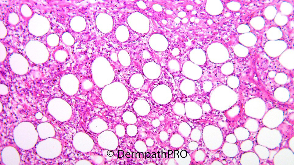
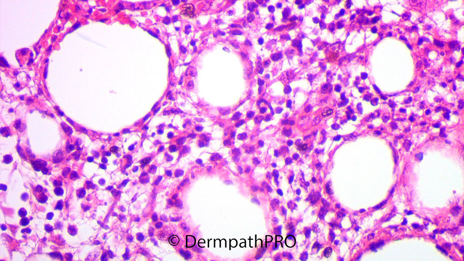
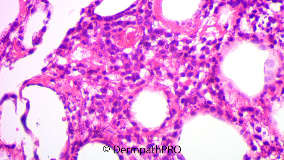
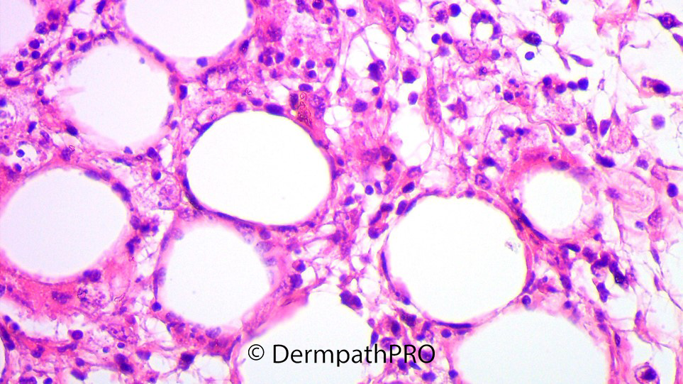
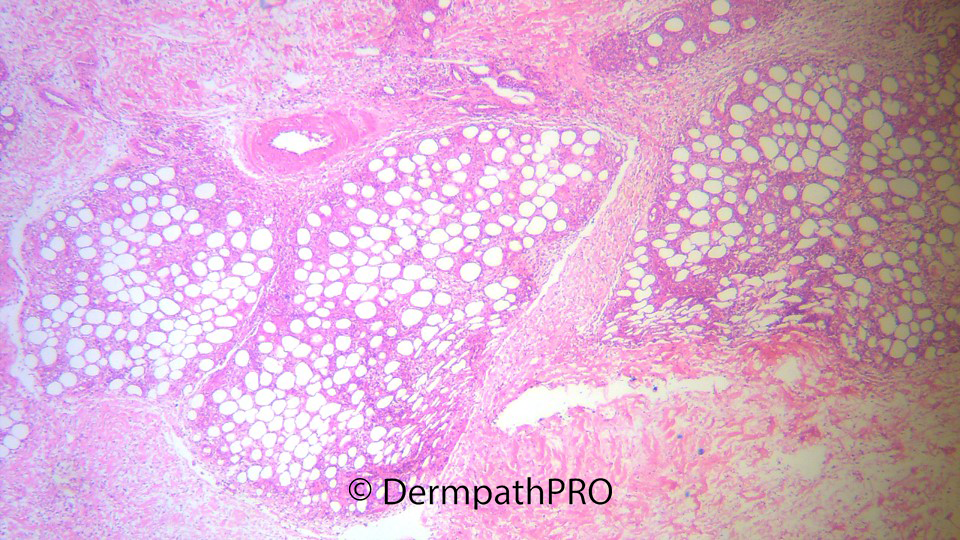
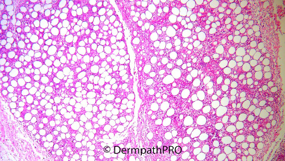
F, 20 Progressive development of indurated subcutaneous plaques over the lower limbs associated with fever, pancytopenia and hepatosplenomegaly.

  Report Record

User Feedback


Anil Patki

Posted

Subcutaneous panniculitis-like T cell lymphoma and lupus panniculitis are the differentials. While the constitutional symptoms suggest the former, it's difficult to rule out LE panniculitis without IHC for T cell markers and serological tests for LE. A considerable overlap between the two has been described. LE profundus may progress to SPTCL and it's likely that the two belong to a spectrum of subcutaneous T cell dyscrasias.

Share this comment


Link to comment
share_externally

Gamma delta TCL would be another consideration given the clinical picture is alarming. Otherwise agree with Anil.

Share this comment


Link to comment
share_externally

I don’t see any fat hyalinization or plasma cells, as expected in lupus panniculitis, so favor lymphoma...but lymphocytic rimming around adipocytes isn’t enough prominent. And I’m afraid there is some hemophagocytic activity. Only 20% of SPTCL are associated with HLH. So favor SPTCL, but agree with Shawn: it need to rule out PCGDTCL.

CD8? BetaF1? CD56?

Share this comment


Link to comment
share_externally

Dr. Mona Abdel-Halim

Posted

This was an interesting case. The condition started 1 year prior to presentation with slowly progressive appearance of subcutaneous indurated plaques over the lower limbs.  1 month prior to presentation, progressive appearance of new lesions started to occur associated with fever. Upon admission, investigations were done and reveled pancytopenia, and hepatosplenomegaly. Bone marrow biopsy revealed hemophagocytosis. Two skin biopsies revealed a process restricted only to subcutaneous fat with no dermal or subepidermal infiltrates. The immunophenotype was: CD3+, CD8+, CD56- and Ki-67 was high with rimming of fat cells with Ki-67+ cells. Unfortunately, we do not have BetaF1 or TCRgamma IHC but clonal rearrangement of the delta chain was negative. We diagnosed this case as SCPTCL associated with hemophagocytosis. SCPTCL usually runs an indolent course with good prognosis unless hemophagocytic syndrome develops. Thank you all! 

Share this comment


Link to comment
share_externally

Anil Patki

Posted

Thanks Mona for sharing this interesting case!

Share this comment


Link to comment
share_externally


Join the conversation

You can post now and register later. If you have an account, sign in now to post with your account.

Guest
Add a comment...

×   Pasted as rich text.   Paste as plain text instead

  Only 75 emoji are allowed.

×   Your link has been automatically embedded.   Display as a link instead

×   Your previous content has been restored.   Clear editor

×   You cannot paste images directly. Upload or insert images from URL.

Loading...

×
×
  • Create New...