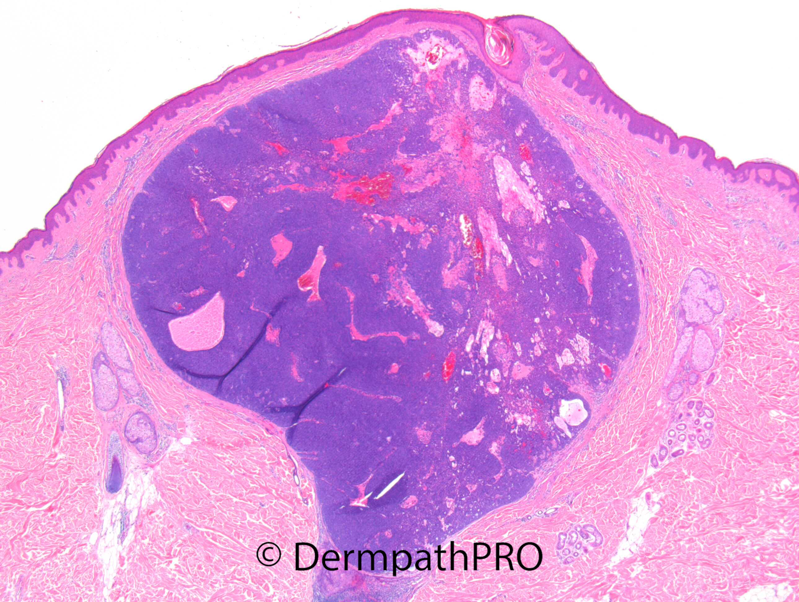
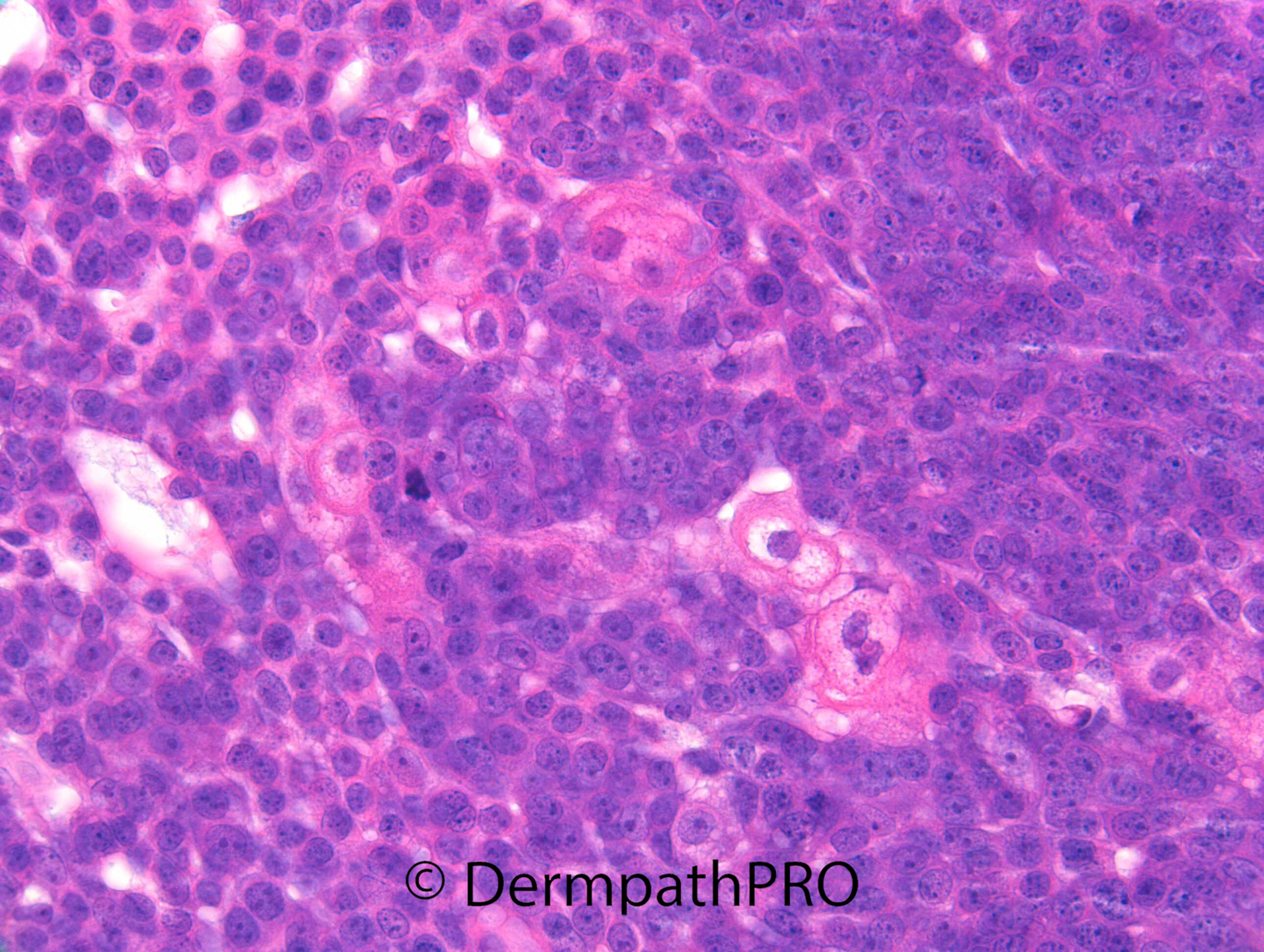
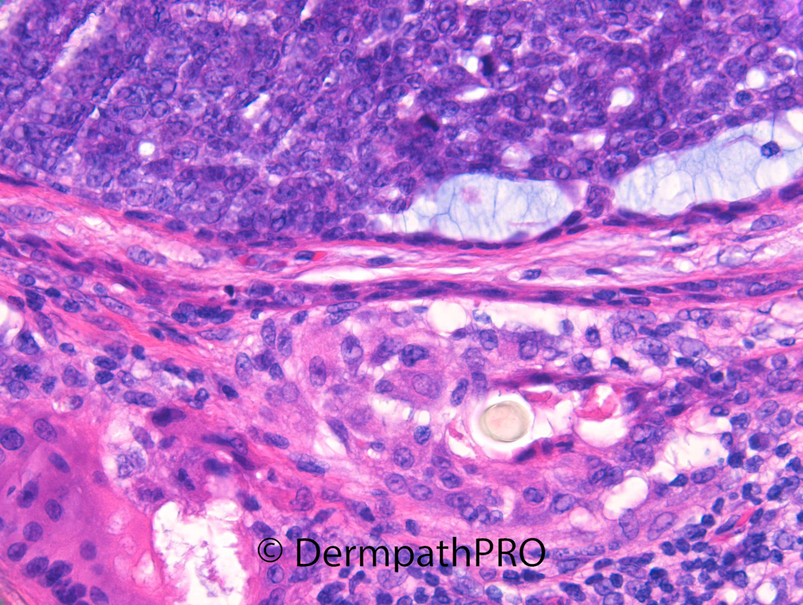
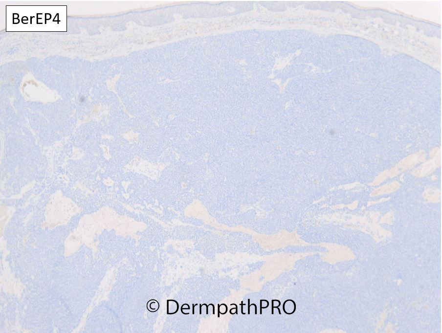
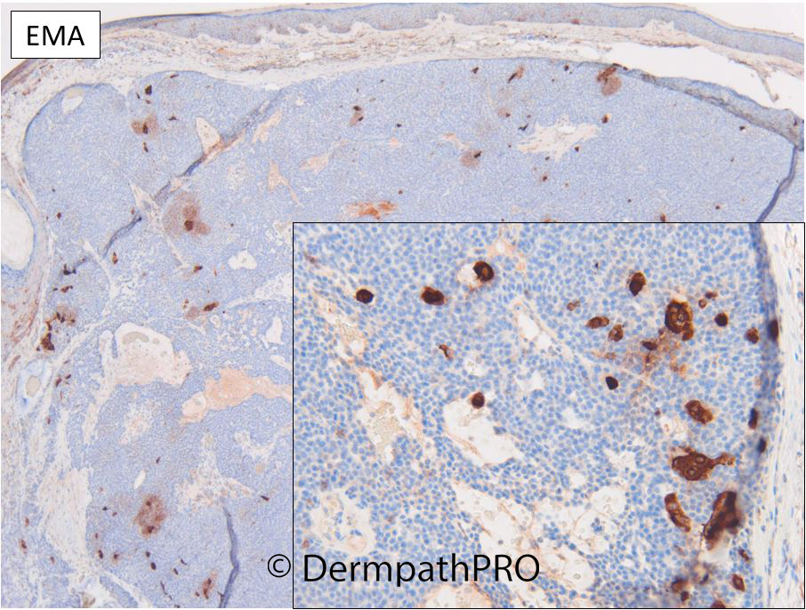
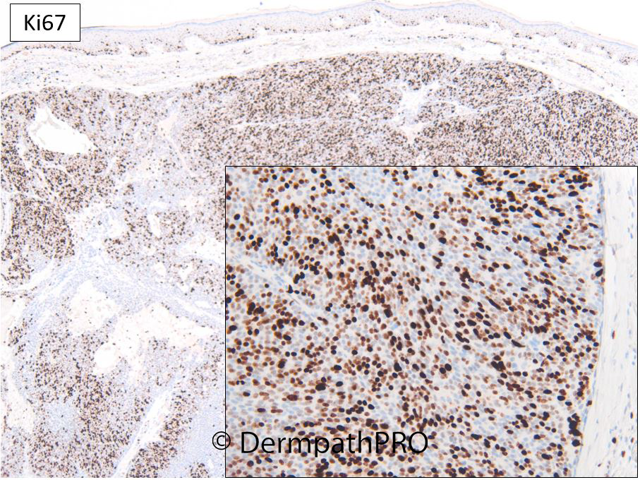
Jump to content
In this section we have spot diagnoses posted on a daily basis since June 2010, now over 4000! You can review the archived cases and read the suggested diagnoses by users and the final comment by the contributors.
Case are uploaded each week day by 10 am UK time with the correct diagnosis will generally be posted at 8 pm UK time. Why not view the most recent spot diagnosis and proffer a diagnosis?

Case Number : Case 3067 - 08 April 2022 Posted By: Dr. Richard Carr

Please read the clinical history and view the images by clicking on them before you proffer your diagnosis.
Submitted Date :
   (0 reviews)

M50. Back / shoulder. ?BCC


  • Like 1
  Report Record

User Feedback


Sebaceoma vs low grade Seb CA ... the threshold in this differential always seems a bit gray to me. If anyone has reproducible criteria please enlighten me!

Share this comment


Link to comment
share_externally

3 hours ago, Shawn said:

Sebaceoma vs low grade Seb CA ... the threshold in this differential always seems a bit gray to me. If anyone has reproducible criteria please enlighten me!

Totally agree. Waiting for diagnosis and enlightening about reproducible criteria concerning dd between low grade sebaceous ca and sebaceoma. 

Share this comment


Link to comment
share_externally

Dr. Richard Carr

Posted

Would be nice to get some comments on the IHC. 

Share this comment


Link to comment
share_externally

I'll give it a shot:

p16 retained in the normal sebaceous glands and lost in the neoplastic proliferation; p53 appears mosaic/wild type; BerEP4 can be positive in sebaceous neoplasms, in this case it's negative (guessing its utility is to r/o an SCC with sebaceous differentiation?)

expected more EMA expression, Ki67 is high. Overall the loss of p16 and high Ki67 would be more in favor of a low grade sebaceous CA for me.

Share this comment


Link to comment
share_externally

Dr. Richard Carr

Posted

Thanks Shawn for having a go. We published in Histoapthology quite a few years ago now the pattern of BerEP4 and EMA in sebaceoma and this tumour fits that pattern perfectly i.e. BerEP4 -ve in germinative cells and EMA largely limited to mature sebocytes. Over the years I've noticed sebaceous carcinomas have variable (not neat / organised like this case) positive staining for EMA and BerEP4 frequently and there is some literature on occular sebaceous carcinomas confirming my personal observations that BerEP4 stains in around 60-80%. BCC with sebaceous differentiation (a very rare tumour in the literarture - I've collected around 4 cases) show the typical pattern of BerEP4+++/EMA- in the basaloid cells and the mature sebocytes EMA+, again I've not published due to academic constipation! An SCC with sebaceous differentiation would classify as a sebaceous carcinoma (correct me if I'm wrong). Nearly all adnexal carcinomas whether sebaceous, ductal, or follicular have squamous differentiation in their group. But of course we could called the tumour with mixed features tumours of the folliclar-sebaceous-apocrine duct unit and not worry about it too much. I'm pretty sure prognosis wise its the grade and degree of infiltration and some tumour biomakers that really matter. 

If you had a tumour that looked like an dermal Spitz tumour architecturally but with perhaps a bit too much mitotic activity with loss of p16 how would you classify it? 

Share this comment


Link to comment
share_externally

So could this be a mitotically active Sebaceoma? But the atypical sebaceoma as subtype of sebaceous tumors doesn’t exist, if I’m not wrong. But I’d like to call it that. Instead the "atypical Spitz tumor" is well recognized nowadays, though still faded. A mitotically active dermal AST, with loss of p16, if BRAF -ve and ROS1 or ALK or NTRK3 +ve could still be an AST. Unlike when you find a braf positivity; in that case Spitz Melanoma become a serious option. But of course these are nightmare situations, in which, unlikely, we find ourselves more and more often. PRAME could be useful. 

Share this comment


Link to comment
share_externally

Dr. Richard Carr

Posted

Dear Vincenzo this is such a good response and I didn't think to do a PRAME!  Although I'd like to see TERT/telomerase studies in these lesions. There are still many concepts to be determined. Indeed why not the range of sebaceous tumours just as complex and challenging on all levels as Spitz/MELTUMPs and KA-spectrum. You may know I'm a fan of admitting uncertainty where it exists but I'm told by some clinicians, and indeed patients, prefer black and white answers (Ackerman approach). I'm not sure philosophers and scientists would agree, biology often confounds us. So how to handle it in practice. We could say don't know and we could go with our gut and experience. For me this is architecturally and in organisation typical of sebaceoma. Pleomorphism is mild to moderate rather than full out malignant phenotype. Mitotic activity and Ki67 are really rather too much for "typical" sebaceoma. We can speculate the lesion may have an isolated p16 knock-out in function with reasonable confidence, normally a signature of malignancy but as you say not definitive as we can have low-risk AST with limited p16 abnormalities and preserved TERT. Maybe this is such a case. So "atypically mitotic sebaceoma-like lesion with a highly aberrant p16 but no clear cut features to indicate metastatic potential based on circumscription and architecture" or a SebTUMP? Kazakov et al reported a group of sebaceous lesions with discordant architectural and cytological features. It would fascinating to work them up with the studies that are routine in the assessment of MELTUMPs / AST as well as many difficult KA-like lesions I'm NOT comfortable are "typical" that I'm labelling SPLUMP KAL (and that is even in the UK RCPath Datasets - I was a co-author!). Note of caution: I've seen a very KA-like tumour with highly aberrant p16 knock-out that did metastasise - PRAME and TERT/telomere studies would be fascinating to further out knowledge and that is the beauty of admitting uncertainty we get to do science and find some more answers to the puzzles! BTW no-one suggested doing MMRs for Muir Torre syn. (MTS) and the mucin in this case was found to be statistically associated with the latter syndrome. Please note KA occur with greater frequency and at a younger at in MTS. MMR stains are pending in this fairly current case. Don't forget to think about cancer syndromes for particular diagnoses. You can save lives this way. 

Kazakov DV, Kutzner H, Spagnolo DV, Rütten A, Mukensnabl P, Michal M. Discordant architectural and cytological features in cutaneous sebaceous neoplasms--a classification dilemma: report of 5 cases. Am J Dermatopathol. 2009;31:31-6.

Konstantinova AM, Kastnerova L, Michal M, Kolm I, Kazakov DV. Sebaceous Tumors of the Skin: A Study of 145 Lesions From 136 Patients Correlating Pathologic Features and DNA Mismatch Repair Staining Pattern. Am J Dermatopathol. 2021 Mar 1;43(3):174-181. doi: 10.1097/DAD.0000000000001691. PMID: 33201015.


Share this comment


Link to comment
share_externally

Dr. Richard Carr

Posted

Interestingly this showed isolated loss of PMS2. Interestingly only 1/89 patient, 1.2% had isolated PMS2 loss in the Konstantinova paper. 

Share this comment


Link to comment
share_externally

msofopoulos

Posted

6 hours ago, Dr. Richard Carr said:

Dear Vincenzo this is such a good response and I didn't think to do a PRAME!  Although I'd like to see TERT/telomerase studies in these lesions. There are still many concepts to be determined. Indeed why not the range of sebaceous tumours just as complex and challenging on all levels as Spitz/MELTUMPs and KA-spectrum. You may know I'm a fan of admitting uncertainty where it exists but I'm told by some clinicians, and indeed patients, prefer black and white answers (Ackerman approach). I'm not sure philosophers and scientists would agree, biology often confounds us. So how to handle it in practice. We could say don't know and we could go with our gut and experience. For me this is architecturally and in organisation typical of sebaceoma. Pleomorphism is mild to moderate rather than full out malignant phenotype. Mitotic activity and Ki67 are really rather too much for "typical" sebaceoma. We can speculate the lesion may have an isolated p16 knock-out in function with reasonable confidence, normally a signature of malignancy but as you say not definitive as we can have low-risk AST with limited p16 abnormalities and preserved TERT. Maybe this is such a case. So "atypically mitotic sebaceoma-like lesion with a highly aberrant p16 but no clear cut features to indicate metastatic potential based on circumscription and architecture" or a SebTUMP? Kazakov et al reported a group of sebaceous lesions with discordant architectural and cytological features. It would fascinating to work them up with the studies that are routine in the assessment of MELTUMPs / AST as well as many difficult KA-like lesions I'm NOT comfortable are "typical" that I'm labelling SPLUMP KAL (and that is even in the UK RCPath Datasets - I was a co-author!). Note of caution: I've seen a very KA-like tumour with highly aberrant p16 knock-out that did metastasise - PRAME and TERT/telomere studies would be fascinating to further out knowledge and that is the beauty of admitting uncertainty we get to do science and find some more answers to the puzzles! BTW no-one suggested doing MMRs for Muir Torre syn. (MTS) and the mucin in this case was found to be statistically associated with the latter syndrome. Please note KA occur with greater frequency and at a younger at in MTS. MMR stains are pending in this fairly current case. Don't forget to think about cancer syndromes for particular diagnoses. You can save lives this way. 

Kazakov DV, Kutzner H, Spagnolo DV, Rütten A, Mukensnabl P, Michal M. Discordant architectural and cytological features in cutaneous sebaceous neoplasms--a classification dilemma: report of 5 cases. Am J Dermatopathol. 2009;31:31-6.

Konstantinova AM, Kastnerova L, Michal M, Kolm I, Kazakov DV. Sebaceous Tumors of the Skin: A Study of 145 Lesions From 136 Patients Correlating Pathologic Features and DNA Mismatch Repair Staining Pattern. Am J Dermatopathol. 2021 Mar 1;43(3):174-181. doi: 10.1097/DAD.0000000000001691. PMID: 33201015.


great insights! thanks!

Share this comment


Link to comment
share_externally

Sorry, I was in Sicily for the Easter. Thanks, Richard! Very learning points! 

Share this comment


Link to comment
share_externally


Join the conversation

You can post now and register later. If you have an account, sign in now to post with your account.

Guest
Add a comment...

×   Pasted as rich text.   Paste as plain text instead

  Only 75 emoji are allowed.

×   Your link has been automatically embedded.   Display as a link instead

×   Your previous content has been restored.   Clear editor

×   You cannot paste images directly. Upload or insert images from URL.

Loading...

×
×
  • Create New...