Jump to content
In this section we have spot diagnoses posted on a daily basis since June 2010, now over 4000! You can review the archived cases and read the suggested diagnoses by users and the final comment by the contributors.
Case are uploaded each week day by 10 am UK time with the correct diagnosis will generally be posted at 8 pm UK time. Why not view the most recent spot diagnosis and proffer a diagnosis?

Case Number : Case 3068 - 11 April 2022 Posted By: Dr. Mona Abdel-Halim

Please read the clinical history and view the images by clicking on them before you proffer your diagnosis.
Submitted Date :
   (0 reviews)

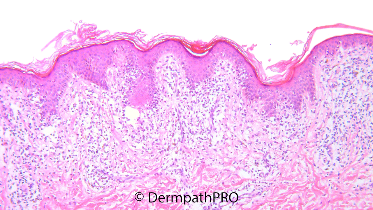
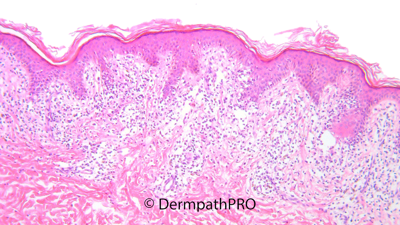
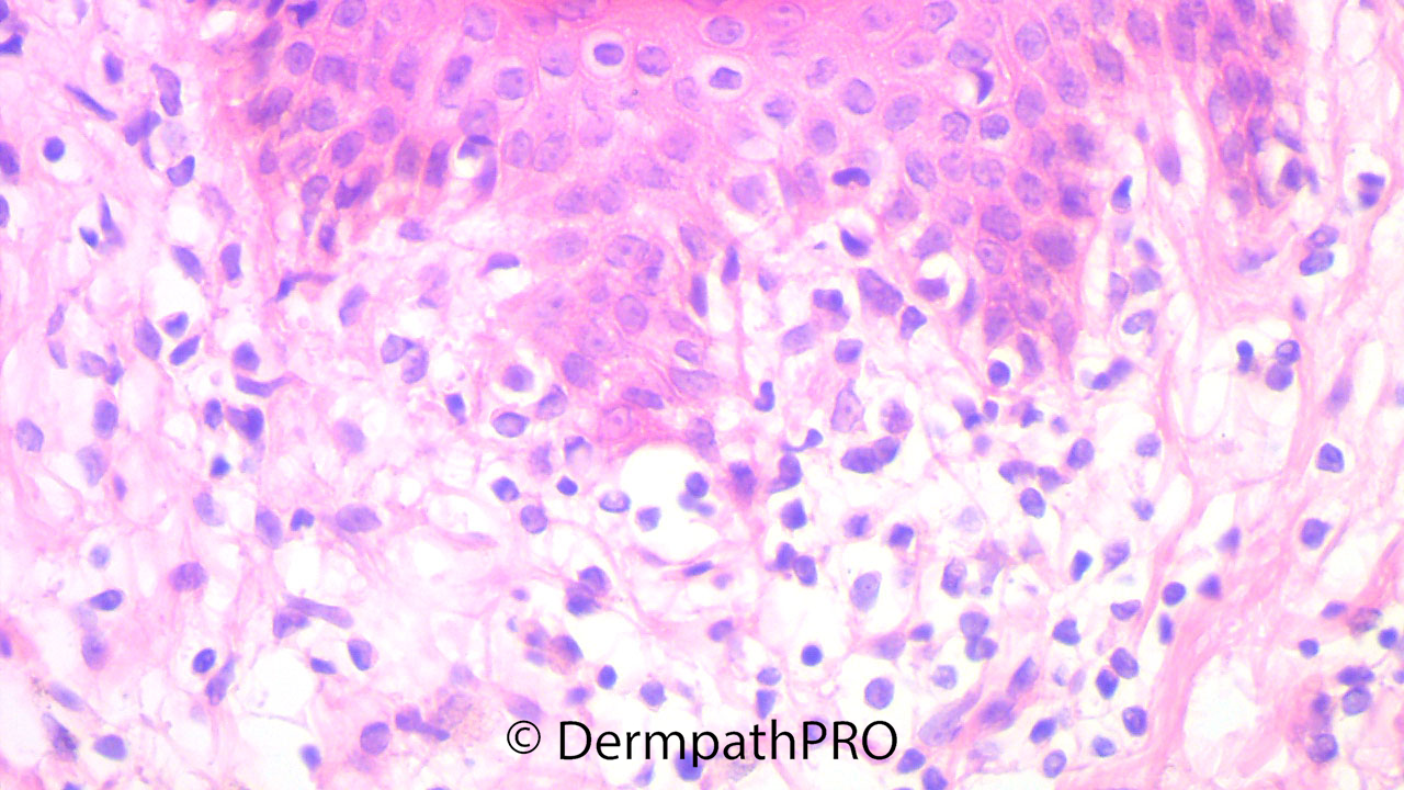
M, 8 years, Annular patches with erythematous/hyperpigmented borders and hypopigmented centers of 3 months duration over the lower abdomen and inguinal folds.


  • Like 1
  Report Record

User Feedback


Anil Patki

Posted

Looks like annular lichen planus. However, I would do a PAS stain to rule out tinea incognito.

Share this comment


Link to comment
share_externally

I don’t see any apoptotic bodies, or other aggressive autoimmune features of dermoepidermal interface. It seems a "passive epidermotropism”. Not sure, but I seem to see some nuclear atypia of lymphocytes. It needs to rule out a hypopigmented variant of MF, or am I wrong?

Share this comment


Link to comment
share_externally

Eman El-Nabarawy

Posted

Annular lichenoid dermatitis of youth.

Share this comment


Link to comment
share_externally

Agree with annular lichenoid dermatitis, action is primarily at the tips of rete, also location fits. Inflammatory vitiligo would be another possibility. Would like to see melanocytic marker (for retained melanocytes r/o vitiligo) and lymphocyte markers/TCR proving polyclonality. 

Share this comment


Link to comment
share_externally

Mohamed khaled

Posted

On 12/04/2022 at 20:12, Shawn said:

Agree with annular lichenoid dermatitis, action is primarily at the tips of rete, also location fits. Inflammatory vitiligo would be another possibility. Would like to see melanocytic marker (for retained melanocytes r/o vitiligo) and lymphocyte markers/TCR proving polyclonality. 

Concur

challinging case

The clinicopathological correlation and follow up of such cases is very important although your discription is in favour of inflammatory vitiligo, three zones of hypopigmentatiin, erythema and hyperpigmentation.

I would like to see the clinical picture of the patient, if possible.

Thanks for sharing.


Share this comment


Link to comment
share_externally

Chirag Desai

Posted

annular lichenoid dermatosis of youth

Share this comment


Link to comment
share_externally

You Guys are right. Age, 3 months duration and clinical features dont’t fit MF and fit instead ALD of youth, or Inflammatory Vitiligo. I didn’t know these lesions, before. Thanks for your comments. 

Share this comment


Link to comment
share_externally


Join the conversation

You can post now and register later. If you have an account, sign in now to post with your account.

Guest
Add a comment...

×   Pasted as rich text.   Paste as plain text instead

  Only 75 emoji are allowed.

×   Your link has been automatically embedded.   Display as a link instead

×   Your previous content has been restored.   Clear editor

×   You cannot paste images directly. Upload or insert images from URL.

Loading...

×
×
  • Create New...