Jump to content
In this section we have spot diagnoses posted on a daily basis since June 2010, now over 4000! You can review the archived cases and read the suggested diagnoses by users and the final comment by the contributors.
Case are uploaded each week day by 10 am UK time with the correct diagnosis will generally be posted at 8 pm UK time. Why not view the most recent spot diagnosis and proffer a diagnosis?

Case Number : Case 3069 - 12 April 2022 Posted By: Uma Sundram

Please read the clinical history and view the images by clicking on them before you proffer your diagnosis.
Submitted Date :
   (0 reviews)

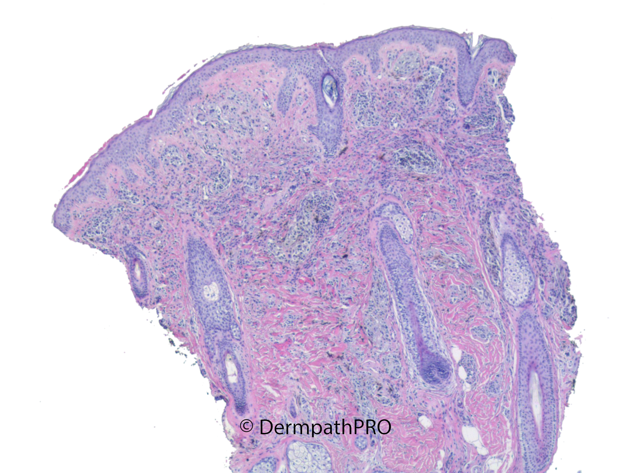
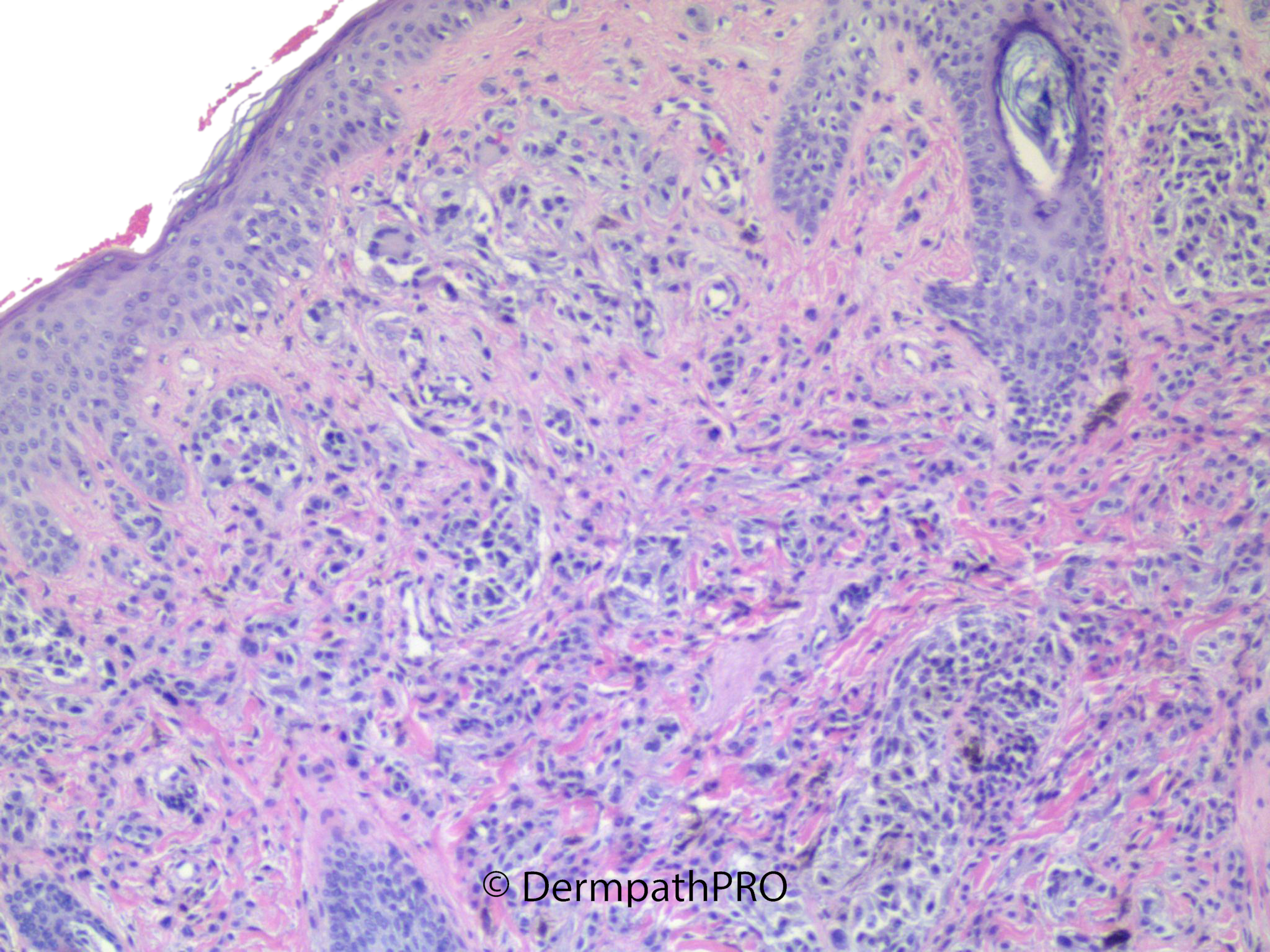
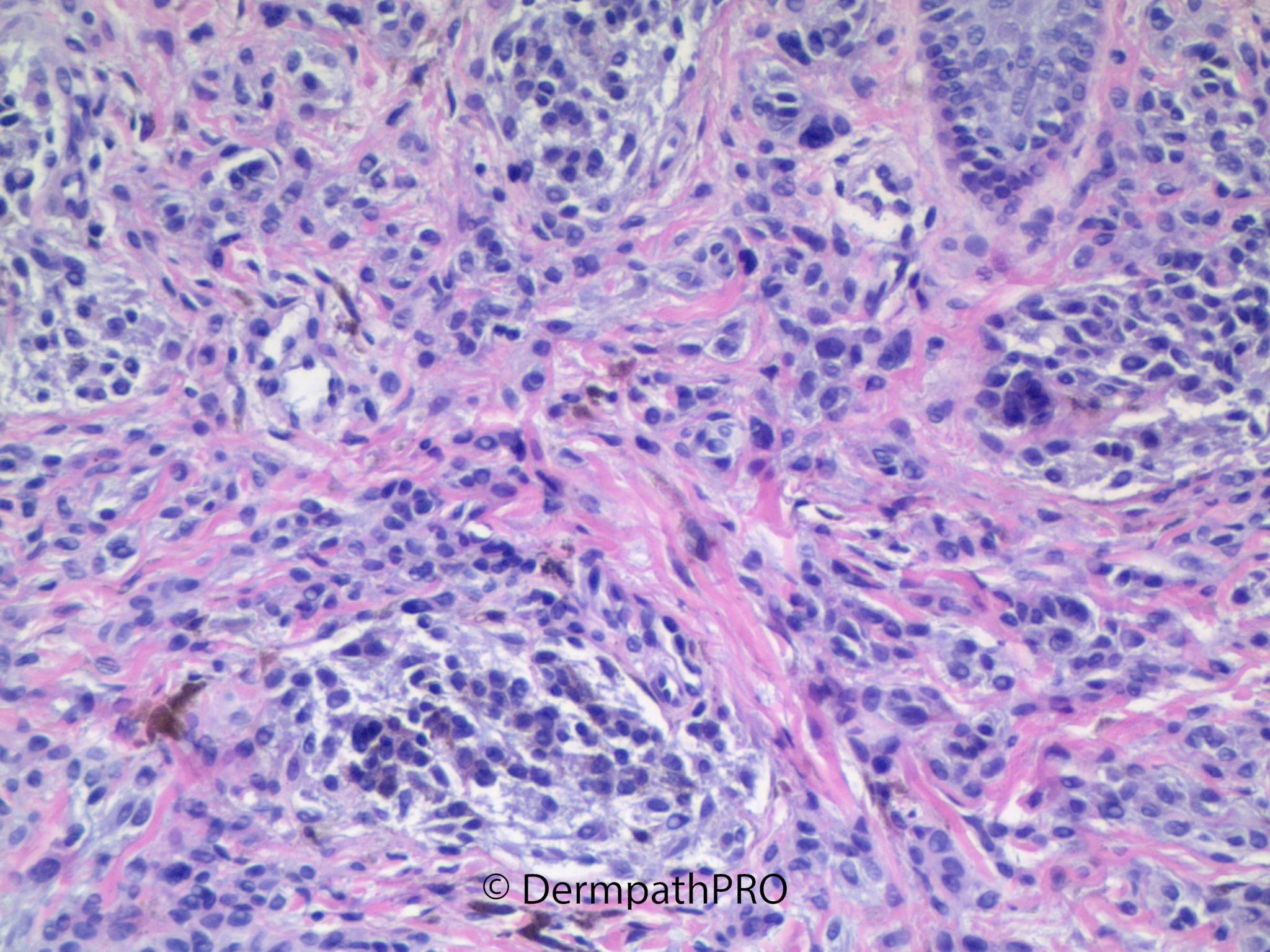
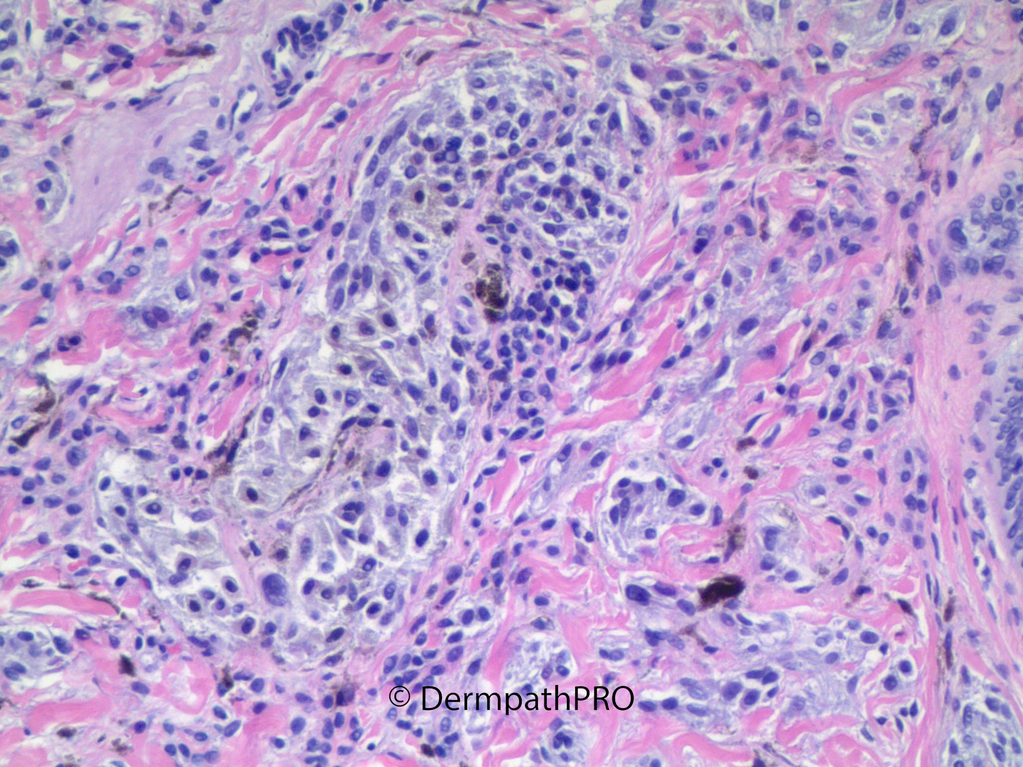
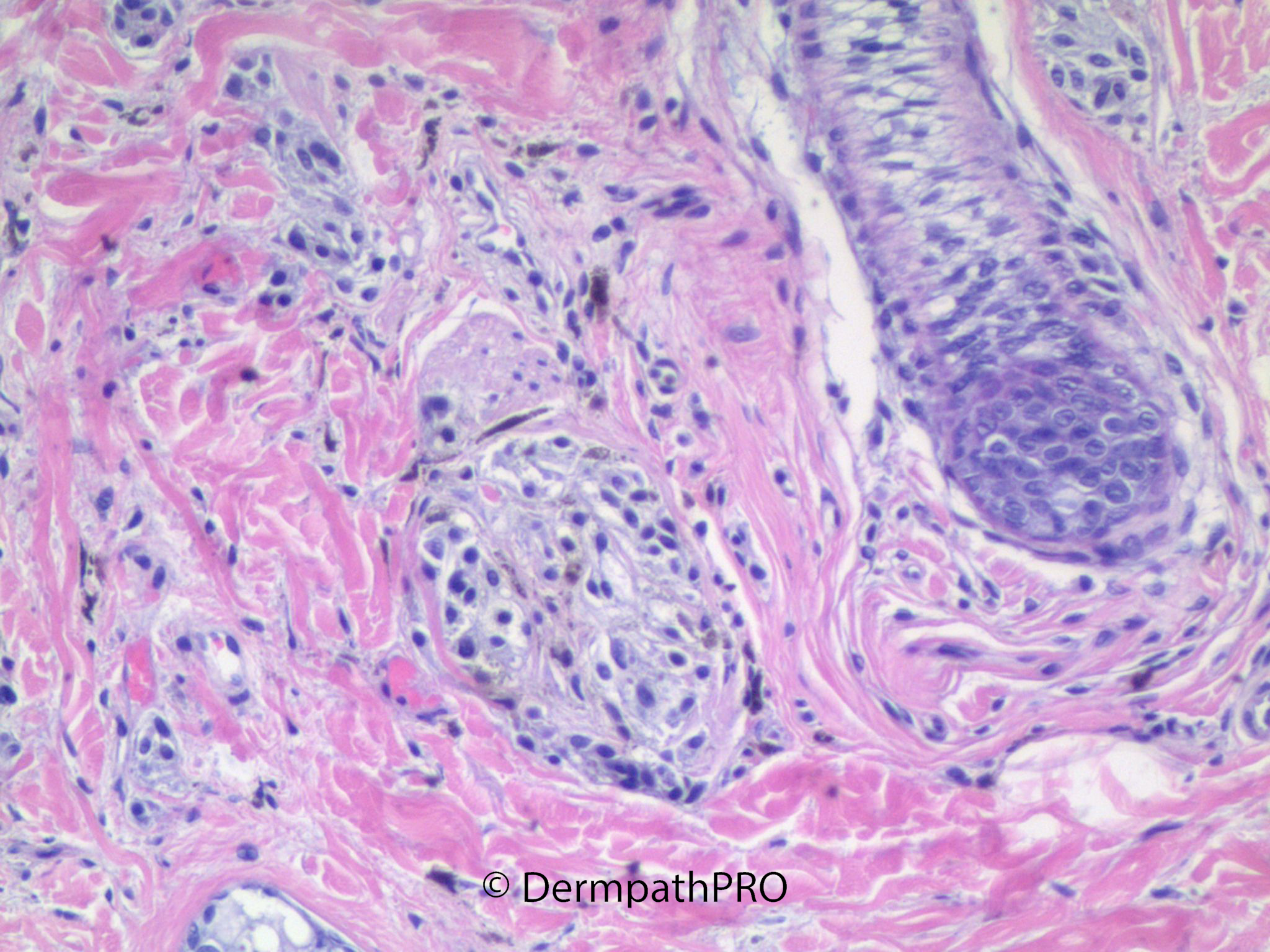
11 year old female with lesion on cheek.


  • Like 1
  Report Record

User Feedback


Dusty cytoplasm, fascicular growth, bland cytology, no mitotic activity. favor DPN, would use beta-catenin for confirmation.

Share this comment


Link to comment
share_externally

Mohamed khaled

Posted (edited)

DPN

Edited by Mohamed khaled

Share this comment


Link to comment
share_externally

Mohamed khaled

Posted

24 minutes ago, Shawn said:

Dusty cytoplasm, fascicular growth, bland cytology, no mitotic activity. favor DPN, would use beta-catenin for confirmation.

Concur

Share this comment


Link to comment
share_externally

Chirag Desai

Posted

9 hours ago, Mohamed khaled said:

DPN

Can it have multinucleated giant cells? 

Share this comment


Link to comment
share_externally

Chirag Desai

Posted

Can DPN have multinucleated giant cells? Sorry for my ignorance; have just seen one case in the past

Share this comment


Link to comment
share_externally

Mohamed khaled

Posted

8 hours ago, Chirag Desai said:

Can DPN have multinucleated giant cells? Sorry for my ignorance; have just seen one case in the past

Although it is not common finding, multinucleated cells are reported in DPN 

Share this comment


Link to comment
share_externally

Chirag Desai

Posted

13 hours ago, Mohamed khaled said:

Although it is not common finding, multinucleated cells are reported in DPN 

thanks

Share this comment


Link to comment
share_externally

Chirag Desai

Posted

10 hours ago, Uma Sundram said:

Deep penetrating nevus.

how do we differentiate this lesion from cellular blue nevus?

Share this comment


Link to comment
share_externally

13 hours ago, Chirag Desai said:

how do we differentiate this lesion from cellular blue nevus?

Cellular blue nevus: look for adjacent common blue nevus component, lacks nuclear beta-catenin staining, less dusty cytoplasm.

Share this comment


Link to comment
share_externally

Chirag Desai

Posted

7 hours ago, Shawn said:

Cellular blue nevus: look for adjacent common blue nevus component, lacks nuclear beta-catenin staining, less dusty cytoplasm.

Ok, Thanks a lot for the explanation. 


Share this comment


Link to comment
share_externally


Join the conversation

You can post now and register later. If you have an account, sign in now to post with your account.

Guest
Add a comment...

×   Pasted as rich text.   Paste as plain text instead

  Only 75 emoji are allowed.

×   Your link has been automatically embedded.   Display as a link instead

×   Your previous content has been restored.   Clear editor

×   You cannot paste images directly. Upload or insert images from URL.

Loading...

×
×
  • Create New...