Jump to content
In this section we have spot diagnoses posted on a daily basis since June 2010, now over 4000! You can review the archived cases and read the suggested diagnoses by users and the final comment by the contributors.
Case are uploaded each week day by 10 am UK time with the correct diagnosis will generally be posted at 8 pm UK time. Why not view the most recent spot diagnosis and proffer a diagnosis?

Case Number : Case 3079 - 22 April 2022 Posted By: Dr. Richard Carr

Please read the clinical history and view the images by clicking on them before you proffer your diagnosis.
Submitted Date :
   (0 reviews)

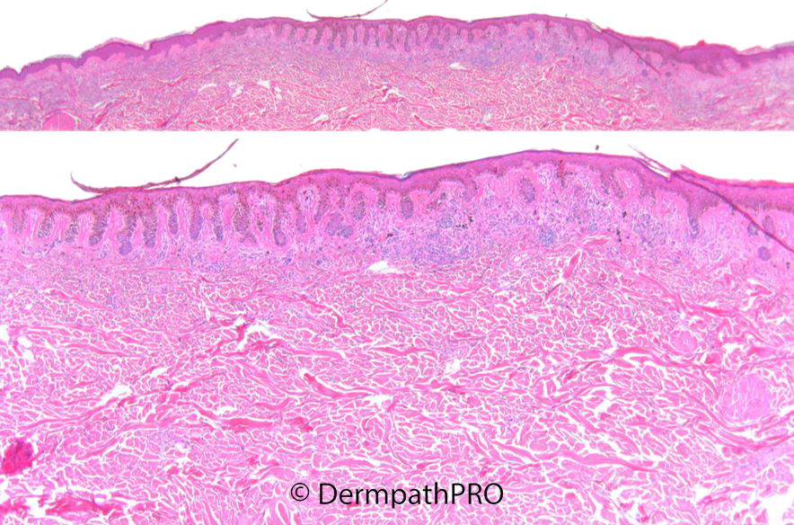
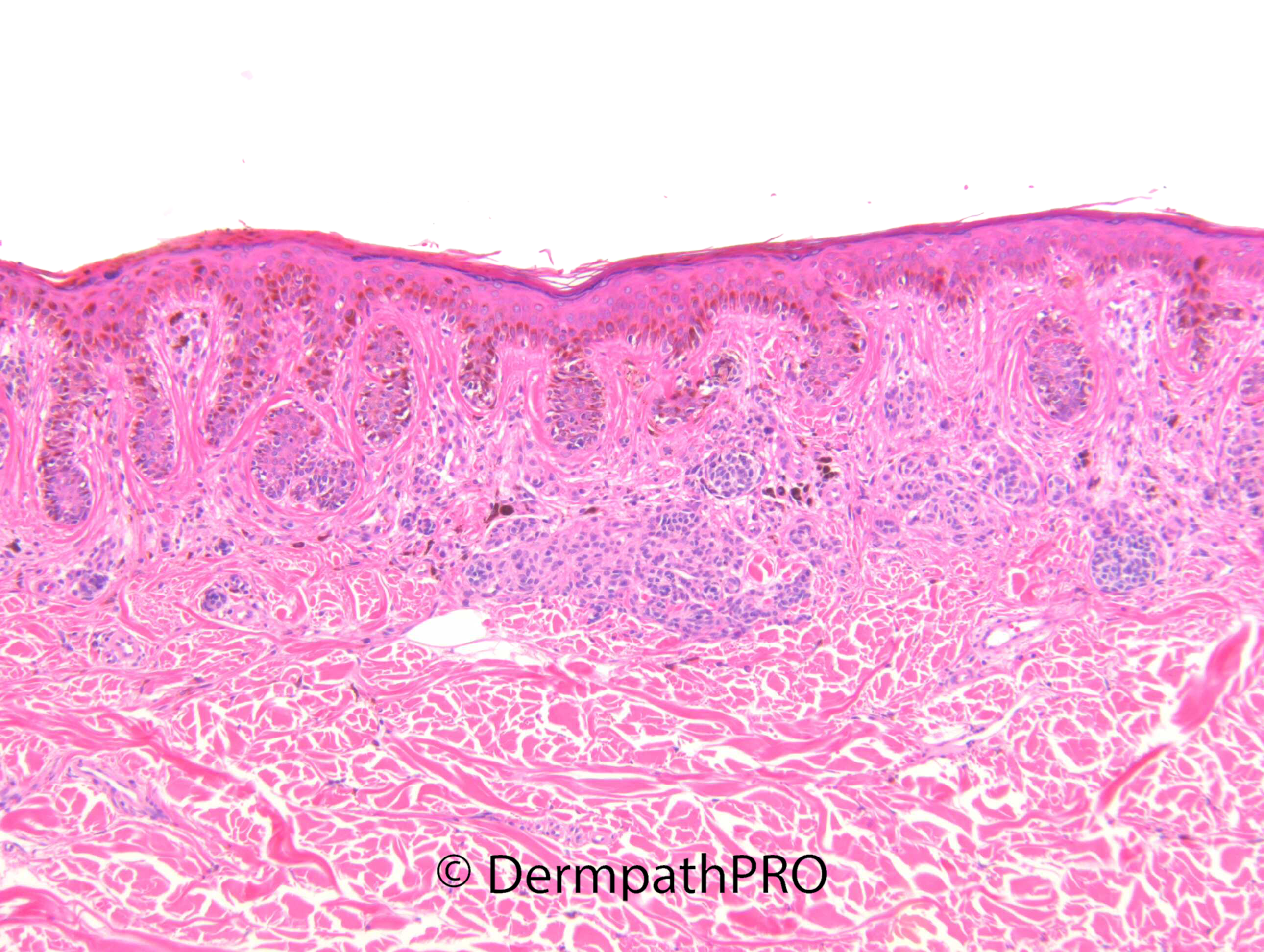
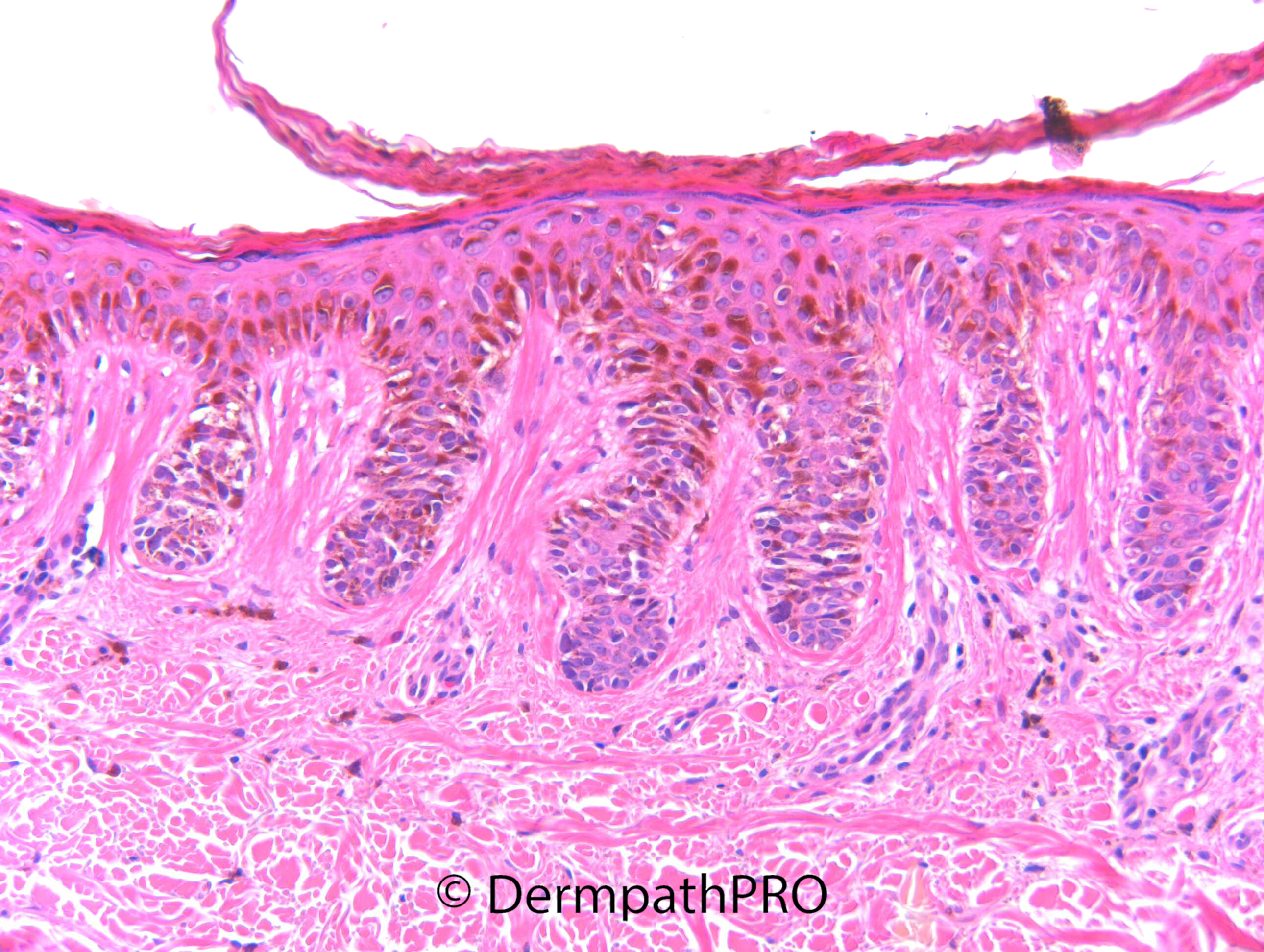
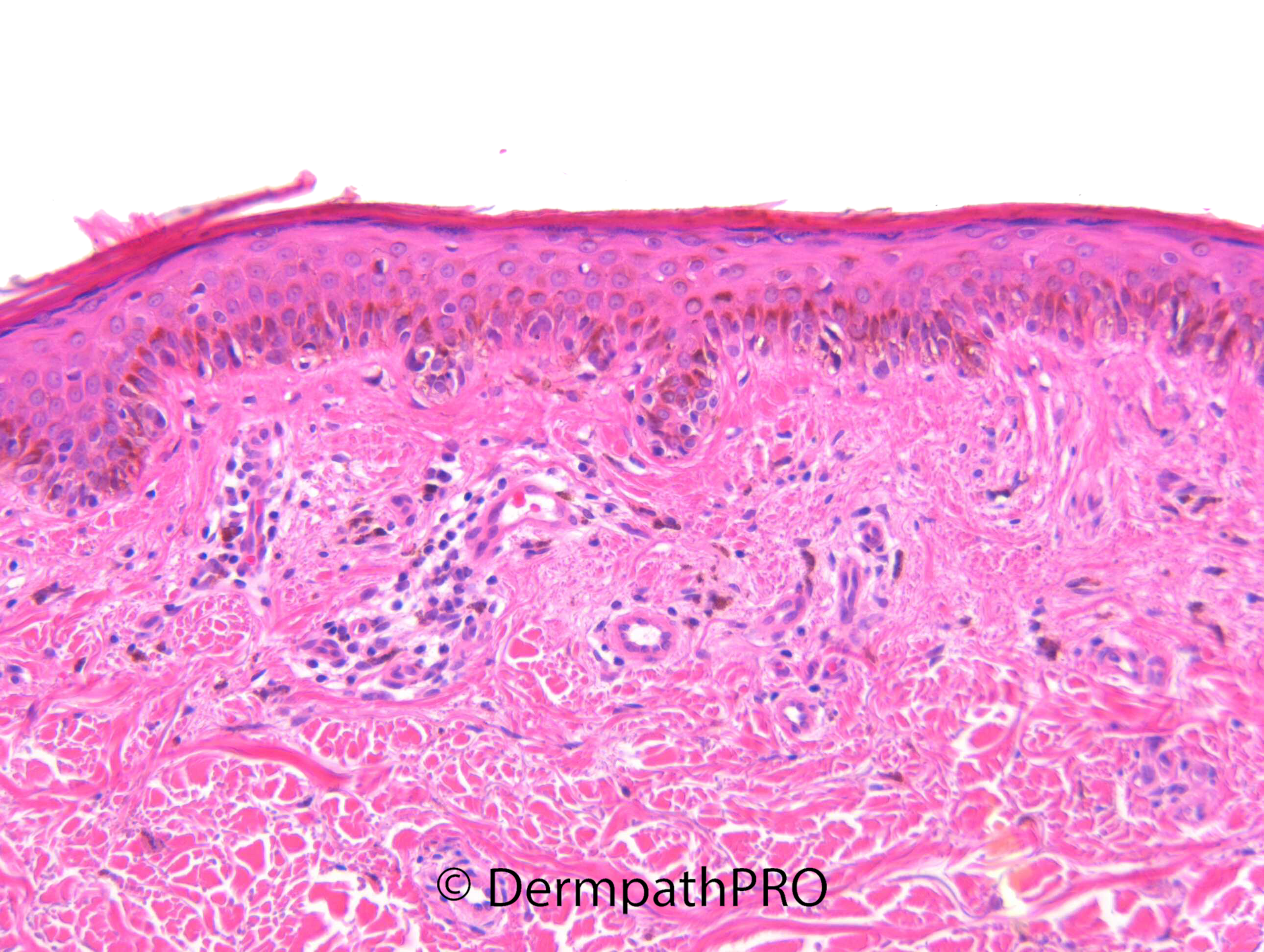
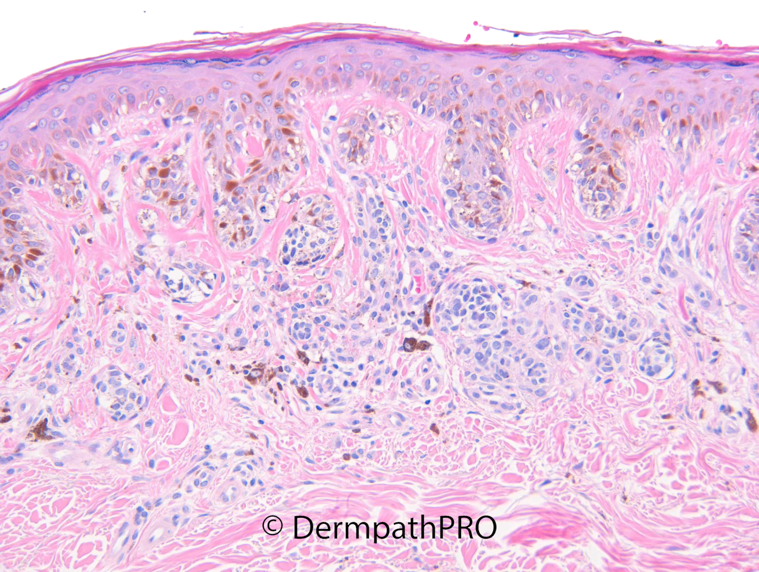
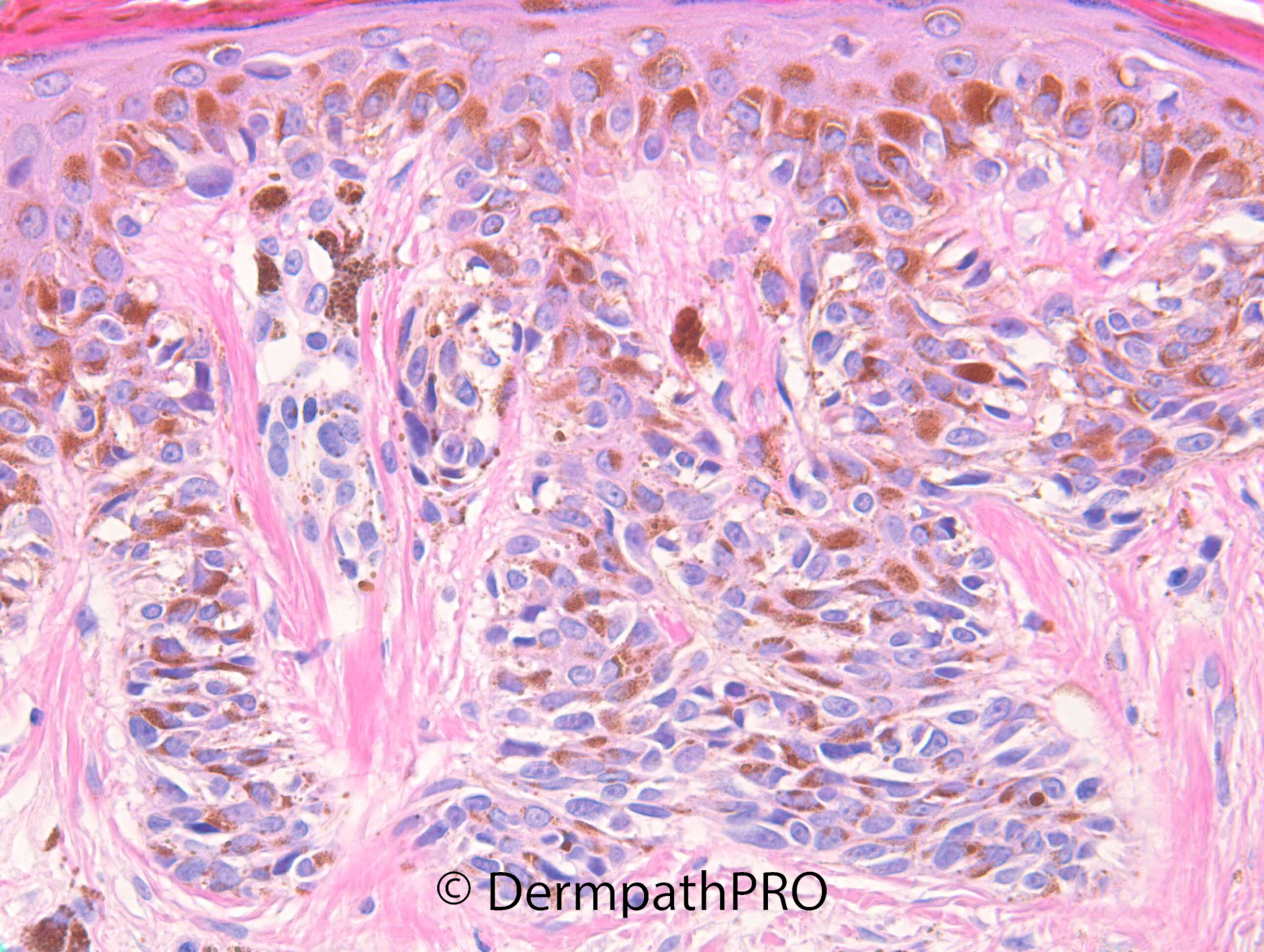
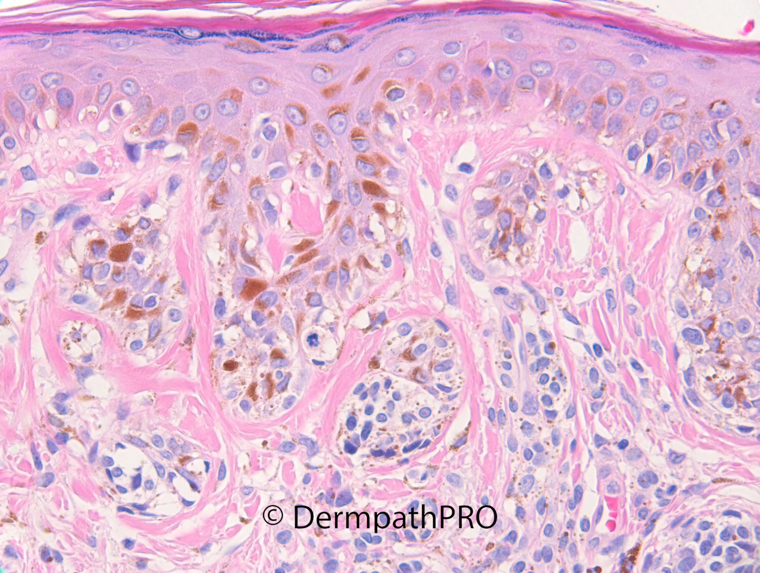
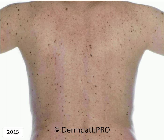
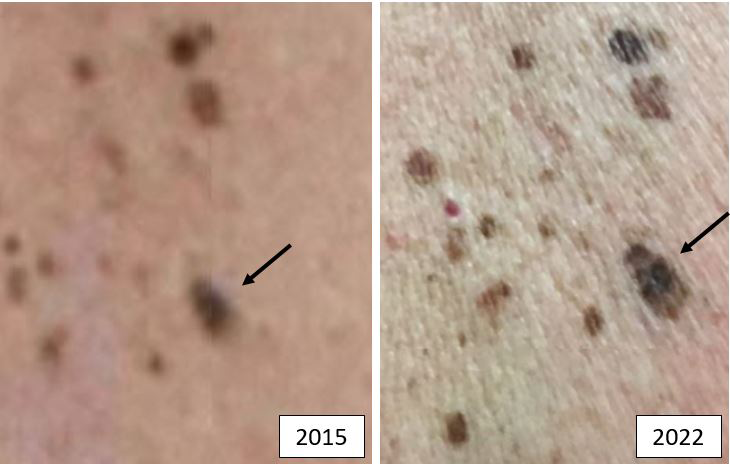
M60. Right mid-back near midline. 10 x 8mm irregularly pigmented lesion. Has atypical moles and is under yearly observation.

  • Like 1
  Report Record

User Feedback


An irritated dysplastic nevus, low grade (mild) atypia. clinical appearance is more or less stable, (limited by resolution of 2015 image)

Share this comment


Link to comment
share_externally

Eman El-Nabarawy

Posted

I thought is dysplasia nevus too..

Share this comment


Link to comment
share_externally

Lentiginous junctional growth pattern, concentric lamellar fibrosis around rete ridge and atypical cells with superficial mitosis, but in a random distribution, of intraepidermal melanocytes. So agree with Colleagues. The sovrapapillary epidermal plate is often free, or am’I wrong?

Share this comment


Link to comment
share_externally

Meenakshi Batrani

Posted

I also favour dysplastic nevus

Share this comment


Link to comment
share_externally

msofopoulos

Posted

I think its a dysplastic nevus. I see parakeratosis probably indicative of irritation?

Share this comment


Link to comment
share_externally

Dr. Richard Carr

Posted

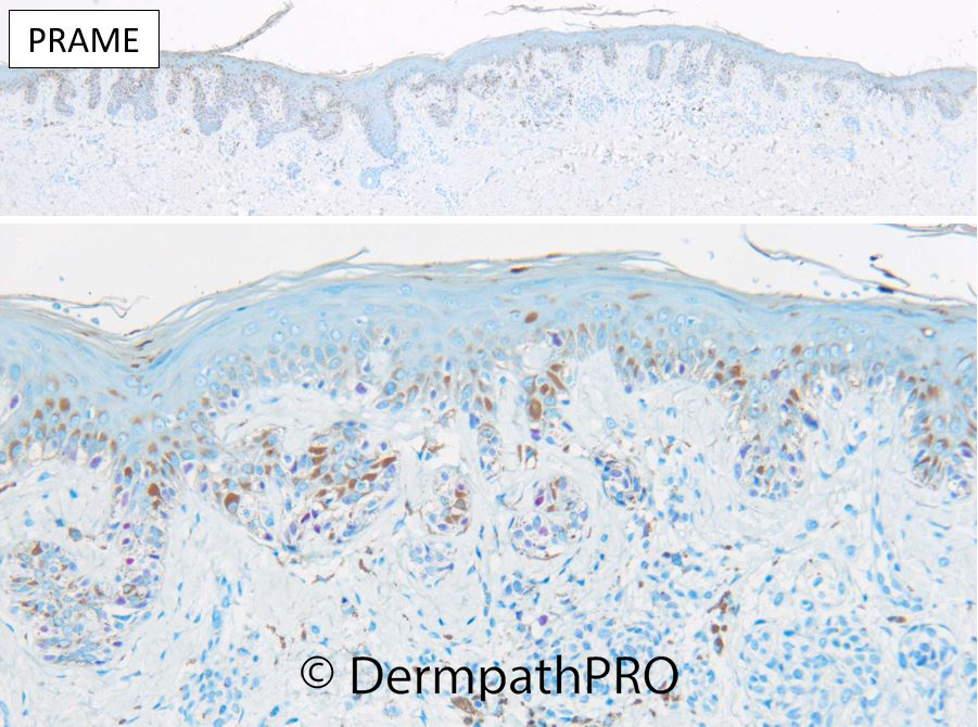
I agree this is a dysplastic naevus (but with only mild atypia) - ?should be called just naevus according to WHO 4th but was the one removed as being clinically atypical. I've tended to to continue to report them as dysplastic naevus with mild atypia (rather than adopting low-grade = moderate atypia). I would worry about lesions with more severe atypia and ensure they were completely excised. We're lucky in the UK as we don't get the partial shave specimens that are common in the U.S. which must be very frustrating. I doubt the moderate atypia is all that reproducible and I've heard stories of lesions recurring occasionally as high grade lesions. Nice that PRAME was negative in this one but would not necessarily do it routinely in such a banal lesion providing it was fully excised.

Share this comment


Link to comment
share_externally


Join the conversation

You can post now and register later. If you have an account, sign in now to post with your account.

Guest
Add a comment...

×   Pasted as rich text.   Paste as plain text instead

  Only 75 emoji are allowed.

×   Your link has been automatically embedded.   Display as a link instead

×   Your previous content has been restored.   Clear editor

×   You cannot paste images directly. Upload or insert images from URL.

Loading...

×
×
  • Create New...