Jump to content
In this section we have spot diagnoses posted on a daily basis since June 2010, now over 4000! You can review the archived cases and read the suggested diagnoses by users and the final comment by the contributors.
Case are uploaded each week day by 10 am UK time with the correct diagnosis will generally be posted at 8 pm UK time. Why not view the most recent spot diagnosis and proffer a diagnosis?

Case Number : Case 3084 - 29 April 2022 Posted By: Dr. Richard Carr

Please read the clinical history and view the images by clicking on them before you proffer your diagnosis.
Submitted Date :
   (0 reviews)

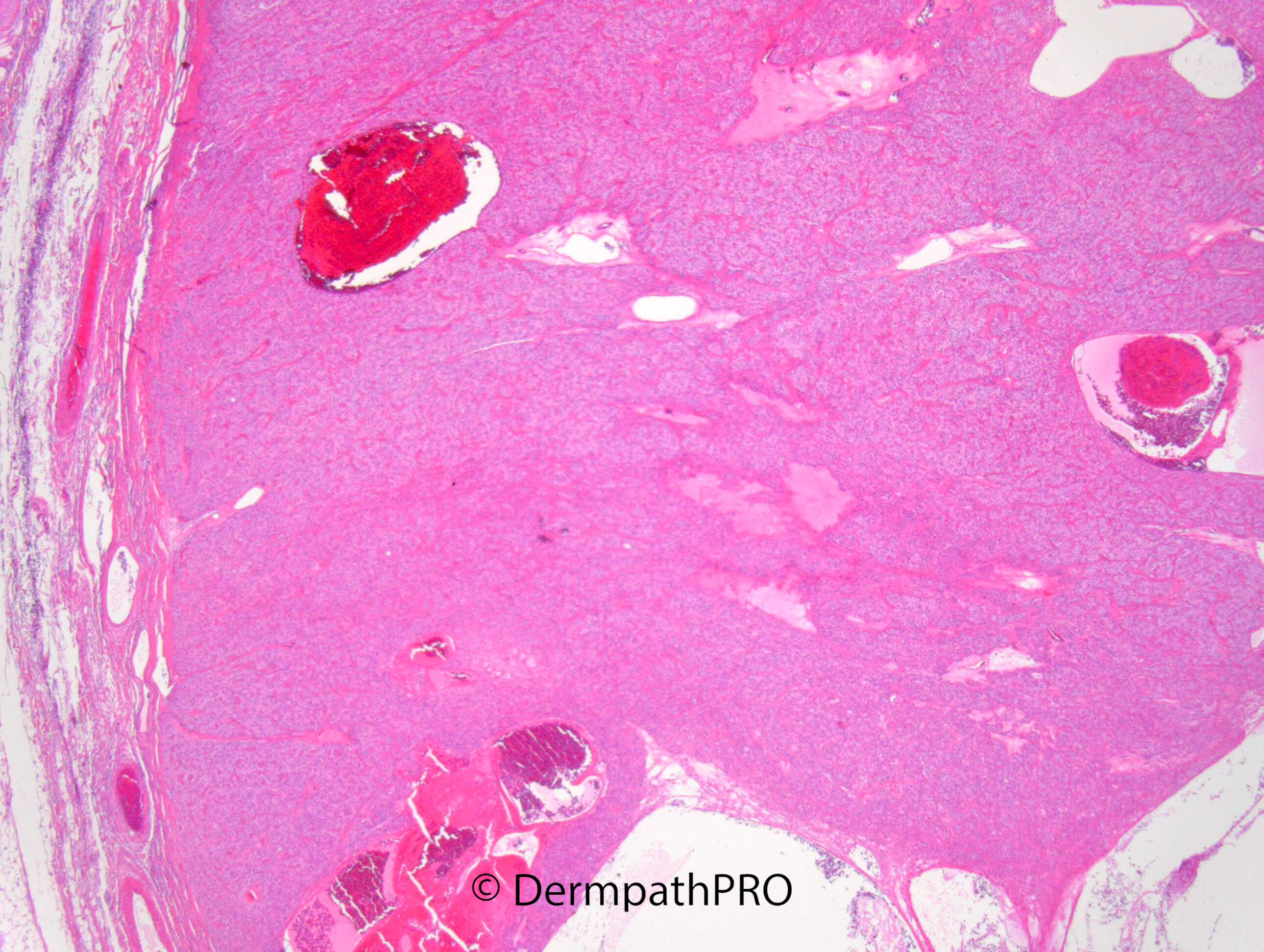
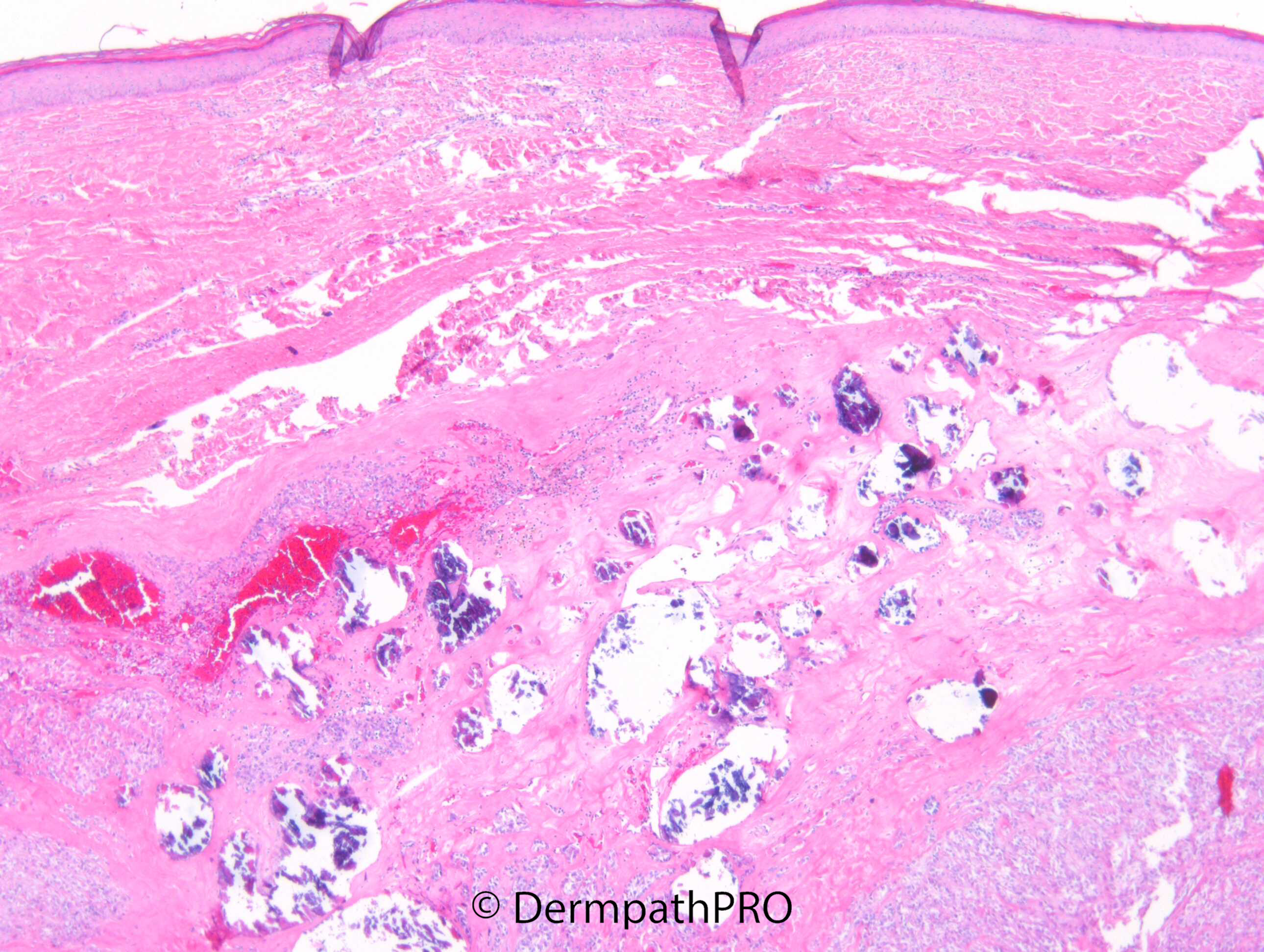
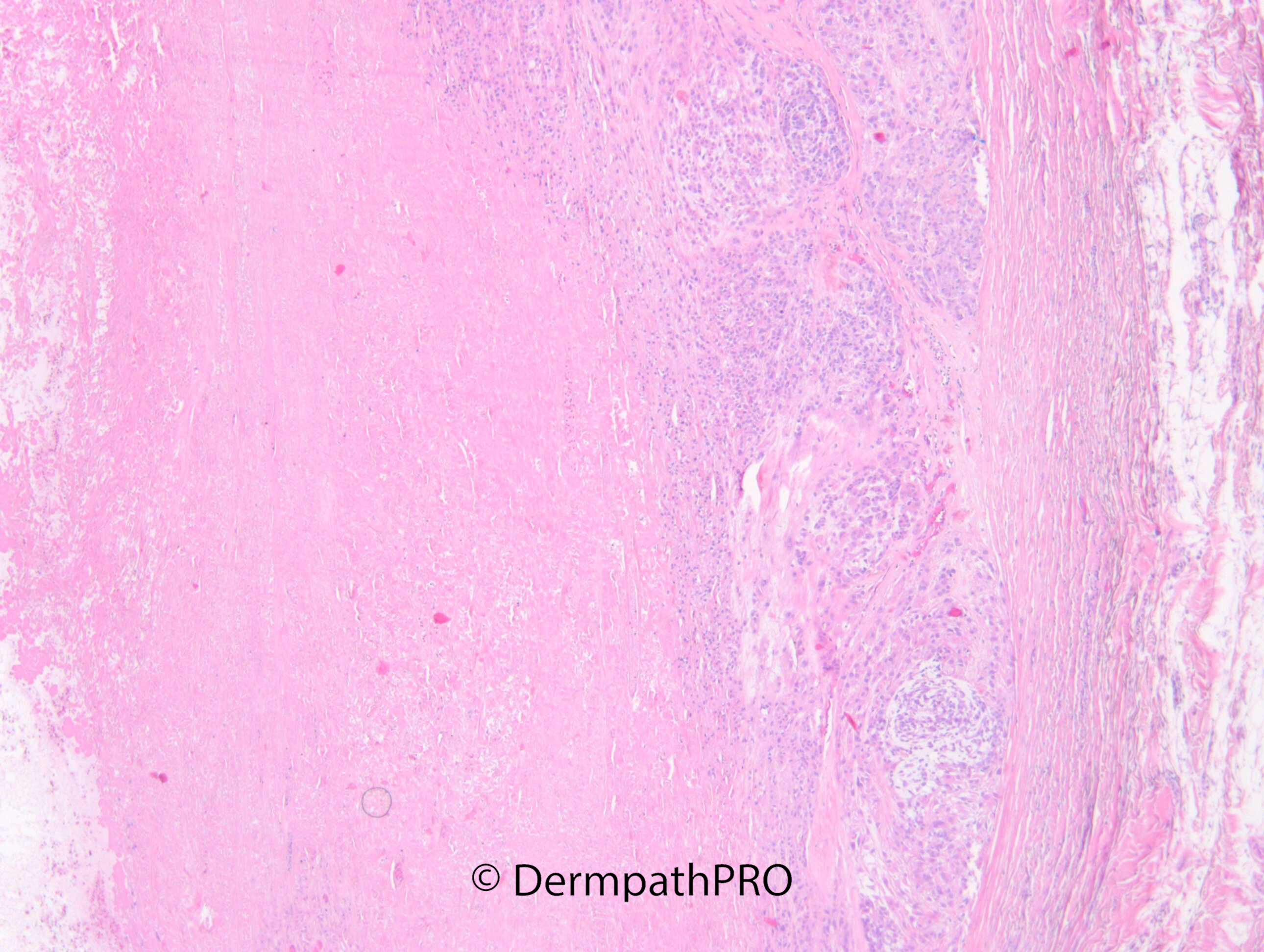
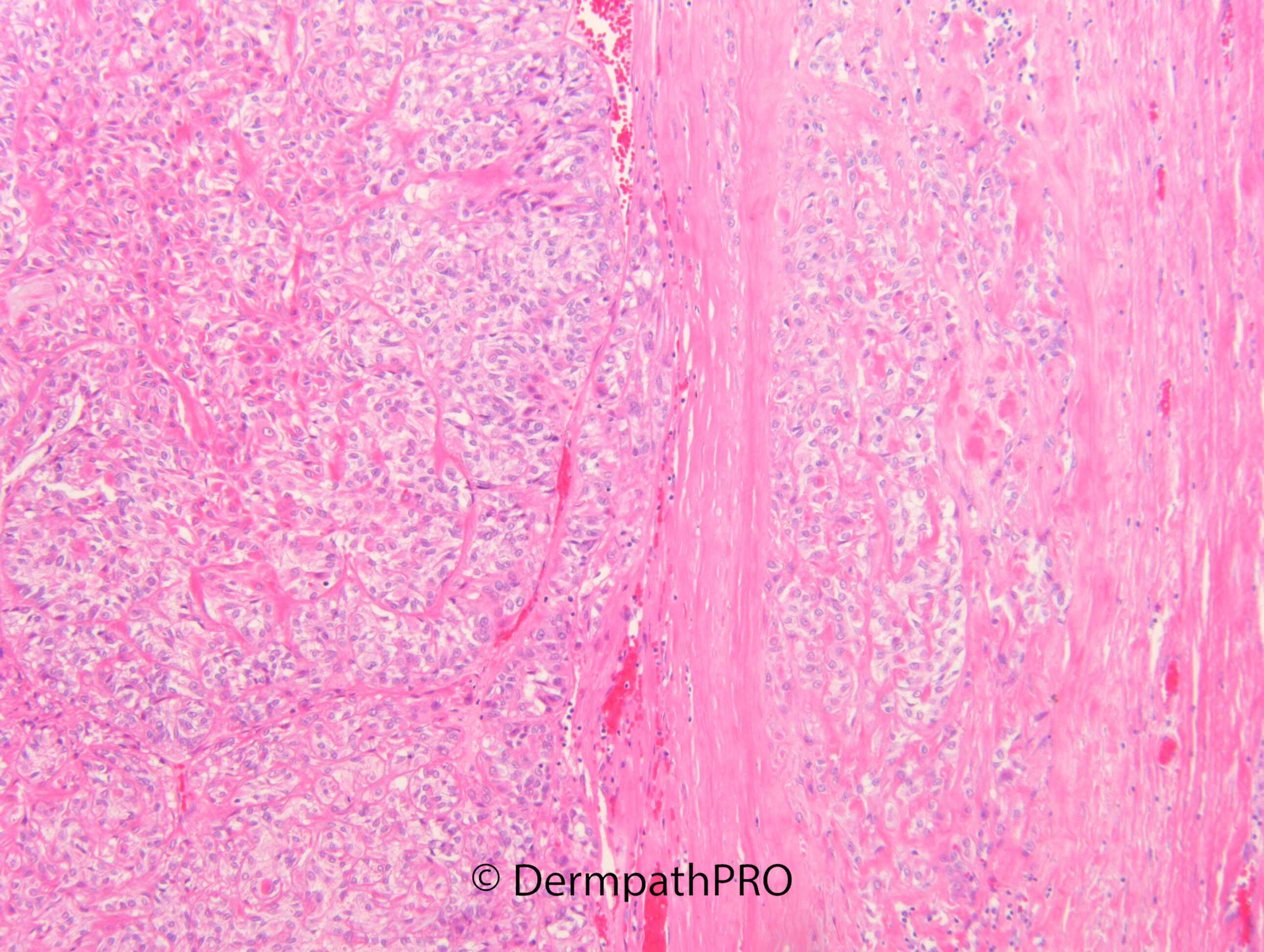
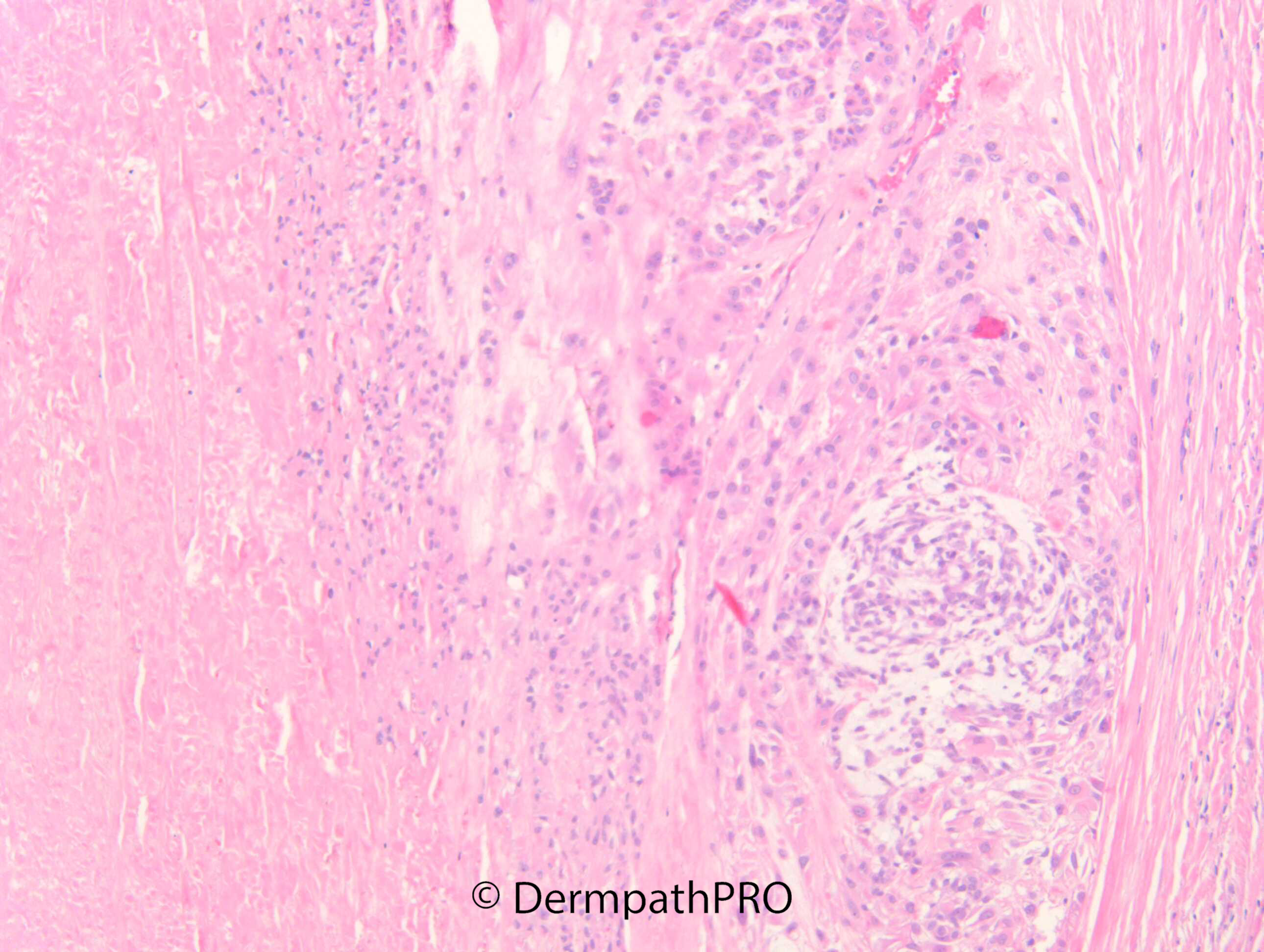
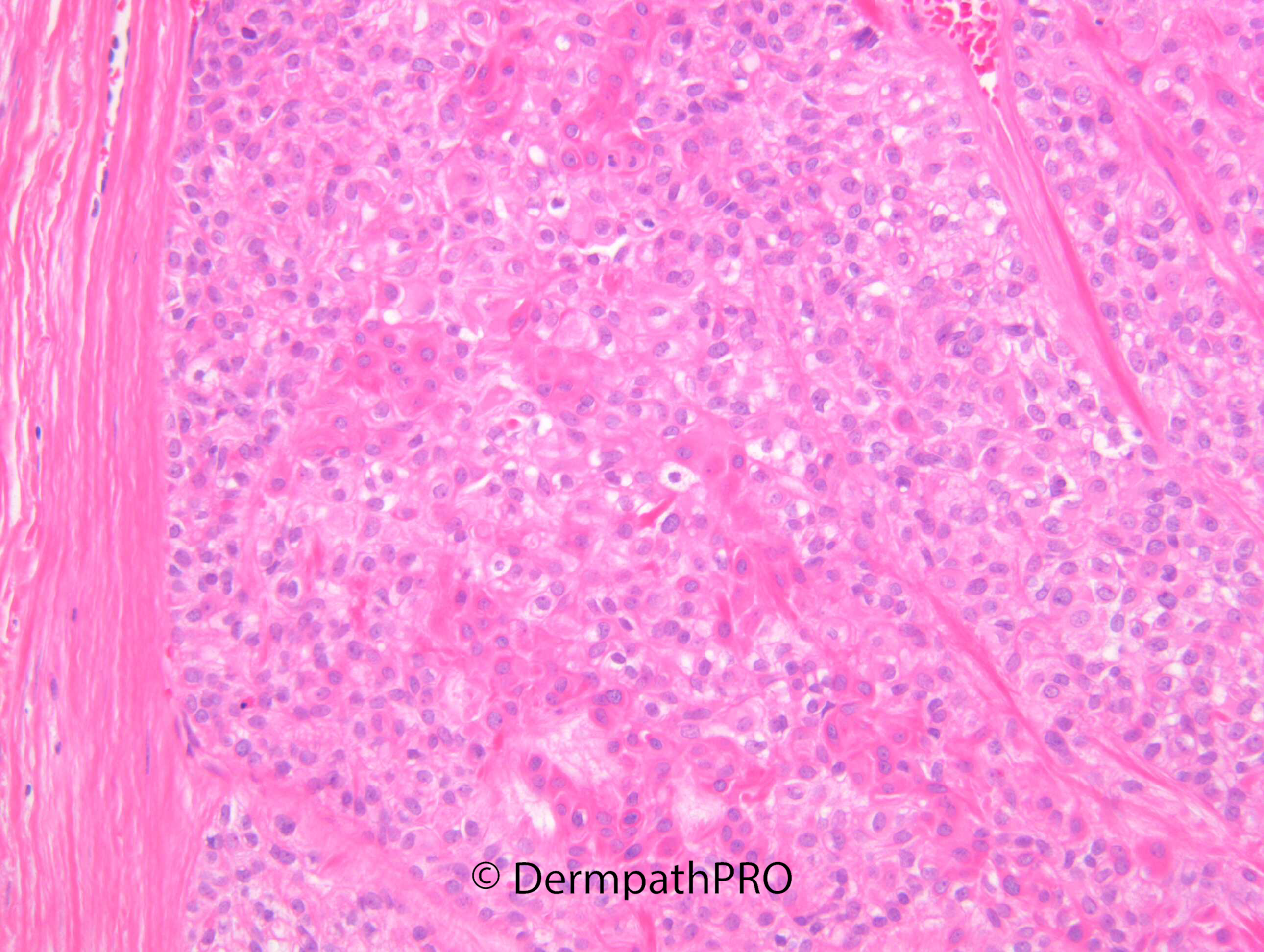
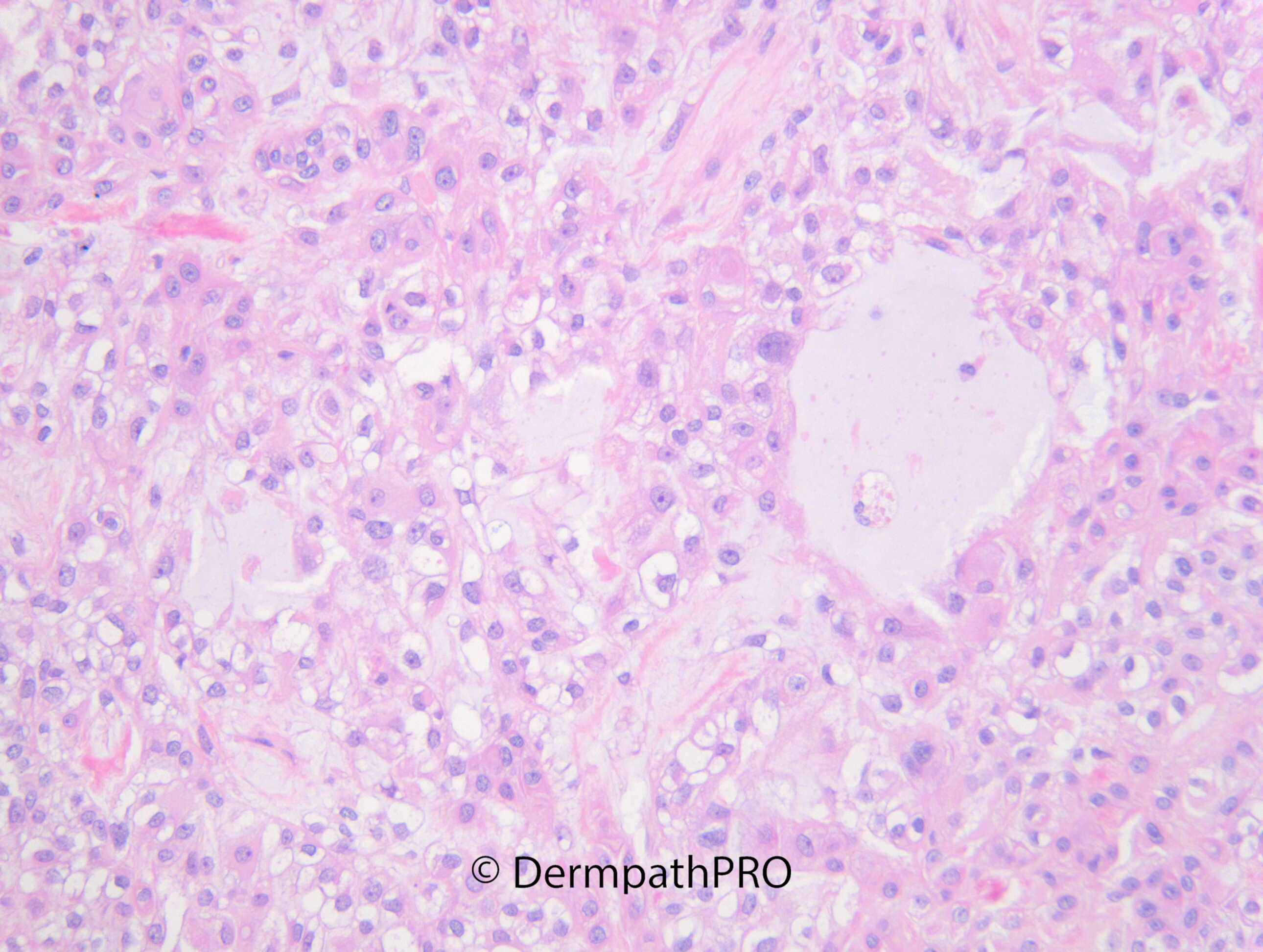
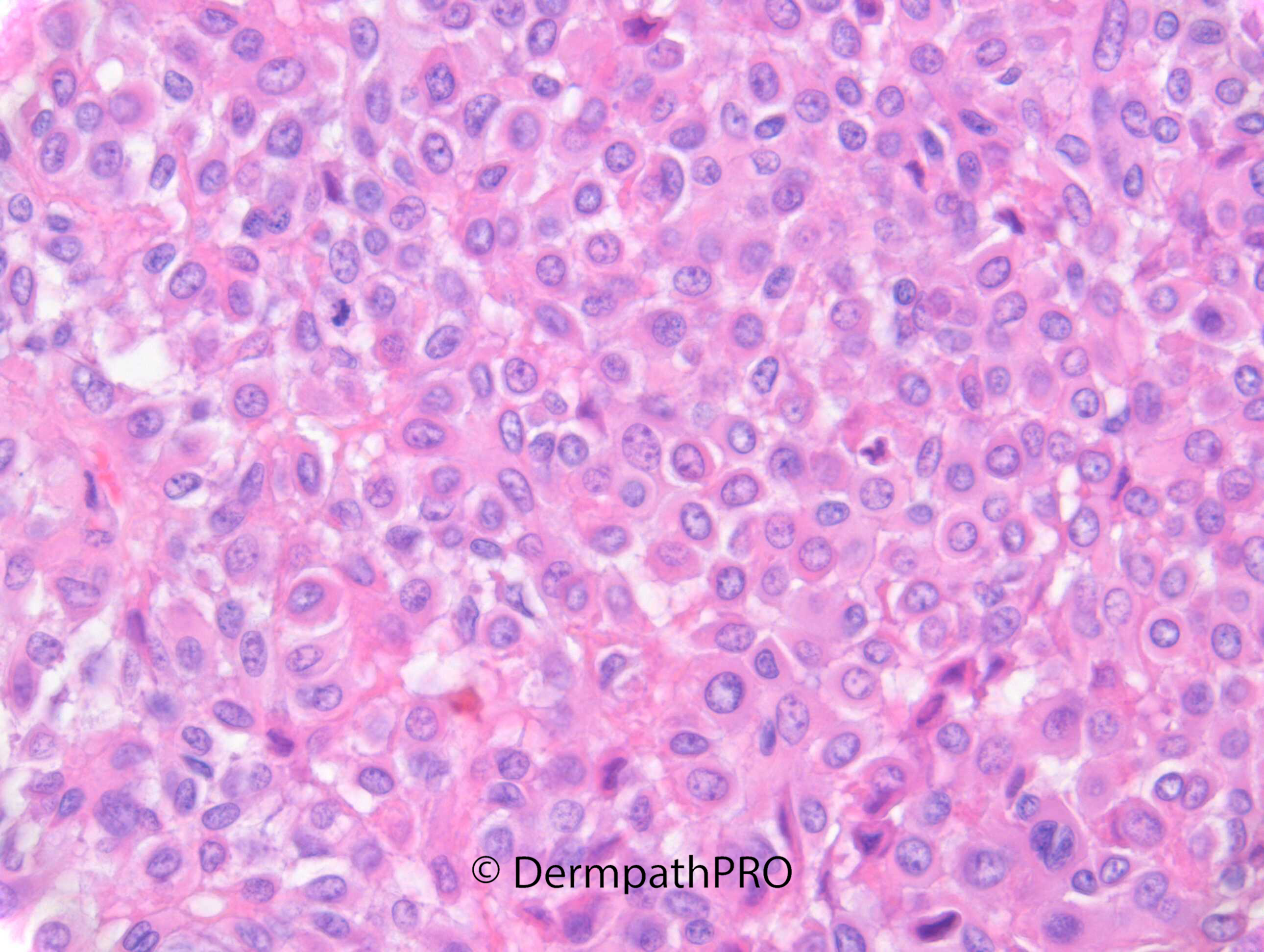
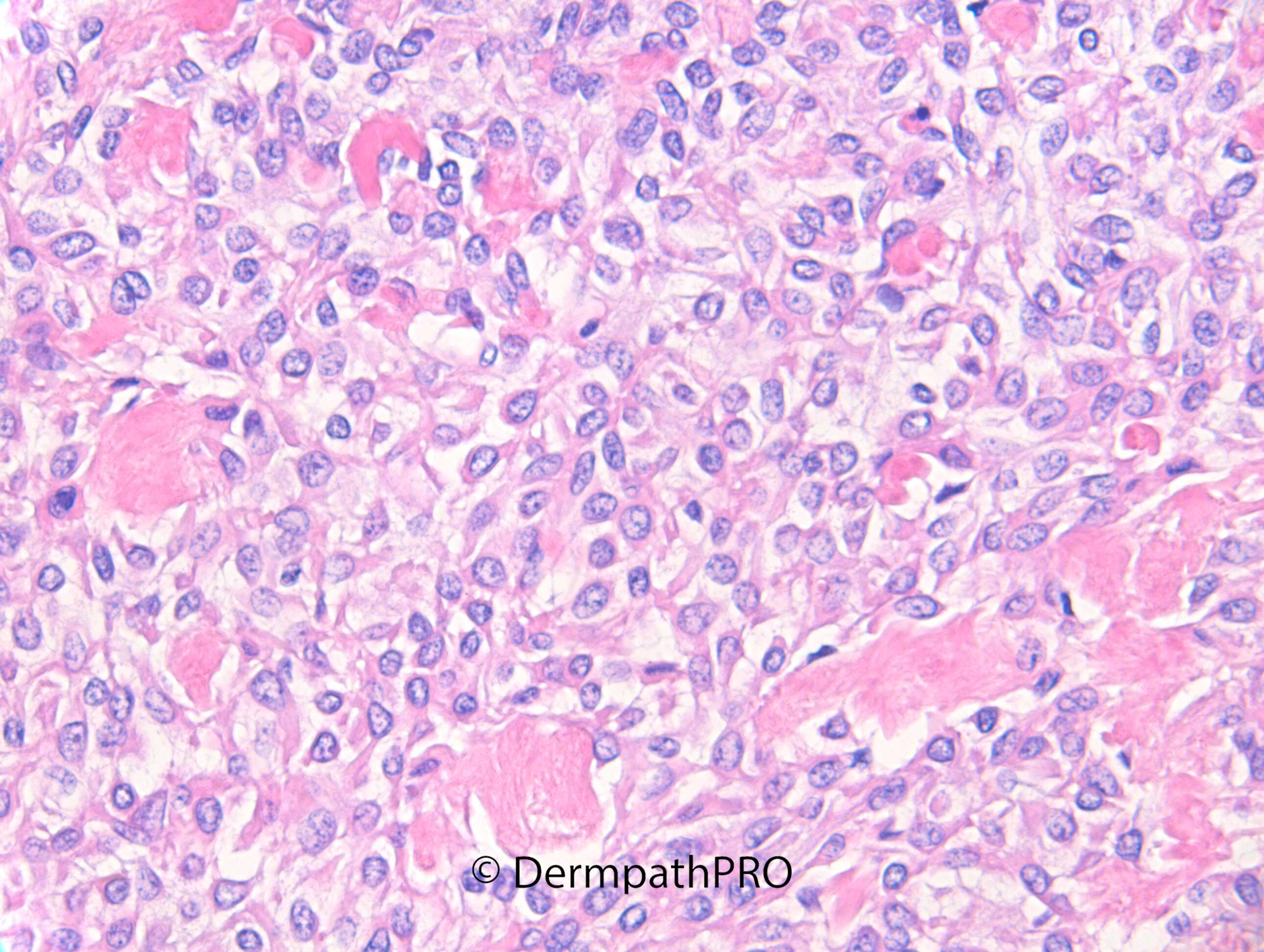
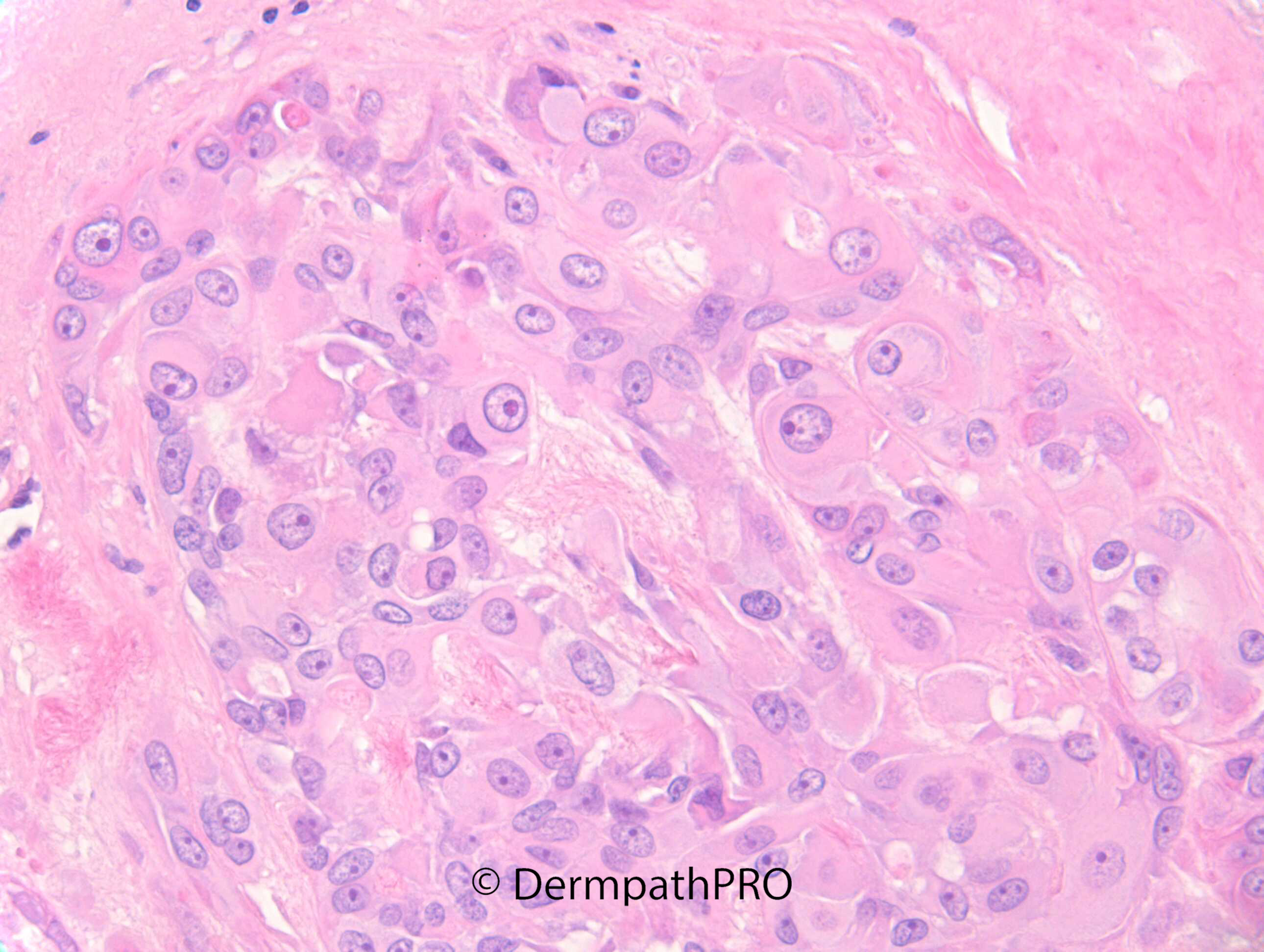
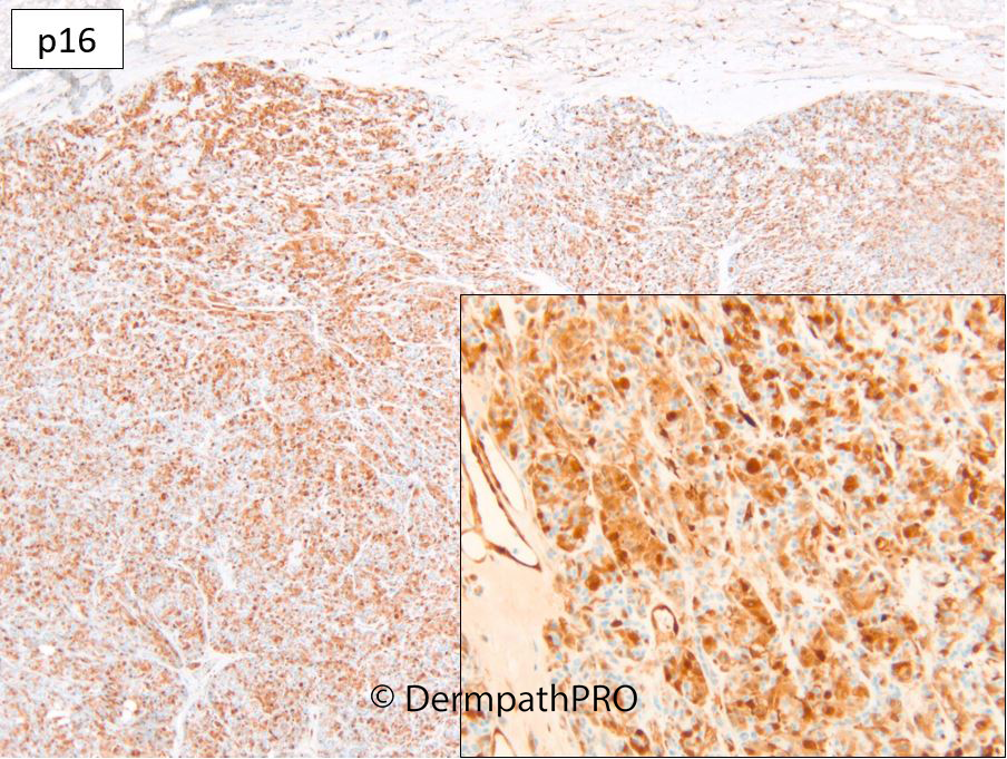
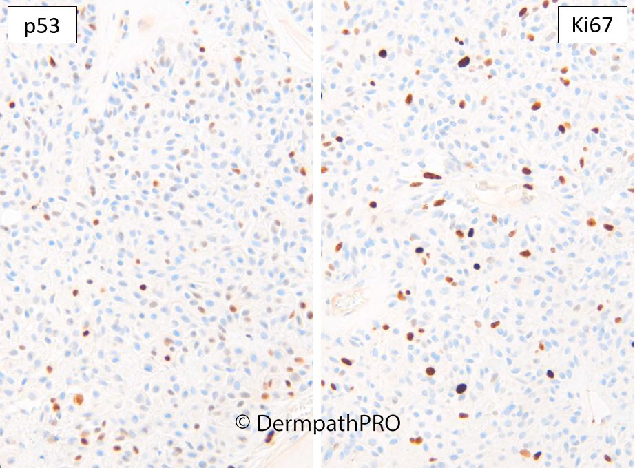
M85. Scalp. Nodule. Slowly enlarging over 18 months. Non-tender, firm consistency, slightly mobile. No bleeding or discharge. Case c/o Dr Andrew O’Keeffe


  • Thanks 1
  Report Record

User Feedback


Meenakshi Batrani

Posted

Looks like a nodular hidradenoma to me

Share this comment


Link to comment
share_externally

vincenzo

Posted

?Proliferating Trichilemmal Cyst, with “ancient” changes.

Share this comment


Link to comment
share_externally

Dr. Richard Carr

Posted

I'll wait for a few more comments. 

Share this comment


Link to comment
share_externally

Meenakshi Batrani

Posted

After IHC, considering myoepithelial neoplasm- not sure of criteria for primary cutaneous myoepithelioma vs myoepithelial carcinoma vs salivary gland metastasis.

Share this comment


Link to comment
share_externally

Dr. Richard Carr

Posted

Strictly speaking the presence of focal ductal differentiation and the squamoid cells favours a myoepithelial-rich chondroid syringoma. Focal confluent necrosis is obviously atypical but there were no other worrying features for malignancy. I shared the case with Luis Requena, Omar Sanqueza and Jason Hornick who are co-authors for myoepithelioma in the next WHO classn who also favoured an essentially benign / low risk lesion. The p16 is mosaic and relatively low Ki67 congruent with p53 were all re-assuring I'm awaiting to hear about IHC for PLAG1 which may  be positive in chrondoid syringoma compared with soft tissue myoepithelioma what often have EWSR1 or FUS re-arrangement. 

Share this comment


Link to comment
share_externally

Meenakshi Batrani

Posted

17 hours ago, Dr. Richard Carr said:

Strictly speaking the presence of focal ductal differentiation and the squamoid cells favours a myoepithelial-rich chondroid syringoma. Focal confluent necrosis is obviously atypical but there were no other worrying features for malignancy. I shared the case with Luis Requena, Omar Sanqueza and Jason Hornick who are co-authors for myoepithelioma in the next WHO classn who also favoured an essentially benign / low risk lesion. The p16 is mosaic and relatively low Ki67 congruent with p53 were all re-assuring I'm awaiting to hear about IHC for PLAG1 which may  be positive in chrondoid syringoma compared with soft tissue myoepithelioma what often have EWSR1 or FUS re-arrangement. 

Thank you for posting some of these rare and challenging cases, and providing the detailed work-up to arrive at a diagnosis. 

Share this comment


Link to comment
share_externally

Dr. Richard Carr

Posted

Update from Jason Hornick: Dear Richard, 

IHC for PLAG1 is positive, supporting the diagnosis of a myoepithelial cell-rich mixed tumor.

Share this comment


Link to comment
share_externally


Join the conversation

You can post now and register later. If you have an account, sign in now to post with your account.

Guest
Add a comment...

×   Pasted as rich text.   Paste as plain text instead

  Only 75 emoji are allowed.

×   Your link has been automatically embedded.   Display as a link instead

×   Your previous content has been restored.   Clear editor

×   You cannot paste images directly. Upload or insert images from URL.

Loading...

×
×
  • Create New...