Jump to content
In this section we have spot diagnoses posted on a daily basis since June 2010, now over 4000! You can review the archived cases and read the suggested diagnoses by users and the final comment by the contributors.
Case are uploaded each week day by 10 am UK time with the correct diagnosis will generally be posted at 8 pm UK time. Why not view the most recent spot diagnosis and proffer a diagnosis?

Case Number : Case 4054 - 04 August 2022 Posted By: Saleem Taibjee

Please read the clinical history and view the images by clicking on them before you proffer your diagnosis.
Submitted Date :
   (0 reviews)

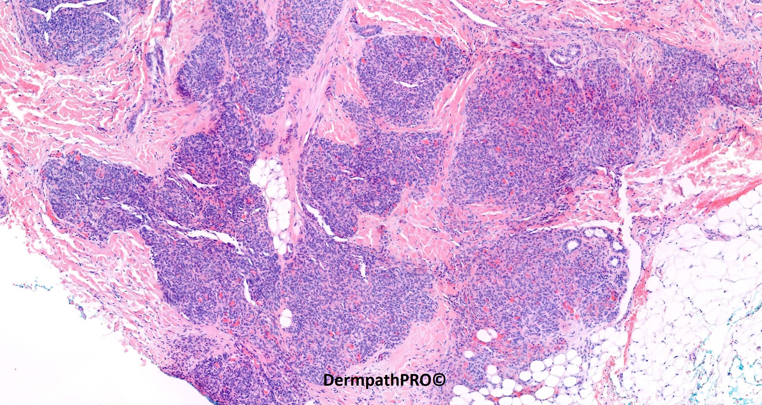
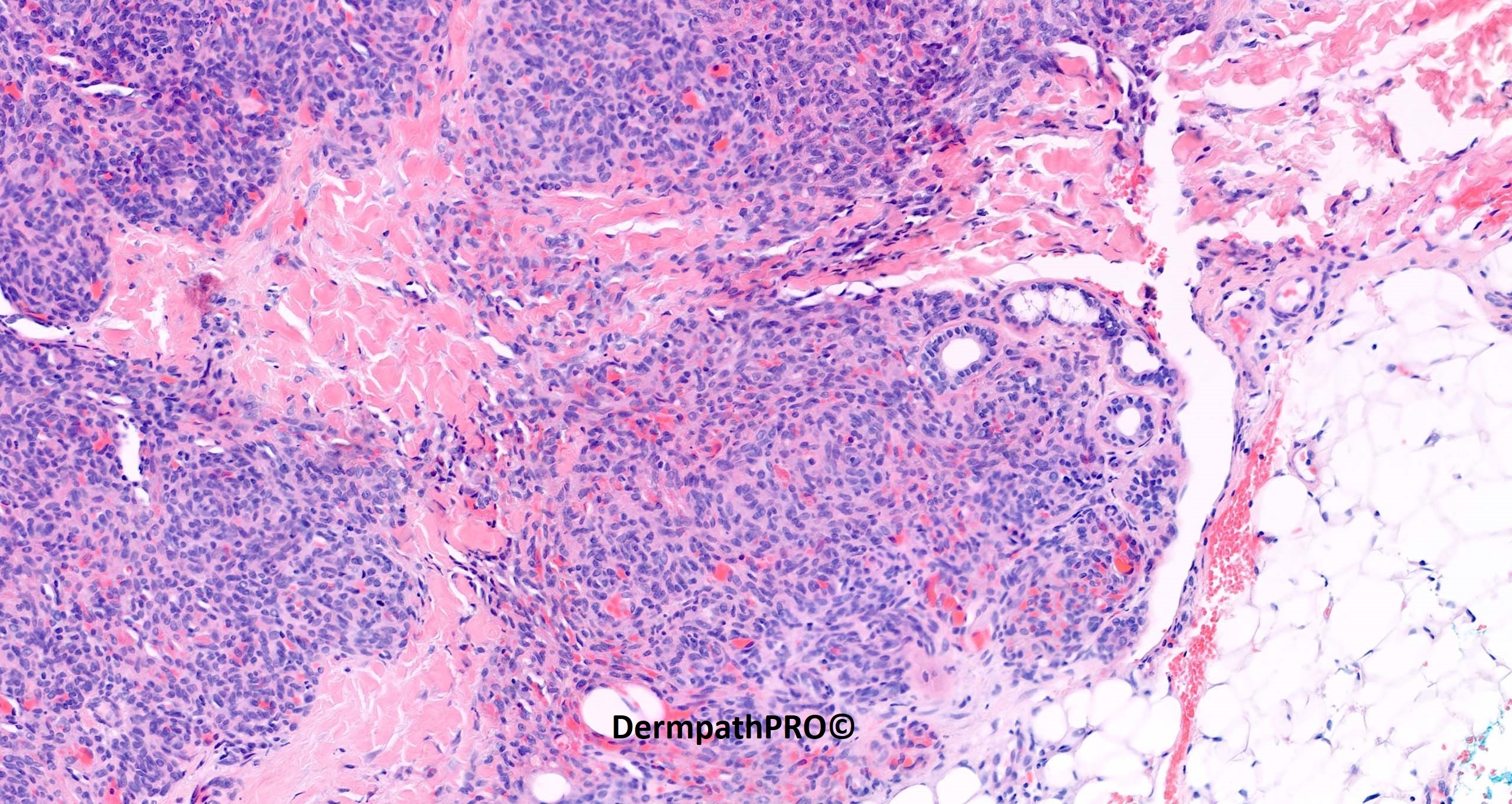
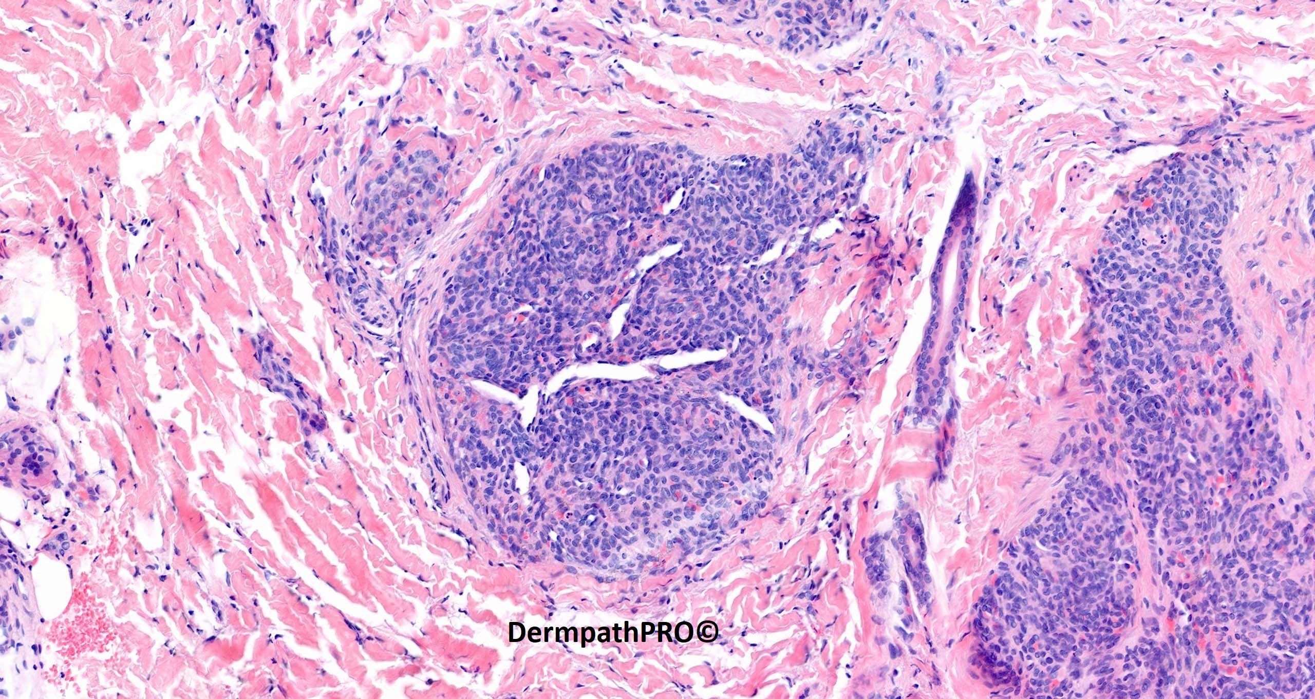
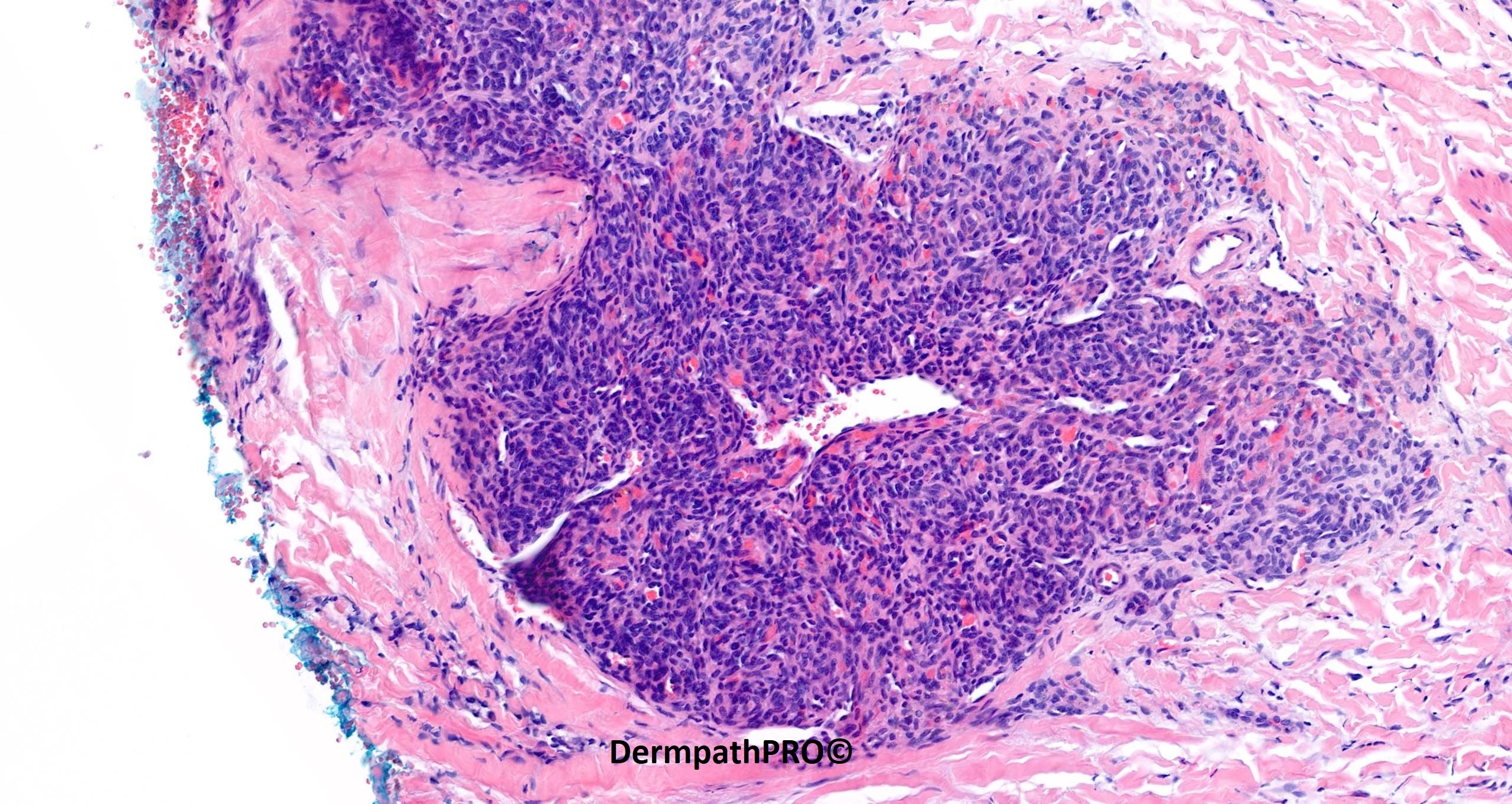
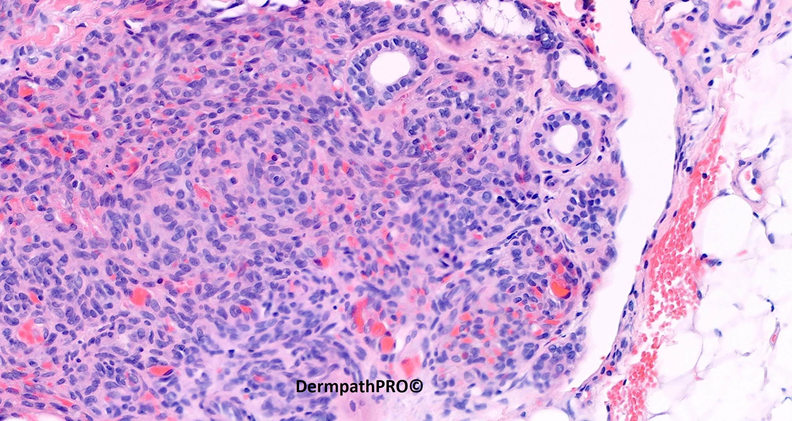
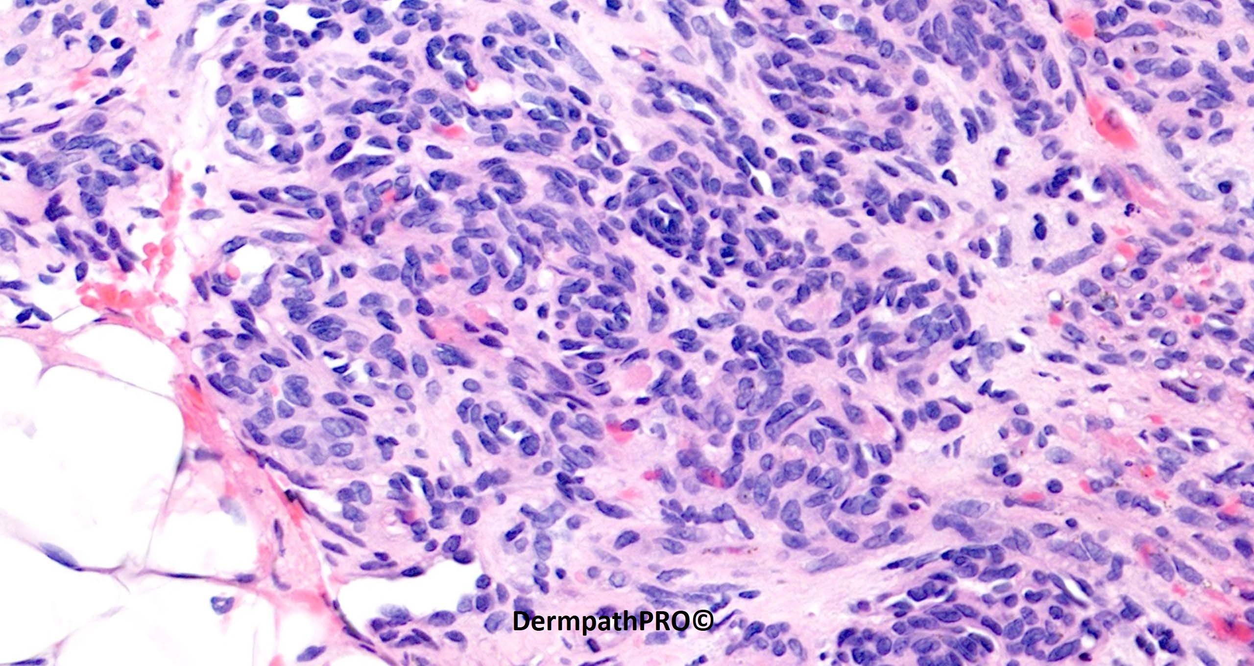
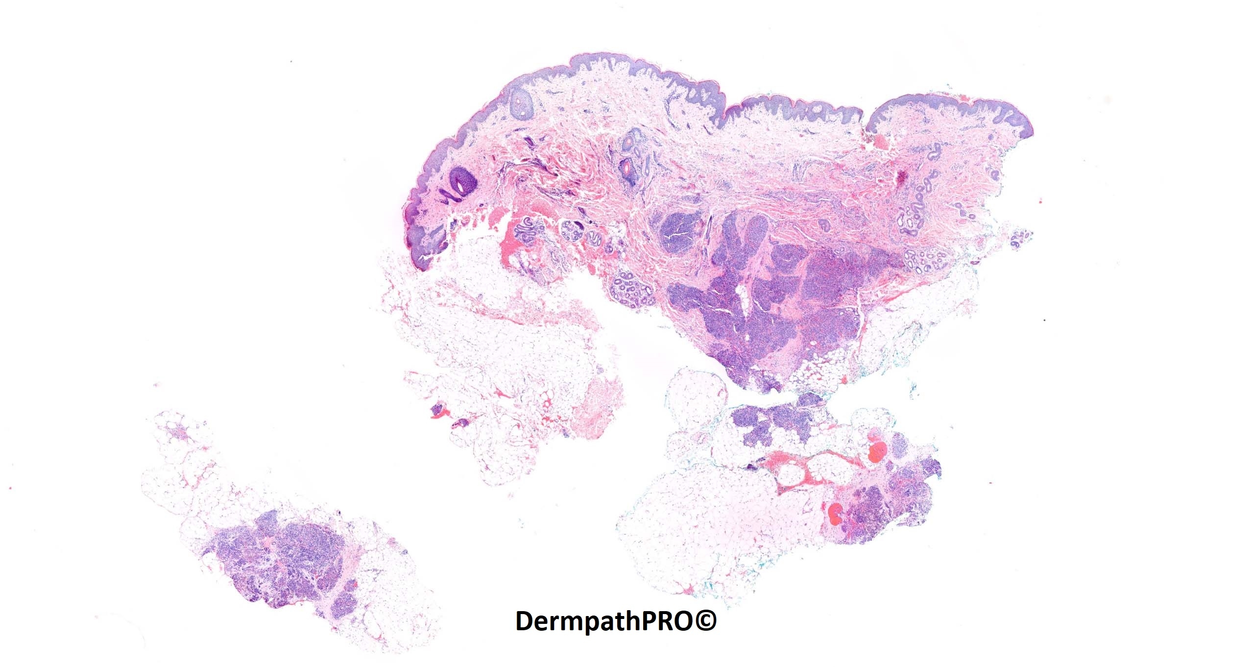
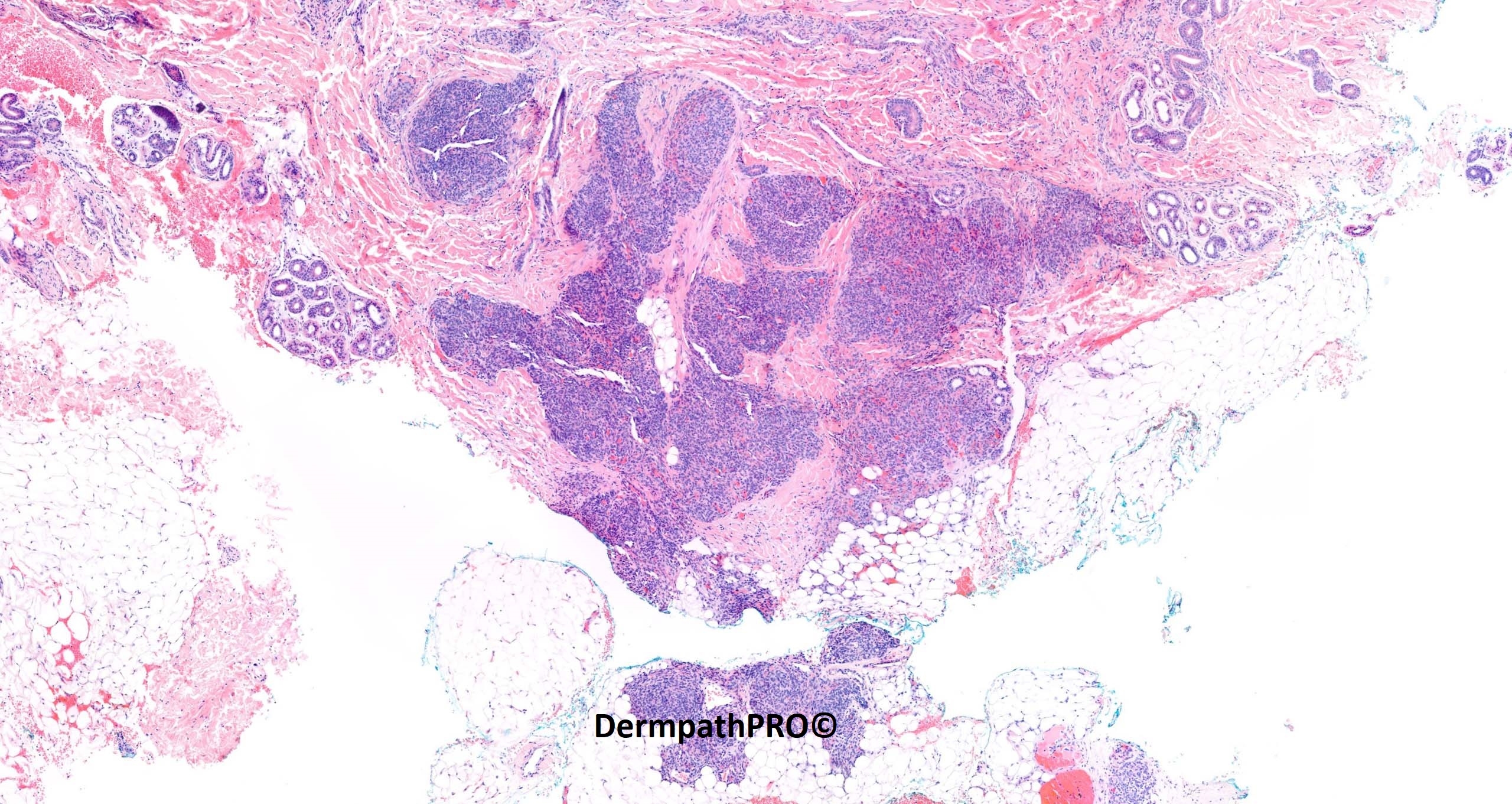
2-year-old boy: Small lump noted at birth on the left chest wall. Over recent months this has become more prominent and is now violaceous and tender.

  • Thanks 1
  Report Record

User Feedback


Eman El-Nabarawy

Posted (edited)

Tufted angioma developing Kasabach-Merrit syndrome (cosumptive coagulopathy). 

Edited by Eman El-Nabarawy

Share this comment


Link to comment
share_externally

How can I differentiate acquired tufted angioma from kaposiform hemangioendothelioma? This is always difficult for me. In this case favor KHE because of many areas with spindled and quasi-fascicular pattern. 

Share this comment


Link to comment
share_externally

Saleem Taibjee

Posted

Yes, very well done. This is a tufted angioma.

Paediatric vascular lesions are not my forte, but Dr Isabel Colmenero kindly reviewed this case and confirmed my impression. Tufted angioma and Kaposiform haemangioendothelioma are likely to be on a spectrum, hence the difficulty of firmly distinguishing in all cases. However, in my limited experience, the clinical scenario tends to be different, with KHE often presenting within the first few days of life/congenital, often very large and dramatic tumours with a high association with Kasabach-Merritt syndrome, compared to a more insidious clinical story with tufted angioma, and perhaps a low grade coagulopathy (Kasabach-Merritt less common).

Both may express lymphatic markers. The D2-40 in this case is shown below.

There has been some initial literature to suggest WT-1 may be helpful in distinguishing vascular tumours from malformations. However, this was negative in this case, and Isabel informs me that this marker is not very robust. Glut-1 was also negative, excluding infantile haemangioma.

BW Saleem

image.jpeg

Share this comment


Link to comment
share_externally


Join the conversation

You can post now and register later. If you have an account, sign in now to post with your account.

Guest
Add a comment...

×   Pasted as rich text.   Paste as plain text instead

  Only 75 emoji are allowed.

×   Your link has been automatically embedded.   Display as a link instead

×   Your previous content has been restored.   Clear editor

×   You cannot paste images directly. Upload or insert images from URL.

Loading...

×
×
  • Create New...