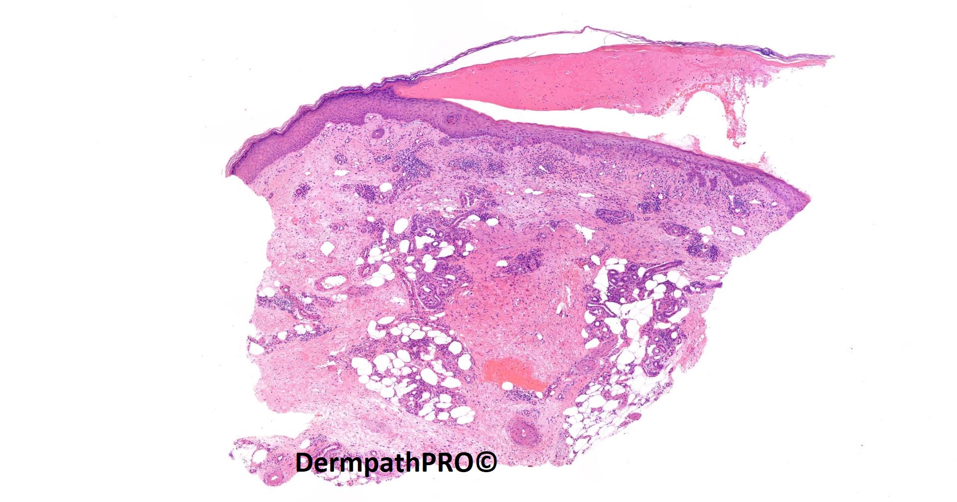
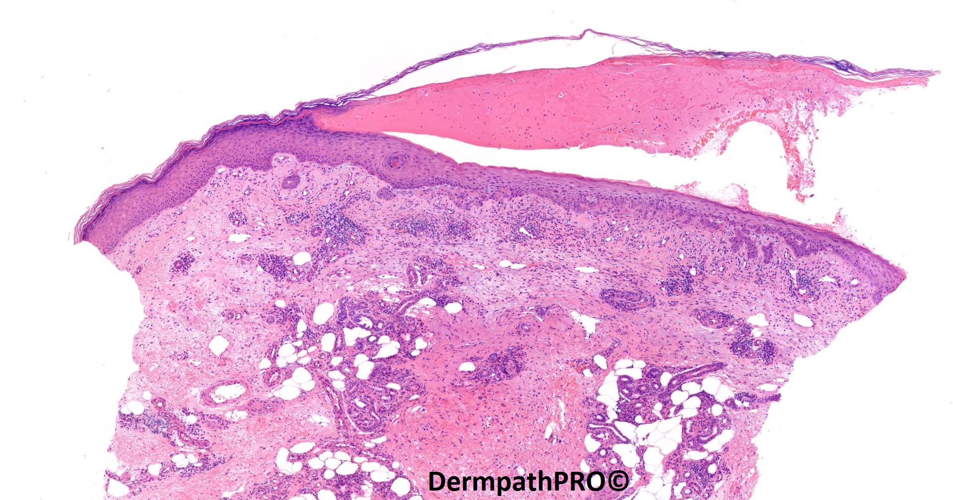
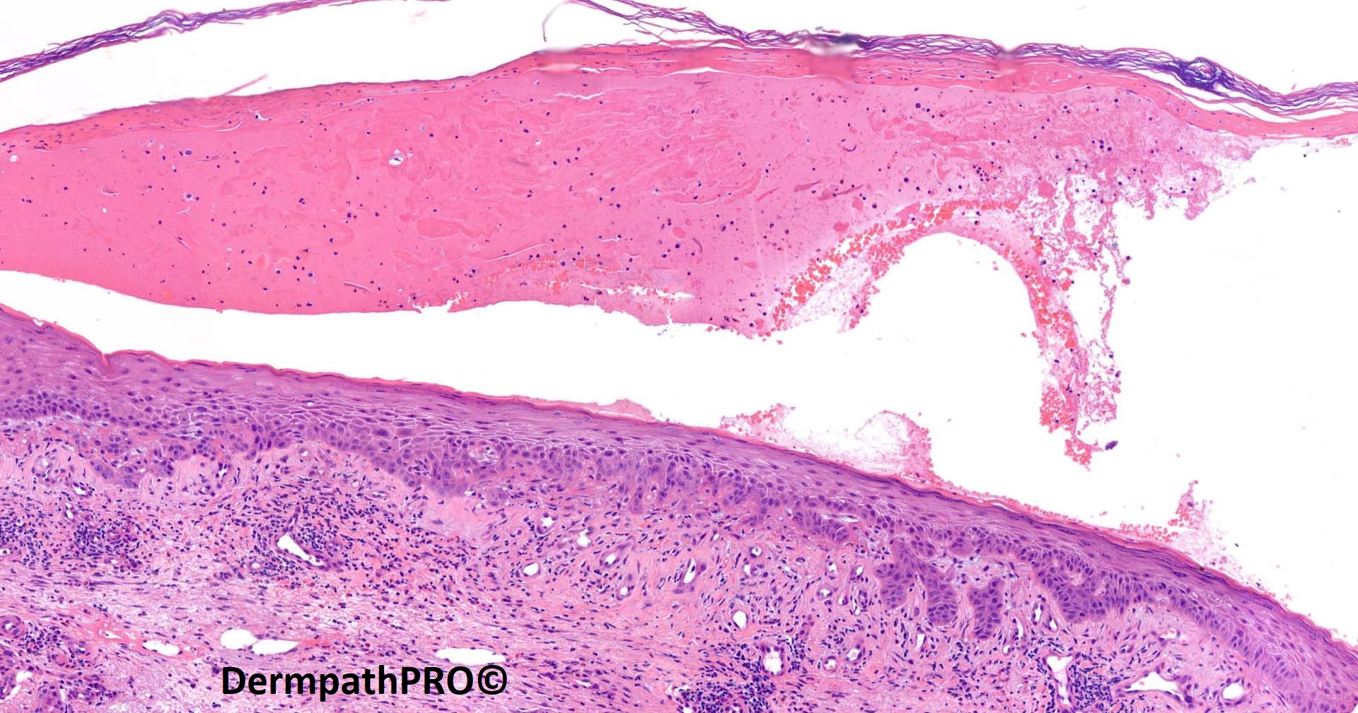
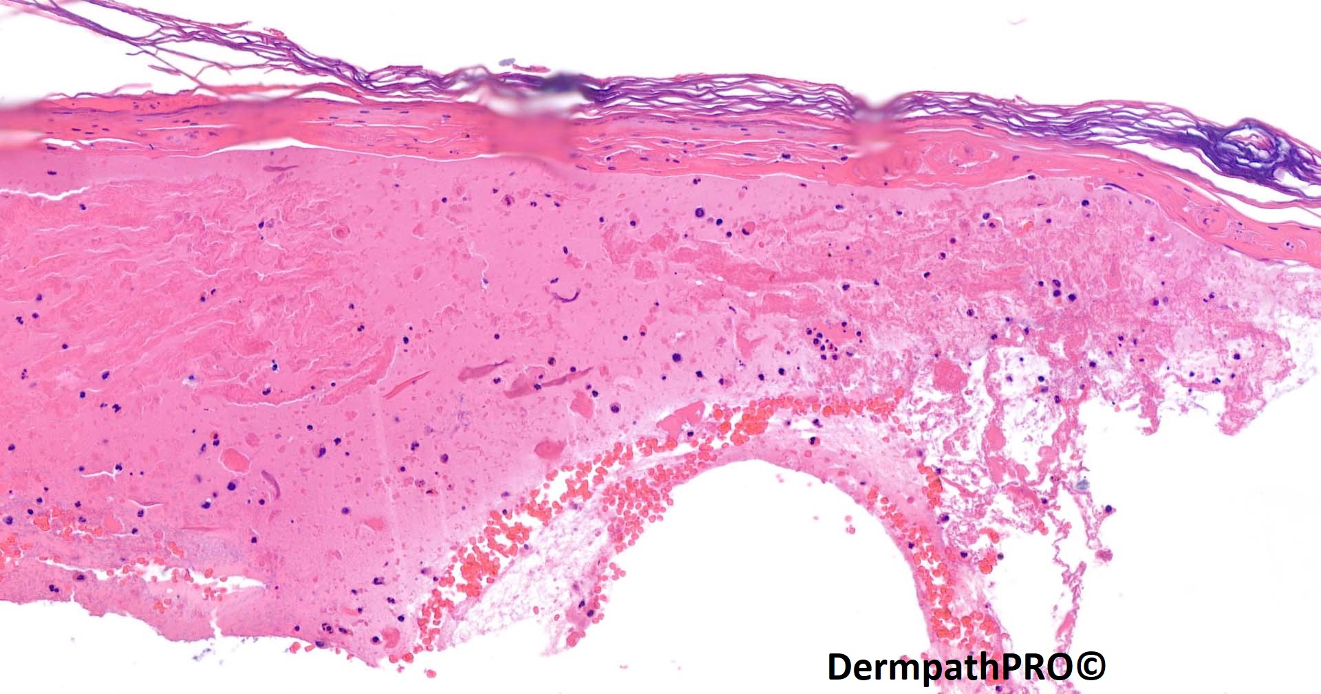
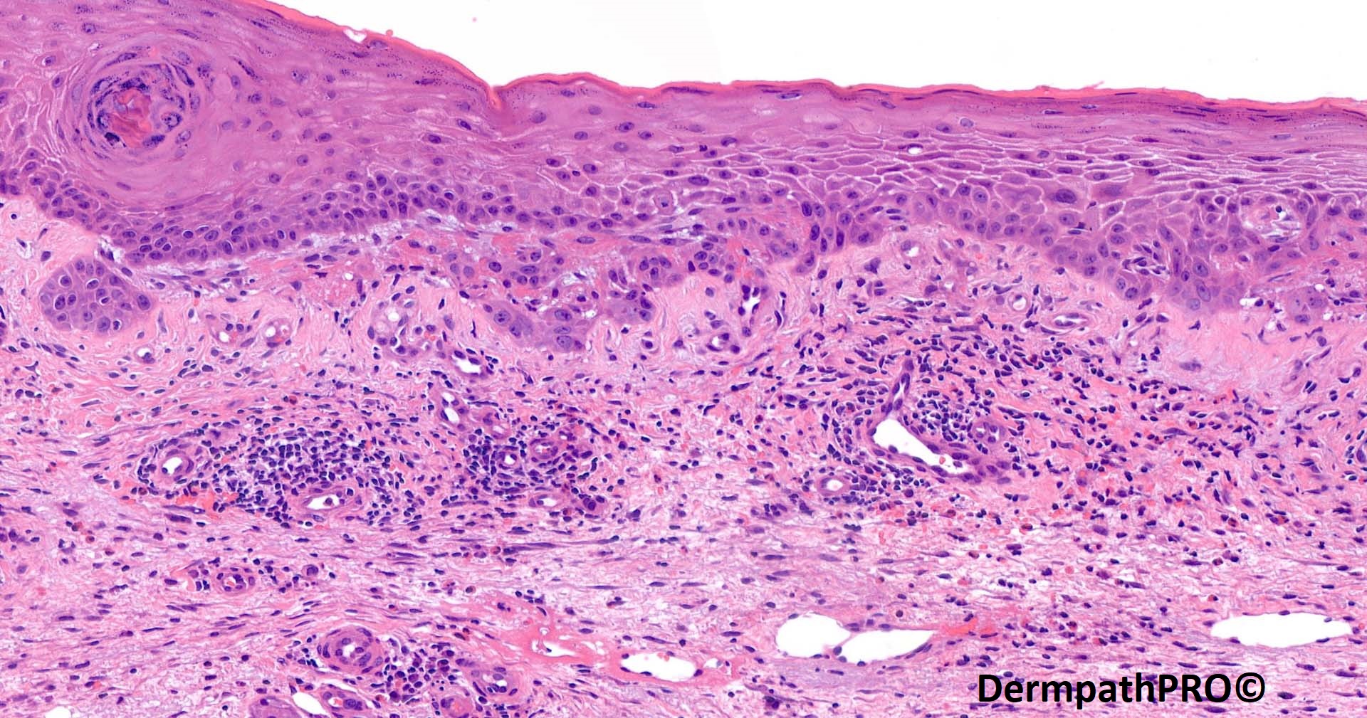
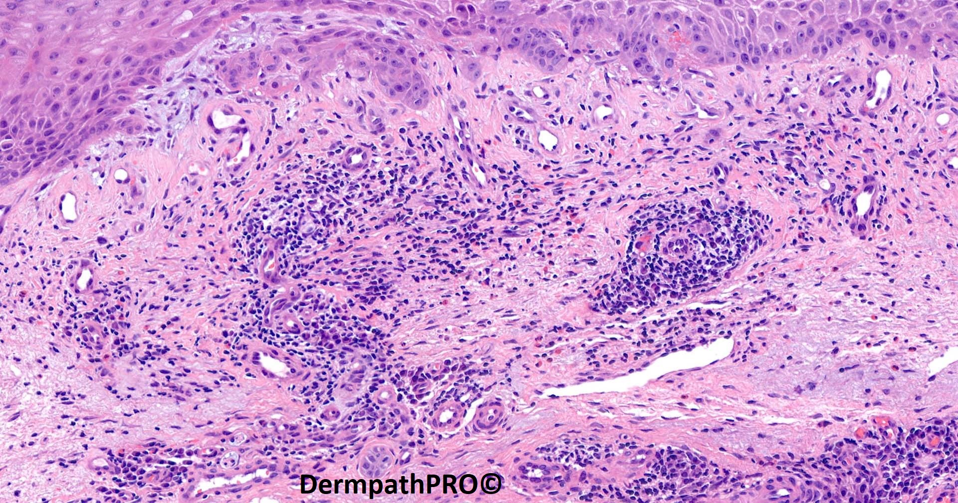
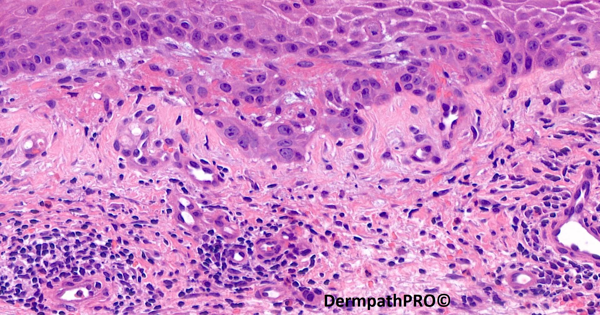
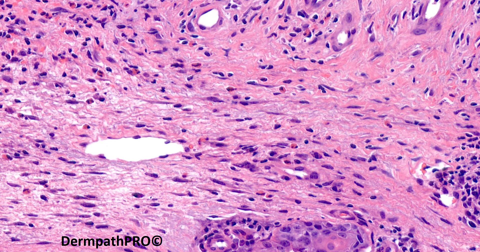
Jump to content
In this section we have spot diagnoses posted on a daily basis since June 2010, now over 4000! You can review the archived cases and read the suggested diagnoses by users and the final comment by the contributors.
Case are uploaded each week day by 10 am UK time with the correct diagnosis will generally be posted at 8 pm UK time. Why not view the most recent spot diagnosis and proffer a diagnosis?

Case Number : Case 4149 - 15 Dec 2022 Posted By: Saleem Taibjee

Please read the clinical history and view the images by clicking on them before you proffer your diagnosis.
Submitted Date :
   (0 reviews)

83F Large blisters to legs & torso last few weeks.


  • Thanks 1
  Report Record

User Feedback


Richard Logan

Posted

There is a large, sub-corneal split, but without the associated neutrophilic infiltrate that would be expected with bullous impetigo, fungal infection, or Sneddon-Wilkinson disease.  The inflammatory infiltrate is mixed, but eosinophils and plasma cells are visible.  I am wondering about pemphigus foliaceous or a drug-induced pemhigus-like problem.

Share this comment


Link to comment
share_externally

Any chance it might be bullous pemphigoid with re-epithelialization?

Share this comment


Link to comment
share_externally

The advanced age, history of large blisters and eosinophils in the infiltrate are suggestive of bullous pemphigoid with re epithelialization. Is the patient a diabetic on any of gliptin group of drugs ? DIF will be decisive. ELIZA for anti BP 180 and anti BP 230 is also available now.

Share this comment


Link to comment
share_externally

Richard Logan

Posted

20 hours ago, sfwenson said:

Any chance it might be bullous pemphigoid with re-epithelialization?

I did consider that possibility, but was put off by the amount of inflammation still present under the epithelium.  I would have thought things might be a bit quieter if this were re-epithelialisaiton.

Share this comment


Link to comment
share_externally

Why not an ischemic damage, like acroangiodermatitis, in a venous insufficiency?

Share this comment


Link to comment
share_externally

A subcorneal split with mixed inflammatory cells, including eosinophils, but lacking acantholytic floating keratinocytes. So I'm not favouring a pemphigus. Also 'large blisters to legs and torso' I think makes pemphigus less likely since these blisters easily rupture and are usually ulcerated/crusted.

I think there are reactive changes in the basal keratinocytes directly underneath the split- perhaps they are dividing to re-epitheliaise the epidermis. So I favour bullous pemphigoid with reepithelialisation.

Share this comment


Link to comment
share_externally

Saleem Taibjee

Posted

Yes, very well done. This is a recent case of bullous pemphigoid. Common things being common. The direct immunofluorescence was confirmatory.

This is an important pitfall - re-epithelialisation giving rise to the false impression of a more superficial level of blistering. I have seen such cases cause great confusion, especially when there is always a risk of false negative IMF.

It emphasises the importance of the clinician trying to select a fresh/new blister to biopsy.... ' the lives of lesions'.

Happy Christmas everyone!

Saleem

Share this comment


Link to comment
share_externally


Join the conversation

You can post now and register later. If you have an account, sign in now to post with your account.

Guest
Add a comment...

×   Pasted as rich text.   Paste as plain text instead

  Only 75 emoji are allowed.

×   Your link has been automatically embedded.   Display as a link instead

×   Your previous content has been restored.   Clear editor

×   You cannot paste images directly. Upload or insert images from URL.

Loading...

×
×
  • Create New...