Jump to content
In this section we have spot diagnoses posted on a daily basis since June 2010, now over 4000! You can review the archived cases and read the suggested diagnoses by users and the final comment by the contributors.
Case are uploaded each week day by 10 am UK time with the correct diagnosis will generally be posted at 8 pm UK time. Why not view the most recent spot diagnosis and proffer a diagnosis?

Case Number : Case 4156 - 26 December 2022 Posted By: Dr. Mona Abdel-Halim

Please read the clinical history and view the images by clicking on them before you proffer your diagnosis.
Submitted Date :
   (0 reviews)

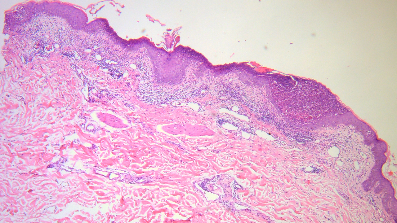
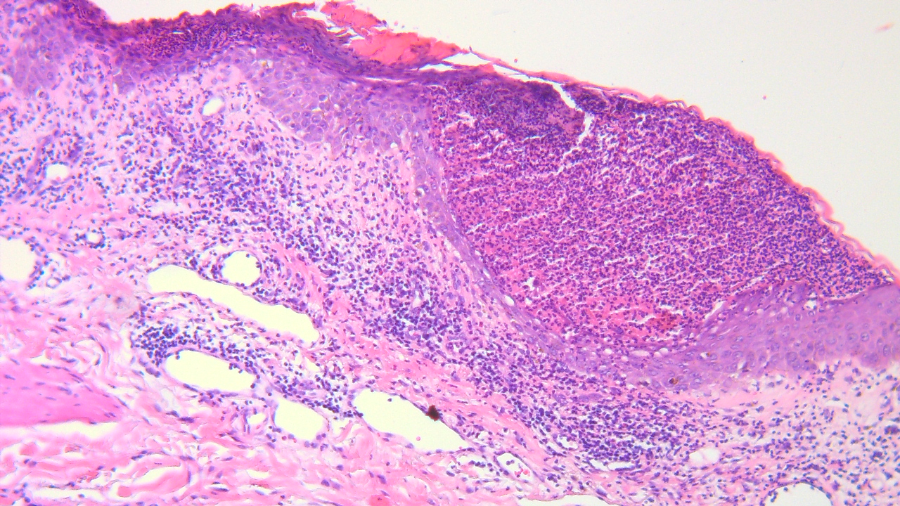
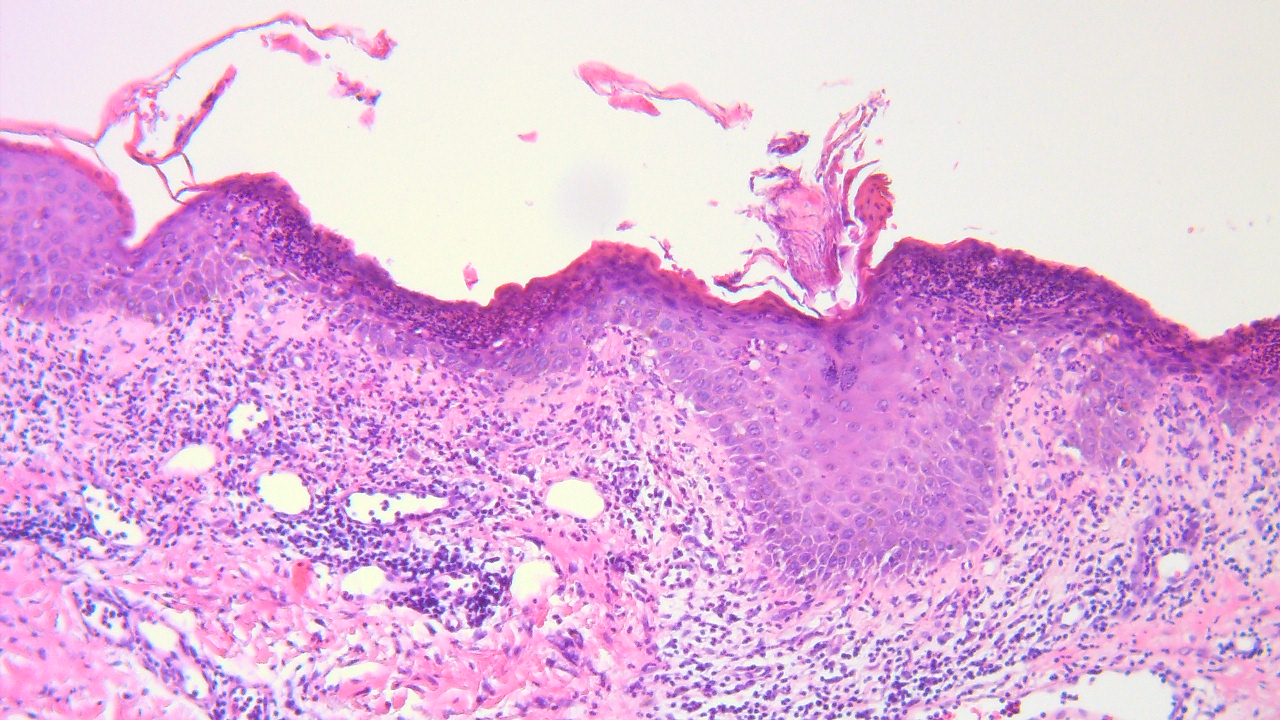
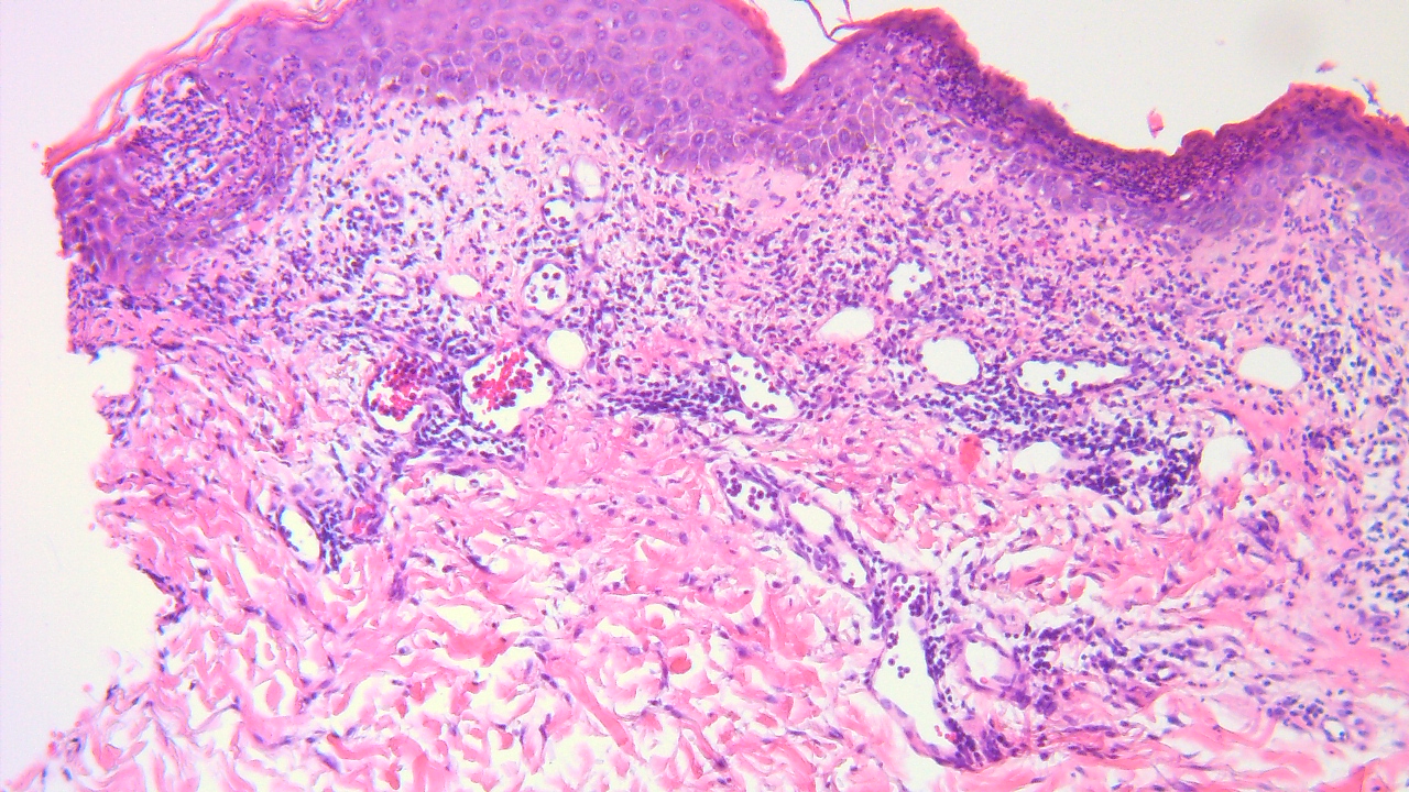
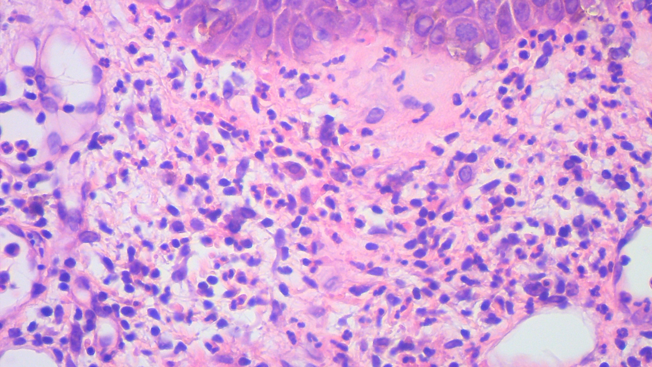
F 45 with SLE, Disseminated pustular rash


  Report Record

User Feedback


Subcorneal and intraepidermal pustules, and I think there are eosinophils in image 5 ~ probably AGEP. In this clinical setting, culprit drug could be hydroxychloroquine 
DDx includes pustular psoriasis, and others. 
PAS to rule out fungus (less likely).

Share this comment


Link to comment
share_externally

PS. Maybe there are some fungal elements in image 3?

Share this comment


Link to comment
share_externally

Favouring AGEP (subcorneal pustules, mixed inflammatory infiltrate in the superficial dermis including eosinophils and possible papillary dermal oedema). However also fungal hyphae seen in 3rd picture...AGEP with superimposed fungal infection?

Share this comment


Link to comment
share_externally

Anil Patki

Posted

Is the patient immunocompromised? Disseminated dermatophytosis is a possibility in relevant clinical situation. Fungal id is another differential.

Share this comment


Link to comment
share_externally

Dr. Mona Abdel-Halim

Posted

More photos should be provided soon

Share this comment


Link to comment
share_externally

Meenakshi Batrani

Posted

Tinea and AGEP are in differential. But I will also add IgA pemphigus which can have subcorneal / intraepidermal pustules while acantholysis may be inconspicuous. PAS and DIF will help. 

Share this comment


Link to comment
share_externally

Saman Fatah

Posted

I think the contributors almost listed various causes of subcorneal/high intraepidermal pustulation other than subcorneal pustular dermatosis of Sneddon and Wilkinson!
Knowledge of whether the pustule is sterile or not is crucial through swab results of an intact pustule, stains and tissue cultures especially in those immunocompromised. Then drug exposure and DIF results.

it will be interesting to see if the pustular dermatosis has a predilection for flexures in this lady with SLE, then Amicrobial Pustulosis of the Folds (APF) requires consideration after exclusion of more common causes of such pattern of pustulosis.

Share this comment


Link to comment
share_externally

On 02/01/2023 at 16:22, Eman El-Nabarawy said:

Fungal infection..

+1. 

Share this comment


Link to comment
share_externally

Dr. Mona Abdel-Halim

Posted

Disseminated dermatophyte infection (Tinea incognito)

Share this comment


Link to comment
share_externally


Join the conversation

You can post now and register later. If you have an account, sign in now to post with your account.

Guest
Add a comment...

×   Pasted as rich text.   Paste as plain text instead

  Only 75 emoji are allowed.

×   Your link has been automatically embedded.   Display as a link instead

×   Your previous content has been restored.   Clear editor

×   You cannot paste images directly. Upload or insert images from URL.

Loading...

×
×
  • Create New...